Towards direct neuronal current imaging via ultra-low-field MR
Pith reviewed 2026-05-25 15:17 UTC · model grok-4.3
The pith
Ultra-low-field MR needs at least double its current SNR to detect neuronal magnetic fields directly.
A machine-rendered reading of the paper's core claim, the machinery that carries it, and where it could break.
Core claim
Phantom measurements simulating long-lasting neuronal activity in the secondary somatosensory cortex demonstrate that the SNR of the ULF MR setup must be increased by at least a factor of 2 to resolve the influence of neuronal magnetic fields on 1H nuclear spin precession.
What carries the argument
The ULF MR measurement setup that senses the faint perturbation of 1H nuclear spin precession caused by simulated neuronal magnetic fields.
Load-bearing premise
The phantom and the chosen long-lasting neuronal activity pattern accurately represent the amplitude, duration, and spatial distribution of real neuronal magnetic fields that would be encountered in vivo.
What would settle it
A direct measurement of the magnetic field strength and time course produced by actual neuronal activity in the secondary somatosensory cortex, compared against the values used in the phantom.
Figures
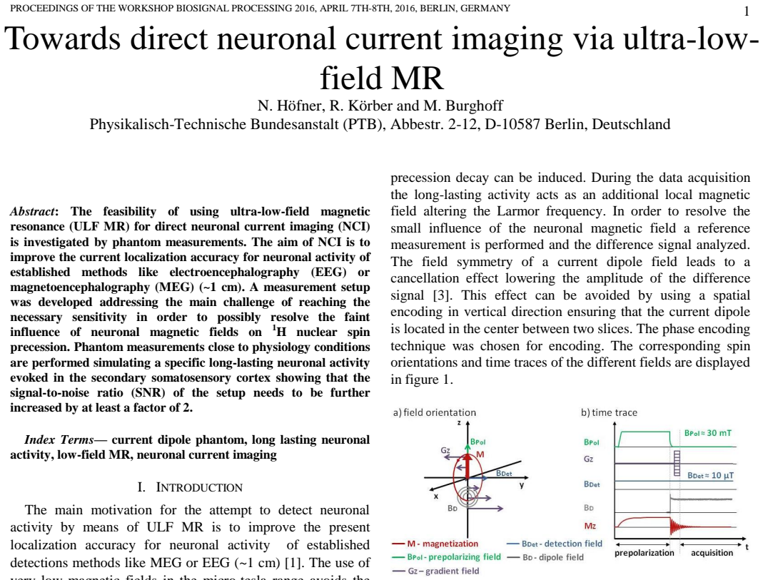

read the original abstract
The feasibility of using ultra-low-field magnetic resonance (ULF MR) for direct neuronal current imaging (NCI) is investigated by phantom measurements. The aim of NCI is to improve the current localization accuracy for neuronal activity of established methods like electroencephalography (EEG) or magnetoencephalography (MEG) (~1 cm). A measurement setup was developed addressing the main challenge of reaching the necessary sensitivity in order to possibly resolve the faint influence of neuronal magnetic fields on 1H nuclear spin precession. Phantom measurements close to physiology conditions are performed simulating a specific long-lasting neuronal activity evoked in the secondary somatosensory cortex showing that the signal-to-noise ratio (SNR) of the setup needs to be further increased by at least a factor of 2.
Editorial analysis
A structured set of objections, weighed in public.
Referee Report
Summary. The paper investigates the feasibility of direct neuronal current imaging (NCI) with ultra-low-field MR by developing a sensitive measurement setup and performing phantom experiments that simulate long-lasting neuronal activity evoked in the secondary somatosensory cortex. The central empirical result is that the SNR of the ULF MR setup must be increased by at least a factor of 2 to resolve the influence of neuronal magnetic fields on 1H precession under the tested conditions.
Significance. If the result holds, the work supplies a practical, phantom-based benchmark for the sensitivity gap that must be closed before ULF MR can be used for NCI, a modality that could in principle improve spatial localization relative to EEG/MEG. The empirical character of the measurement (rather than a purely theoretical scaling argument) is a positive feature.
major comments (2)
- [Abstract] Abstract: the headline claim that SNR must be increased by a factor of at least 2 is presented without error bars, raw time-series, or an explicit statement of how the neuronal-field amplitude was scaled into the phantom currents. Because this factor is the load-bearing quantitative result, the absence of these details prevents assessment of its precision and reproducibility.
- [Phantom measurements] Phantom measurements (abstract): the experiments employ a specific long-lasting activity pattern in S2. No quantitative comparison is supplied to the ~nT-scale, millisecond-scale dipolar fields measured by MEG for typical transient neuronal currents, so it is unclear whether the inferred SNR requirement generalizes to the in-vivo NCI use case the paper aims to address.
minor comments (1)
- [Abstract] Abstract: a short clause indicating the number of repeated measurements or the statistical procedure used to arrive at the factor-of-2 figure would improve clarity.
Simulated Author's Rebuttal
We thank the referee for the constructive feedback on our manuscript investigating the feasibility of ULF MR for direct neuronal current imaging. We address each major comment below, indicating where revisions will be incorporated.
read point-by-point responses
-
Referee: [Abstract] Abstract: the headline claim that SNR must be increased by a factor of at least 2 is presented without error bars, raw time-series, or an explicit statement of how the neuronal-field amplitude was scaled into the phantom currents. Because this factor is the load-bearing quantitative result, the absence of these details prevents assessment of its precision and reproducibility.
Authors: The abstract is length-constrained, but the manuscript provides the explicit scaling procedure from neuronal-field amplitudes to phantom currents in the Methods section, along with raw time-series and analysis in the Results. The factor of 2 is obtained from the threshold at which the induced phase accumulation exceeds the noise floor under the tested conditions. We agree that a brief qualifier referencing these details would improve the abstract and will revise it to include such a statement along with mention of the supporting figures. revision: yes
-
Referee: [Phantom measurements] Phantom measurements (abstract): the experiments employ a specific long-lasting activity pattern in S2. No quantitative comparison is supplied to the ~nT-scale, millisecond-scale dipolar fields measured by MEG for typical transient neuronal currents, so it is unclear whether the inferred SNR requirement generalizes to the in-vivo NCI use case the paper aims to address.
Authors: The experiments target long-lasting activity patterns in S2 because these produce sustained fields amenable to the phantom setup under near-physiological conditions, providing a practical benchmark for that regime. The paper does not claim the factor-of-2 requirement applies directly to transient dipolar fields; instead it focuses on the long-lasting case as a first feasibility step. We will add a short discussion paragraph contrasting the field timescales and amplitudes with typical MEG transients and noting that integration time differences would alter the SNR needs for the transient case. revision: partial
Circularity Check
No circularity: empirical phantom study with direct measurements
full rationale
The paper reports physical phantom measurements that simulate a long-lasting neuronal activity pattern and directly measure the resulting SNR limitation on 1H precession. No equations, fitted parameters, or self-citations are invoked to derive the factor-of-2 SNR requirement; the result is an experimental observation. The central claim therefore does not reduce to its inputs by construction and contains independent empirical content.
Axiom & Free-Parameter Ledger
axioms (1)
- domain assumption The magnetic field produced by the chosen long-lasting neuronal activity pattern can be faithfully reproduced by the phantom hardware.
Reference graph
Works this paper leans on
-
[1]
C. Babiloni, V. Pizzella, C.D. Gratta, A. Ferretti, G.L. Romani, Fundamentals of Electroencefalography, Magnetoencefalography, and Functional Magnetic Resonance Imaging, International Review of Neurobiology 86, 67–80, 2009
work page 2009
-
[2]
R.H. Kraus Jr., P. Volegov, A. Matlachov, M. Espy, Towards direct neural current imaging by resonant mechanism at ultra-low field, Neuroimage 39, 310–317, 2008
work page 2008
-
[3]
R. Körber, J.O. Nieminen, N. Höfner, V. Jazbinšek. H.-J. Scheer, K. Kim, M. Burghoff, An advanced phantom study assessing the feasibility of neuronal current imaging by ultra-low-field NMR, Journal of Magnetic Resonance 237, 182-190, 2013
work page 2013
-
[4]
V.S. Zotev, A.N. Matlashov, I.M. Savukov, T. Owens, P.L. Volegov J.J. Gomez, M.A. Espy, SQUID-Based Microtesla MRI for In Vivo Relaxometry of the Human Brain. TASC 19(3), 823-6, 2009
work page 2009
-
[5]
M. Burghoff, H.H. Albrecht, S. Hartwig, R. Körber, T.S. Thömmes, H.J. Scheer, J. Voigt, L. Trahms, Squid system for meg and low field magnetic resonance. Metrol. Meas. Syst. XVI, 371-5, 2009
work page 2009
discussion (0)
Sign in with ORCID, Apple, or X to comment. Anyone can read and Pith papers without signing in.