Structure and dynamics of dynorphin peptide and its receptor
Pith reviewed 2026-05-25 13:48 UTC · model grok-4.3
The pith
X-ray, EM and NMR data together map the dynamic binding of dynorphin to the kappa opioid receptor.
A machine-rendered reading of the paper's core claim, the machinery that carries it, and where it could break.
Core claim
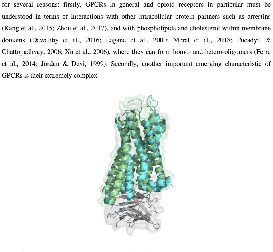
X-ray and EM structures of the four opioid receptors in inactive states, together with active-state structures of the mu and kappa receptors, are integrated with NMR restraints on dynorphin and receptor flexibility. This combination supplies complementary information on the conformational changes that accompany binding and activation at the kappa opioid receptor.
What carries the argument
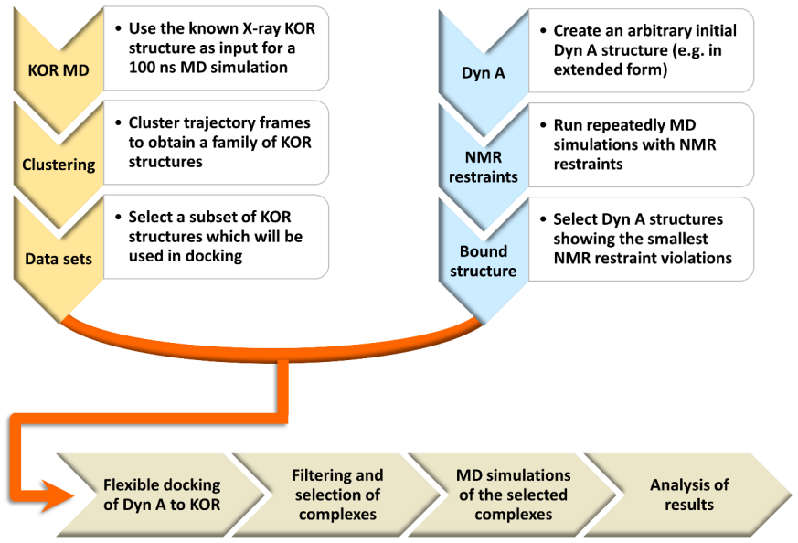
The dynorphin-KOP receptor complex constructed by docking the peptide into available crystal structures under NMR-derived distance and dynamics restraints.
If this is right
- Molecular models of the complex now incorporate both static atomic positions and measured dynamics of the peptide and receptor.
- These models guide the design of ligands that stabilize or destabilize specific conformational states during activation.
- The approach demonstrates how NMR data can refine static crystal structures to explain activation mechanisms in other peptide-activated receptors.
Where Pith is reading between the lines
- The same integration of crystallography with NMR could be applied to other G-protein-coupled receptors that bind short peptides.
- Ligands developed from these dynamics-based models might show reduced side effects if they avoid certain receptor conformations linked to addiction pathways.
- Time-resolved NMR or cryo-EM experiments on the same system would directly test whether the modeled intermediate states occur in solution.
Load-bearing premise
The published X-ray and EM structures of the receptors capture the actual binding orientation and the conformational shifts that dynorphin induces in living cells.
What would settle it
A high-resolution experimental structure of dynorphin bound to KOP that shows a peptide pose or receptor conformation inconsistent with the NMR-restrained models.
Figures

read the original abstract
Dynorphin is a neuropeptide involved in pain, addiction and mood regulation. It exerts its activity by binding to the kappa opioid receptor (KOP) which belongs to the large family of G-protein coupled receptors. The dynorphin peptide was discovered in 1975, while its receptor was cloned in 1993. This review will describe: a) the activities and physiological functions of dynorphin and its receptor, b) early structure-activity relationship studies performed before cloning of the receptor (mostly pharmacological and biophysical studies of peptide analogues), c) structure-activity relationship studies performed after cloning of the receptor via receptor mutagenesis and the development of recombinant receptor expression systems, d) structural biology of the opiate receptors culminating in X-ray structures of the four opioid receptors in their inactive state and structures of MOP and KOP receptors in their active state. X-ray and EM structures are combined with NMR data, which gives complementary insight into receptor and peptide dynamics. Molecular modelling greatly benefited from the availability of atomic resolution 3D structures of receptor-ligand complexes and an example of the strategy used to model a dynorphin-KOP receptor complex using NMR data will be described. These achievements have led to a better understanding of the complex dynamics of KOP receptor activation and to the development of new ligands and drugs.
Editorial analysis
A structured set of objections, weighed in public.
Referee Report
Summary. This review summarizes the discovery and functions of dynorphin and the kappa opioid receptor (KOP), pre- and post-cloning structure-activity relationship studies (pharmacological, biophysical, and mutagenesis), the structural biology of opioid receptors (X-ray structures of inactive states for all four receptors and active states for MOP and KOP), integration of these with NMR data on dynamics, an example of NMR-informed molecular modeling of the dynorphin-KOP complex, and the resulting advances in understanding KOP activation dynamics and ligand development.
Significance. As a literature synthesis, the review usefully collates complementary X-ray/EM and NMR insights on receptor-peptide dynamics for the GPCR and opioid fields. It correctly timelines key milestones (dynorphin 1975, KOP cloning 1993) and notes how atomic structures have enabled modeling. No original data or derivations are presented, so significance rests on the accuracy and balance of the cited summaries rather than novel claims.
minor comments (2)
- [Abstract] Abstract, final paragraph: the phrasing 'these achievements have led to ... the development of new ligands and drugs' is a field-level observation; adding one or two concrete post-structure ligand examples (with citations) would make the causal link more traceable without altering the review's scope.
- [Modeling section] The description of the dynorphin-KOP modeling strategy (mentioned in the abstract) would be clearer if the key NMR restraints or distance constraints used were listed explicitly rather than summarized at a high level.
Simulated Author's Rebuttal
We thank the referee for their positive assessment of the manuscript, accurate summary of its scope, and recommendation to accept. We appreciate the recognition that the review usefully collates X-ray/EM and NMR insights on receptor-peptide dynamics.
Circularity Check
Review article with no original derivations or models
full rationale
This is a literature review summarizing prior X-ray/EM structures, NMR data, SAR studies, and modeling strategies from the field. No equations, quantitative predictions, fitted parameters, or novel derivations are presented that could reduce to the paper's own inputs by construction. All statements are descriptive citations of external work, so the content is self-contained against external benchmarks with no load-bearing self-citation chains or self-definitional steps.
Axiom & Free-Parameter Ledger
Reference graph
Works this paper leans on
-
[1]
doi:10.1185/03007995.2010.545379 Al-Hasani, R., & Bruchas, M. R. (2011). Molecular Mechanisms of Opioid Receptor -Dependent Signaling and Behavior. Anesthesiology, 115, 1363–1381. Aldrich, J. V. , & McLaughlin, J. P. (2009). Peptide kappa opioid receptor ligands: potential for drug development. The AAPS journal, 11(2), 312-322. doi:10.1208/s12248-009-9105...
-
[2]
doi:10.1021/jm100240h Casiraghi, M., Damian, M., Lescop, E., Point, E., Moncoq, K., Morellet, N., . . . Catoire, L. J. (2016). Functional Modulation of a G Protein-Coupled Receptor Conformational Landscape in a Lipid Bilayer. Journal of the American Chemical Society, 138 (35), 11170 -11175. doi:10.1021/jacs.6b04432 Chavkin, C. (2013). Dynorphin –Still an ...
-
[3]
A model for rec eptor activation
binding to extracellular loop 2 of the kappa -opioid receptor. A model for rec eptor activation. Journal of medicinal chemistry, 40 20, 3254-3262. Patra, M. C., Kumar, K., Pasha, S., & Chopra, M. (2012). Comparative modeling of human kappa opioid receptor and docking analysis with the peptide YFa. Journal of Molecular Graphics and Modelling, 33, 44 - 51. ...
-
[4]
doi:10.1016/j.pnpbp.2005.08.011 Rosenbaum, D. M., Cherezov, V., Hanson, M. A., Rasmussen, S. G. F., Thian, F. S., Kobilka, T. S., . . . Kobilka, B. K. (2007). GPCR engineering yields high -resolution structural insights into beta(2)-adrenergic receptor function. Science, 318 (5854), 1266 -1273. doi:10.1126/science.1150609 Rosenbaum, D. M., Rasmussen, S. G...
discussion (0)
Sign in with ORCID, Apple, or X to comment. Anyone can read and Pith papers without signing in.