Technical Report: Time-Activity-Curve Integration in Lu-177 Therapies in Nuclear Medicine
Pith reviewed 2026-05-24 20:52 UTC · model grok-4.3
The pith
Particle filtering reduces noise in voxel time-activity curves for Lu-177 dosimetry.
A machine-rendered reading of the paper's core claim, the machinery that carries it, and where it could break.
Core claim
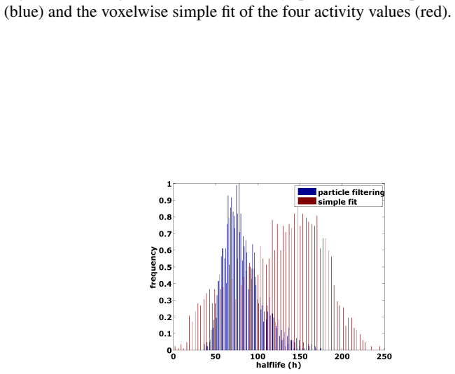
When a particle filter based on a mono-exponential decay model is applied to voxel-wise time-activity curves from four serial SPECT acquisitions, the resulting time-integrated activities produce half-life distributions closer to organ averages and contain fewer implausibly long values than those from independent mono-exponential fits to each voxel.
What carries the argument
Particle filter that regularizes each voxel's time-activity curve using a mono-exponential decay evolution model
Load-bearing premise
The time-activity curves are assumed to follow a mono-exponential decay, which is used both as the particle filter's evolution model and as the comparison method.
What would settle it
Obtain or simulate SPECT data with known true time-activity curves per voxel and measure whether the particle filter recovers the true half-lives more accurately than direct fitting.
Figures
read the original abstract
Currently, there is a high interest in Lu-177 targeted radionuclide therapies, which could be attributed to favourable results obtained from Lu-177 compounds targeting neuroendocrine and prostate tumours. Generally, it has been recognized that a transition from dosimetry based on planar images towards based on fully-quantitative SPECT is beneficial in terms of increased accuracy. SPECT based dosimetry could not only be used for achieving accurate absorbed dose per-organ, but even for deriving dose values for individual voxels. However, a voxel-wise determination of TACs is problematic since several confounding factors exist, such as e.g. poor count-statistics or registration inaccuracies. A particle filter (PF) is a class of methods which applies regularization based on a model of a state's evolution over time. We applied PFs for de-noising the TACs of 26 patients, who underwent Lu-177-DOTATOC or -PSMA therapy. The TACs were obtained from four serial SPECT(/CT) data. The model used in the PF was a mono-exponential decay. The time-integrated activities (TIA) resulting from the PF were compared to the results of a monoexponential fit of the individual voxels in several volumes of interest. Optimal values for noise of observations and noise of the model were 0.25 and 0.5, respectively. The distribution of voxel-wise halflives resulting from the PFFit method were considerably closer to the organ average value and the number of implausibly long halflives was reduced. However, one has to admit that voxel-wise fitting generally lead to considerable deviations from the organ-average TIA as obtained by conventional whole-organ evaluation. Unfortunately, we did not have ground-truth TIA of our patient data and proper ground-truth could even be impossible to obtain. Nevertheless, there are strong indicators that Particle Filtering can be used for reducing voxel-wise TAC noise.
Editorial analysis
A structured set of objections, weighed in public.
Referee Report
Summary. The manuscript proposes applying particle filters (PF) with a mono-exponential decay evolution model to denoise voxel-wise time-activity curves (TACs) extracted from four serial SPECT/CT scans in 26 patients receiving Lu-177-DOTATOC or -PSMA therapy. Noise parameters are optimized to 0.25 (observations) and 0.5 (model); the resulting half-life distributions and time-integrated activities (TIAs) are compared to direct mono-exponential voxel fits, with the PF reported to yield values closer to organ averages and fewer implausibly long half-lives. The authors acknowledge the absence of ground-truth TIA values.
Significance. If the denoising effect can be confirmed independently, the approach would address a practical barrier to reliable voxel-level dosimetry in Lu-177 therapies, where poor count statistics and registration errors degrade direct TAC fitting. The work correctly identifies the regularization potential of state-evolution models but currently provides only indirect distributional evidence.
major comments (2)
- [Abstract (parameter optimization and evaluation)] The noise parameters (0.25 observation, 0.5 model) were selected to optimize closeness of voxel half-lives to organ averages on the identical 26-patient cohort used for all reported comparisons (abstract). Because the PF and the baseline comparator both enforce the same mono-exponential model, this procedure risks circularity: the metric used for optimization is also the primary evidence of improvement.
- [Abstract (results and discussion)] The central claim that PF reduces voxel-wise TAC noise rests on distributional shifts (closer to organ-average half-lives, fewer long half-lives) without any ground-truth TIA values, as the authors explicitly state. This leaves open whether the observed changes reflect genuine noise suppression or simply stronger enforcement of the shared mono-exponential assumption relative to unconstrained voxel fits.
minor comments (2)
- [Abstract] The term 'PFFit method' appears without prior definition; clarify whether it denotes the particle-filtered fit or another procedure.
- The manuscript would benefit from explicit reporting of the number of particles, resampling strategy, and any implementation details of the PF that are not already covered in the full text.
Simulated Author's Rebuttal
We thank the referee for the constructive and detailed comments. We address each major comment below and indicate where revisions will be made to the manuscript.
read point-by-point responses
-
Referee: [Abstract (parameter optimization and evaluation)] The noise parameters (0.25 observation, 0.5 model) were selected to optimize closeness of voxel half-lives to organ averages on the identical 26-patient cohort used for all reported comparisons (abstract). Because the PF and the baseline comparator both enforce the same mono-exponential model, this procedure risks circularity: the metric used for optimization is also the primary evidence of improvement.
Authors: The referee correctly identifies a limitation in our parameter selection process. The noise parameters were indeed tuned on the same 26-patient dataset to minimize deviation from organ-level averages. This choice was made because organ averages from conventional dosimetry provide an independent reference point, separate from voxel-wise fitting. However, we acknowledge the risk of circularity in using the same data for both optimization and evaluation. In the revised manuscript, we will explicitly state this limitation and recommend that future studies validate the parameters on an independent cohort. The primary comparison remains between the PF approach and direct mono-exponential fitting, both applied to the same TACs, showing reduced outliers with PF. revision: partial
-
Referee: [Abstract (results and discussion)] The central claim that PF reduces voxel-wise TAC noise rests on distributional shifts (closer to organ-average half-lives, fewer long half-lives) without any ground-truth TIA values, as the authors explicitly state. This leaves open whether the observed changes reflect genuine noise suppression or simply stronger enforcement of the shared mono-exponential assumption relative to unconstrained voxel fits.
Authors: We agree that the evidence presented is indirect, as we have already noted in the manuscript that ground-truth voxel TIA values are unavailable. The observed shifts in half-life distributions and reduction in implausible values provide supportive but not definitive evidence of denoising. It is possible that part of the effect comes from the PF's stronger incorporation of the mono-exponential model through its state evolution. We will revise the discussion section to more explicitly discuss this ambiguity and the reliance on distributional evidence rather than direct validation. revision: yes
Circularity Check
Moderate circularity from fitting noise parameters to the evaluation metric used for the noise-reduction claim
specific steps
-
fitted input called prediction
[Abstract]
"Optimal values for noise of observations and noise of the model were 0.25 and 0.5, respectively. The distribution of voxel-wise halflives resulting from the PFFit method were considerably closer to the organ average value and the number of implausibly long halflives was reduced."
The noise parameters were selected as optimal on the patient cohort (using closeness to organ-average halflives as the optimization target); the reported improvement in halflife distribution is therefore partly enforced by construction of the parameter choice rather than emerging as an independent validation of the PF's denoising effect.
full rationale
The paper selects observation/model noise parameters (0.25/0.5) as optimal on the patient cohort and then reports that the PF (mono-exponential evolution) produces halflife distributions closer to organ averages than direct mono-exponential voxel fits. Because the same mono-exponential model is used both inside the PF and as the comparator, and because parameters were tuned to improve exactly the reported closeness metric, the 'strong indicators' of denoising reduce in part to the fitting process rather than an independent test. The paper itself notes the absence of ground-truth TIA, so the distributional comparison supplies only partial independent content. This matches the fitted-input-called-prediction pattern at moderate severity; no self-citation or uniqueness claims are involved.
Axiom & Free-Parameter Ledger
free parameters (2)
- noise of observations =
0.25
- noise of the model =
0.5
axioms (1)
- domain assumption Time-activity curves follow a mono-exponential decay
Reference graph
Works this paper leans on
-
[1]
R. Douc and O. Cappe. Comparison of resampling schemes for particle filtering. pages 64 – 69, 2005
work page 2005
- [2]
-
[3]
A. Eberle. Markoc Processes, 2017
work page 2017
-
[4]
G.S. Fishman. Monte Carlo - Concepts, Algorithms and Applications . Springer, 1996
work page 1996
-
[5]
N.J. Gordon, D.J. Salmond, and A.F.M. Smith. Novel approach to nonlinear/non-gaussianbayesian state estimation. Radar and Signal Process- ing, IEEE Proceedings, 140(2):107–113, 1993
work page 1993
-
[6]
E. Grassi, F. Fioroni, V . Ferri, E. Mezzenga, M.A. Sarti, T. Paulus, N. Lan- conelli, A. Filice, A. Versari, and M. Iori. Quantitative comparison between the commercial software stratos R⃝ by philips and a homemade software for voxel-dosimetry in radiopeptide therapy. Physica Medica: European Jour- nal of Medical Physics, 31(1):72–79, 2015
work page 2015
-
[7]
Heribert Hänscheid, Constantin Lapa, Andreas K Buck, Michael Lassmann, and Rudolf A Werner. Dose mapping after endoradiotherapy with 177lu- dotatate/dotatoc by a single measurement after 4 days. Journal of nuclear medicine: official publication, Society of Nuclear Medicine , 59(1):75–81, 2018
work page 2018
-
[8]
Michael S Hofman, John Violet, Rodney J Hicks, Justin Ferdinandus, Sue Ping Thang, Tim Akhurst, Amir Iravani, Grace Kong, Aravind Ravi Kumar, Declan G Murphy, Peter Eu, Price Jackson, Mark Scalzo, Scott G Williams, and Shahneen Sandhu. [177lu]-psma-617 radionuclide treatment in patients with metastatic castration-resistant prostate cancer (lupsma trial): ...
work page 2018
-
[9]
M. Isard and A. Blake. CONDENSATION– conditional density propagation of visual tracking. International Journal of Computer Vision , 29(1):5–28, 2001
work page 2001
-
[10]
P. Jackson, J.M. Beauregard, M.S. Hofman, T. Kron, A. Hogg, and R.J. Hicks. An automated voxelized dosimetry tool for radionuclide therapy based on serial quantitative spect/ct imaging.Medical physics, 40(11), 2013
work page 2013
-
[11]
R.E. Kalman. A new approach to linear filtering and prediction problems. J. Basic Engineering, pages 35–45, 1960
work page 1960
-
[12]
Lutetium-labelled pep- tides for therapy of neuroendocrine tumours
BLR Kam, JJM Teunissen, Eric P Krenning, Wouter W de Herder, Saima Khan, EI Van Vliet, and Dirk Jan Kwekkeboom. Lutetium-labelled pep- tides for therapy of neuroendocrine tumours. European journal of nuclear medicine and molecular imaging, 39(1):103–112, 2012
work page 2012
-
[13]
K. Kanazawa, D. Koller, and S. Russell. Stochastic simulation algorithms for dynamic probabilistic networks. In Proceedings of the Eleventh Confer- ence on Uncertainty in Artificial Intelligence , UAI’95, pages 346–351, San Francisco, CA, USA, 1995. Morgan Kaufmann Publishers Inc
work page 1995
-
[14]
S. D. Kost, Y .K. Dewaraja, R. G. Abramson, and M. G. Stabin. Vida: A voxel-based dosimetry method for targeted radionuclide therapy using geant4. Cancer Biotherapy and Radiopharmaceuticals, 30(1):16–26, 2015. PMID: 25594357
work page 2015
-
[15]
D.J.C. MacKay. Information Theory, Inference and Learning Algorithms . Cambridge University Press, 2003
work page 2003
-
[16]
S. Marcatili, C. Pettinato, S. Daniels, G. Lewis, P. Edwards, S. Fanti, and E. Spezi. Development and validation of raydose: a geant4-based application for molecular radiotherapy. Physics in Medicine and Biology , 58(8):2491, 2013
work page 2013
-
[17]
A. O’Hagan and J. Forster. Kendall’s Advanced Theory od Statistics, Vol. 2B: Bayesian Inference. Arnold Publishers, 1999
work page 1999
-
[18]
A. Prideaux, H. Song, R. Hobbs, B. He, E. Frey, P. Ladenson, R. Wahl, and Sgouros G. 3D radiobiologic dosimetry: application of radiobiologic modelling to patient-specific 3D imaging-based internal dosimetry. J. Nucl. Med,, 48:1008–1016, 2007
work page 2007
-
[19]
P. Ritt, J. Hornegger, and T. Kuwert. Technik und physikalische aspekte der spect/ct. Der Nuklearmediziner, 34(01):9–20, 2011. 39
work page 2011
-
[20]
J.C. Sanders, T. Kuwert, J. Hornegger, and Ph. Ritt. Quantitative SPECT/CT imaging of 177-lu with In Vivo Validation in Patients Undergoing Peptide Receptor Radionuclide Therapy. Mol. Imaging Biol, 2014
work page 2014
-
[21]
Individualized dosimetry in patients undergoing therapy with 177 lu-dota-d-phe 1-tyr 3-octreotate
Mattias Sandström, Ulrike Garske, Dan Granberg, Anders Sundin, and Hans Lundqvist. Individualized dosimetry in patients undergoing therapy with 177 lu-dota-d-phe 1-tyr 3-octreotate. European journal of nuclear medicine and molecular imaging, 37(2):212–225, 2010
work page 2010
-
[22]
Mattias Sandström, Ulrike Garske-Román, Dan Granberg, Silvia Johansson, Charles Widström, Barbro Eriksson, Anders Sundin, Hans Lundqvist, and Mark Lubberink. Individualized dosimetry of kidney and bone marrow in patients undergoing 177lu-dota-octreotate treatment. Journal of Nuclear Medicine, 54(1):33–41, 2013
work page 2013
-
[23]
D. Sarrut, J.N. Badel, A. Halty, G. Garin, D. Perol, P. Cassier, J.Y . Blay, D. Kryza, and A.L. Giraudet. 3d absorbed dose distribution estimated by monte carlo simulation in radionuclide therapy with a monoclonal antibody targeting synovial sarcoma. EJNMMI Physics, 4(1):6, Jan 2017
work page 2017
- [24]
-
[25]
G. Sgouros, K. S. Kolbert, A. Sheikh, K. S. Pentlow, et al. Patient- specific dosimetry for 131-i thyroid cancer therapy using 124-i pet and 3- dimensional-internal dosimetry (3d-id) software. The Journal of Nuclear Medicine, 45(8):1366, 2004
work page 2004
-
[26]
J.A. Siegel, S.R. Thomas, J.B. Stubbs, M.G. Stabin, M.T. Hays, K.F. Koral, J.S. Robertson, R.W. Howell, D. R. Wessels, B. W .and Fisher, et al. Mird pamphlet no. 16: techniques for quantitative radiopharmaceutical biodistri- bution data acquisition and analysis for use in human radiation dose esti- mates. Journal of Nuclear Medicine, 40(2):37S, 1999
work page 1999
-
[27]
Jonathan Strosberg, Ghassan El-Haddad, Edward Wolin, Andrew Hendi- far, James Yao, Beth Chasen, Erik Mittra, Pamela L. Kunz, Matthew H. Kulke, Heather Jacene, David Bushnell, Thomas M. O Dorisio, Richard P. Baum, Harshad R. Kulkarni, Martyn Caplin, Rachida Lebtahi, Timothy Hob- day, Ebrahim Delpassand, Eric Van Cutsem, Al Benson, Rajaventhan Srira- jaskan...
- [28]
-
[29]
Christiane Wehrmann, Stefan Senftleben, Carolin Zachert, Dirk Müller, and Richard P Baum. Results of individual patient dosimetry in peptide recep- tor radionuclide therapy with 177lu dota-tate and 177lu dota-noc. Cancer biotherapy & radiopharmaceuticals, 22(3):406–416, 2007
work page 2007
-
[30]
S.J. Wilderman, A.M. Avram, J. Kritzman, R. Ackerman, and Y .K. De- waraja. Dosimetry in 131i internal emitter therapy using voxel dependent integrated time-activities derived from multiple, registered spect and ct im- ages. In Nuclear Science Symposium Conference Record, 2006. IEEE, vol- ume 6, pages 3492–3496. IEEE, 2006
work page 2006
-
[31]
P.A. Yushkevich, J. Piven, H. Cody Hazlett, R. Gimpel Smith, S. Ho, J.C. Gee, and G. Gerig. User-guided 3D active contour segmentation of anatomi- cal structures: Significantly improved efficiency and reliability.Neuroimage, 31(3):1116–1128, 2006
work page 2006
-
[32]
John J. Zaknun, L. Bodei, J. Mueller-Brand, M. E. Pavel, R. P. Baum, D. Hörsch, M. S. O’Dorisio, T. M. O’Dorisiol, J. R. Howe, M. Cremonesi, and D. J. Kwekkeboom. The joint iaea, eanm, and snmmi practical guidance on peptide receptor radionuclide therapy (prrnt) in neuroendocrine tumours. European Journal of Nuclear Medicine and Molecular Imaging, 40(5):8...
work page 2013
discussion (0)
Sign in with ORCID, Apple, or X to comment. Anyone can read and Pith papers without signing in.