Scanning through space and time: past, present, and future of time-resolved scanning transmission soft X-ray microscopy
Pith reviewed 2026-05-19 23:14 UTC · model grok-4.3
The pith
Time-resolved scanning transmission soft X-ray microscopy has tracked magnetic dynamics at the nanoscale since 2006.
A machine-rendered reading of the paper's core claim, the machinery that carries it, and where it could break.
Core claim
The authors describe time-resolved scanning transmission soft X-ray microscopy as a method introduced in 2006 that uses synchronized pump and probe pulses to image magnetic dynamics with high spatial and temporal precision, and they outline its implementations, applications, and potential enhancements from diffraction-limited light sources.
What carries the argument
The pump-probe protocol, in which an excitation pulse triggers the sample dynamics and a timed X-ray probe pulse records the state at chosen delays.
If this is right
- Magnetic switching events and spin waves can be imaged directly at relevant length and time scales.
- Device behavior in data storage and spintronics becomes observable under operating conditions.
- Higher photon flux from new sources will shorten measurement times or raise image quality.
- The approach supports extension to faster dynamics once source brightness increases.
Where Pith is reading between the lines
- The same timing approach might apply to non-magnetic processes such as structural or chemical changes at the nanoscale.
- Data from repeated pump-probe cycles could feed into models that reconstruct full time sequences from sparse samples.
- Alignment precision between pump and probe beams may set a practical limit on achievable time resolution in real experiments.
Load-bearing premise
The review assumes that the described pump-probe implementations accurately represent current experimental capabilities and that synchrotron upgrades to diffraction-limited sources will directly enable the expected improvements in time-resolved imaging.
What would settle it
An experimental result showing that existing time-resolved scanning transmission soft X-ray microscopy setups cannot resolve magnetic processes below one nanosecond or that upgraded synchrotrons produce no gain in resolution or speed would disprove the claimed performance and outlook.
Figures


read the original abstract
Time-resolved microscopy with the pump-probe protocol is one of the most important techniques for the investigation of dynamical processes at the nanoscale, thanks to the possibility of combining nanometric resolution imaging with sub-nanosecond temporal resolutions. Amongst the ensemble of time-resolved microscopy techniques, time-resolved scanning transmission X-ray microscopy has been, since its inception in 2006, extensively utilized for the study of magneto-dynamical processes. In this review, an overview of the concept and experimental implementations of the pump-probe protocol in time-resolved scanning transmission X-ray microscopy imaging will be presented together with some examples of recent applications of the technique. Possible future developments aimed at meeting the new opportunities and challenges offered by the upgrade of synchrotrons to diffraction limited lightsources will also be discussed.
Editorial analysis
A structured set of objections, weighed in public.
Referee Report
Summary. This review traces the development and applications of time-resolved scanning transmission soft X-ray microscopy (STXM) employing the pump-probe protocol. It states that the technique, introduced in 2006, has been extensively used for magneto-dynamical processes at the nanoscale, combining nanometric spatial resolution with sub-nanosecond temporal resolution. The manuscript outlines the underlying concept and experimental implementations, provides selected recent application examples, and discusses prospective advances enabled by upgrades of synchrotrons to diffraction-limited light sources.
Significance. If the cited experimental examples and historical summary are accurate, the review would provide a useful consolidation of the field for researchers working on nanoscale dynamics. It explicitly credits the established utilization of time-resolved STXM since 2006 and the body of published pump-probe work. The forward-looking section on diffraction-limited sources is framed as identifying opportunities rather than guaranteeing specific gains, which aligns with the review format and avoids overcommitment. The stress-test concern on technical barriers does not appear to undermine the central historical claim.
minor comments (3)
- [Abstract] The abstract states that 'some examples of recent applications' will be presented; specifying the approximate number or thematic range of these examples would help readers gauge the scope of the applications section.
- [Future developments] In the discussion of future developments, the text could clarify whether any quantitative estimates (e.g., expected improvements in temporal resolution) are drawn from cited references or remain qualitative projections.
- [Experimental implementations] Ensure that all cited implementations of the pump-probe protocol include the original publication years and key parameters (such as repetition rates or detector types) for easy cross-referencing by readers.
Simulated Author's Rebuttal
We thank the referee for their positive assessment of the manuscript and for recommending minor revision. The report provides a concise summary of the review's scope and notes that the historical and application sections appear accurate, with the forward-looking discussion appropriately framed. No specific major comments were raised.
Circularity Check
No significant circularity in this review paper
full rationale
This is a review article that summarizes the historical development, experimental implementations of the pump-probe protocol, selected application examples, and forward-looking discussion of synchrotron upgrades for time-resolved scanning transmission soft X-ray microscopy. No new derivations, equations, fitted parameters, or quantitative predictions are introduced that could reduce to the paper's own inputs by construction. All claims rest on citations to prior published experimental work, with the future section framed as opportunities enabled by upgrades rather than guaranteed results derived from self-referential assumptions. The paper is self-contained against external benchmarks and contains no load-bearing self-citations or ansatzes that would trigger circularity under the defined criteria.
Axiom & Free-Parameter Ledger
Lean theorems connected to this paper
-
IndisputableMonolith/Foundation/RealityFromDistinction.leanreality_from_one_distinction unclear?
unclearRelation between the paper passage and the cited Recognition theorem.
Time-resolved scanning transmission X-ray microscopy has been, since its inception in 2006, extensively utilized for the study of magneto-dynamical processes... pump-probe protocol... single pump-multiple probe... time-of-arrival detection
What do these tags mean?
- matches
- The paper's claim is directly supported by a theorem in the formal canon.
- supports
- The theorem supports part of the paper's argument, but the paper may add assumptions or extra steps.
- extends
- The paper goes beyond the formal theorem; the theorem is a base layer rather than the whole result.
- uses
- The paper appears to rely on the theorem as machinery.
- contradicts
- The paper's claim conflicts with a theorem or certificate in the canon.
- unclear
- Pith found a possible connection, but the passage is too broad, indirect, or ambiguous to say the theorem truly supports the claim.
Reference graph
Works this paper leans on
- [1]
-
[2]
E. Albisetti, S. Tacchi, R. Silvani, G. Scaramuzzi, S. Finizio, S. Wintz, C. Rinaldi, M. Can- toni, J. Raabe, G. Carlotti, R. Bertacco, E. Riedo, and D. Petti, Optically inspired nano- magnonics with nonreciprocal spin waves in synthetic antiferromagnets, Advanced Materials 32, 1906439 (2020)
work page 2020
-
[3]
D. Girardi, S. Finizio, C. Donnelly, G. Rubini, S. Mayr, V. Levati, S. Cuccurullo, F. Maspero, J. Raabe, D. Petti, and E. Albisetti, Three-dimensional spin wave dynamics, localization and interference in a synthetic antiferromagnet, Nature Communications15, 3057 (2024)
work page 2024
-
[4]
N. Tr¨ ager, P. Gruszecki, F. Lisiecki, G. F, J. F¨ orster, M. Weigand, H. Glowinski, P. Kuswik, J. Dubowik, G. Sch¨ utz, M. Krawczyk, and J. Gr¨ afe, Real-space observation of magnon in- teraction with driven space-time crystals, Physical Review Letters126, 057201 (2021)
work page 2021
-
[5]
M. Baumgartner, K. Garello, J. Mendil, C. O. Avci, E. Grimaldi, C. Murer, J. Feng, M. Gabu- reac, C. Stamm, Y. Ackermann, S. Finizio, S. Wintz, J. Raabe, and P. Gambardella, Spatially 37 and time-resolved magnetization dynamics driven by spin-orbit torques, Nature Nanotech- nology12, 980 (2017)
work page 2017
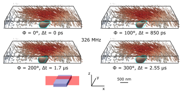
-
[6]
S. Finizio, S. Wintz, K. Zeissler, A. V. Sadovnikov, S. Mayr, S. A. Nikitov, C. H. Marrows, and J. Raabe, High resolution dynamic imaging of the delay- and tilt-free motion of n´ eel domain walls in perpendicularly magnetized superlattices, Nano Letters19, 375 (2019)
work page 2019
-
[7]
A. Bisig, M. St¨ ark, M.-A. Mawass, C. Moutafis, J. Rhensius, J. Heidler, F. B¨ uttner, M. Noske, M. Weigand, S. Eisebitt, T. Tyliszczak, B. Van Waeyenberge, H. Stoll, G. Sch¨ utz, and M. Kl¨ aui, Correlation between spin structure oscillations and domain wall velocities, Nature Communications4, 2328 (2013)
work page 2013
-
[8]
J. Rhensius, L. Heyne, D. Backes, S. Krzyk, L. J. Heyderman, L. Joly, F. Nolting, and M. Kl¨ aui, Imaging of domain wall inertia in permalloy half-ring nanowires by time-resolved photoemission electron microscopy, Physical Review Letters104, 067201 (2010)
work page 2010
- [9]
-
[10]
K. Litzius, I. Lemesh, B. Kr¨ uger, P. Bassirian, L. Caretta, K. Richter, F. B¨ uttner, K. Sato, O. A. Tretiakov, J. F¨ orster, R. M. Reeve, M. Weigand, I. Bykova, H. Stoll, G. Sch¨ utz, G. S. D. Beach, and M. Kl¨ aui, Skyrmion hall effect revealed by direct time-resolved x-ray microscopy, Nature Physics13, 170 (2017)
work page 2017
-
[11]
F. B¨ uttner, C. Moutafis, M. Schneider, B. Kr¨ uger, C. M. G¨ unther, J. Geilhufe, C. von Korff Schmising, J. Mohanty, B. Pfau, S. Schaffert, A. Bisig, M. Foerster, T. Schulz, C. A. F. Vaz, J. H. Franken, H. J. M. Swagten, M. Kl¨ aui, and S. Eisebitt, Dynamics and inertia of skyrmionic spin structures, Nature Physics11, 225 (2015)
work page 2015
-
[12]
S. Finizio, K. Zeissler, S. Wintz, S. Mayr, T. Weßels, A. J. Huxtable, G. Burnell, C. H. Marrows, and J. Raabe, Deterministic field-free skyrmion nucleation at a nano-engineered injector device, Nano Letters19, 7246 (2019)
work page 2019
-
[13]
N. Nagaosa and Y. Tokura, Topological properties and dynamics of magnetic skyrmions, Nature Nanotechnology8, 899 (2013)
work page 2013
-
[14]
S. Woo, K. Litzius, B. Kr¨ uger, M.-Y. Im, L. Caretta, K. Richter, M. Mann, A. Krone, R. M. Reeve, M. Weigand, P. Agrawal, I. Lemesh, M.-A. Mawass, P. Fischer, M. Kl¨ aui, and G. S. D. Beach, Observation of room-temperature magnetic skyrmions and their current- 38 driven dynamics in ultrathin metallic ferromagnets, Nature Materials15, 501 (2016)
work page 2016
-
[15]
S. Woo, K.-M. Song, X. Zhang, M. Ezawa, Y. Zhou, X. Liu, M. Weigand, S. Finizio, J. Raabe, M.-C. Park, K.-Y. Lee, J. W. Choi, B.-C. Min, H. C. Koo, and J. Chang, Deterministic creation and deletion of a single magnetic skyrmion observed by direct time-resolved x-ray microscopy, Nature Electronics1, 288 (2018)
work page 2018
-
[16]
J. Zazvorka, F. Jakobs, D. Heinze, N. Keil, S. Kromin, S. Jaiswal, G. Jakob, P. Virnau, D. Pinna, K. Eveschor-Sitte, L. Rosza, a. Donges, U. Nowak, and M. Kl¨ aui, Thermal skyrmion diffusion used in a reshuffler device, Nature Nanotechnology14, 658 (2019)
work page 2019
-
[17]
C. Klose, F. B¨ uttner, W. Hu, C. Mazzoli, K. Litzius, R. Battistelli, S. Zayko, I. Lemesh, J. M. Bartell, M. Huang, C. M. G¨ unther, M. Schneider, A. Barbour, S. B. Wilkins, G. S. D. Beach, S. Eisebitt, and B. Pfau, Coherent correlation imaging for resolving fluctuating states of matter, Nature614, 256 (2023)
work page 2023
-
[18]
B. L. Henke, E. M. Gullikson, and J. C. Davis, X-ray interactions: photoabsorption, scatter- ing, transmission, and reflection at e=50-30000 ev, z=1-92, Atomic Data and Nuclear Data Tables54, 181 (1993)
work page 1993
-
[19]
G. Sch¨ utz, W. Wagner, W. Wilhelm, P. Kienle, R. Zeller, R. Frahm, and G. Materlik, Absorption of circularly polarized x-rays in iron, Physical Review Letters58, 737 (1987)
work page 1987
- [20]
-
[21]
J. St¨ ohr and M. G. Samant, Liquid crystal alignment by rubbed polymer surfaces: a micro- scopic bond orientation model, Journal of Electron Spectroscopy and Related Phenomena 98, 189 (1999)
work page 1999
-
[22]
G. Sch¨ onhense, Imaging of magnetic structures by photoemission electron microscopy, Jour- nal of Physics: Condensed Matter11, 9517 (1999)
work page 1999
-
[23]
P. Fischer, G. Sch¨ utz, G. Schmal, P. Guttmann, and D. Raasch, Imaging of magnetic domains with the x-ray microscope at bessy using x-ray magnetic circular dichroism, Zeitschrift f¨ ur Physik B: Condensed Matter101, 313 (1996)
work page 1996
-
[24]
J. B. Kortright, S.-K. Kim, H. Ohldag, G. Meighs, and A. Warwick, Magnetization imaging using scanning transmission x-ray microscopy, X-ray Microscopy Proceedings507, 49 (2000)
work page 2000
-
[25]
D. V. Christensenet al., 2024 roadmap on magnetic microscopy techniques and their appli- cations in materials science, Journal of Physics: Materials7, 032501 (2024)
work page 2024
-
[26]
Y. Martin and H. K. Wickramasinghe, Magnetic imaging by force microscopy with a 1000 ˚ a 39 resolution, Applied Physics Letters1455, 50 (1987)
work page 1987
-
[27]
M. R. Koblischka and U. Hartmann, Recent advances in magnetic force microscopy, Ultra- microscopy97, 103 (2003)
work page 2003
-
[28]
R. Wiesendanger and M. Bode, Nano- and atomic-scale magnetism studied by spin-polarized scanning tunneling microscopy and spectroscopy, Solid State Communications119, 341 (2001)
work page 2001
-
[29]
M. E. Hale, H. W. Fuller, and H. Rubinstein, Magnetic domain observations by electron microscopy, Journal of Applied Physics30, 789 (1959)
work page 1959
-
[30]
J. N. Chapman and M. R. Scheinfein, Transmission electron microscopies of magnetic nanos- tructures, Journal of Magnetism and Magnetic Materials200, 729 (1999)
work page 1999
-
[31]
T. Weßels, S. D¨ aster, Y. Murooka, B. Zingsem, V. Migunov, M. Kruth, S. Finizio, P.-H. Lu, A. Kovacs, A. Oelsner, K. M¨ uller-Caspary, Y. Acremann, and R. E. Dunin-Borkowski, Continuous illumination picosecond imaging using a delay line detector in a transmission electron microscope, Ultramicroscopy233, 113392 (2022)
work page 2022
-
[32]
J. McCord, Progress in magnetic domain observation by advanced magneto-optical mi- croscopy, Journal of Physics D: Applied Physics48, 333001 (2015)
work page 2015
-
[33]
W. K. Hiebert, A. Stankiewicz, and M. R. Freeman, Direct observation of magnetic relaxation in a small permalloy disk by time-resolved scanning kerr microscopy, Physical Review Letters 79, 1134 (1997)
work page 1997
-
[34]
M. R. Freeman and J. F. Smyth, Picosecond time-resolved magnetization dynamics of thin- film heads, Journal of Applied Physics79, 5898 (1996)
work page 1996
-
[35]
P. Maletinsky, S. Hong, M. S. Grinolds, B. Hausmann, M. D. Lukin, R. L. Walsworth, M. Loncar, and A. Yacoby, A robust scanning diamond sensor for nanoscale imaging with single nitrogen-vacancy centres, Nature Nanotechnology7, 320 (2012)
work page 2012
-
[36]
B. J. Maertz, A. P. Wijnheijmer, G. D. Fuchs, M. E. Nowakowski, and D. D. Awschalom, Vector magnetic field microscopy using nitrogen vacancy centers in diamond, Applied Physics Letters96, 092504 (2010)
work page 2010
-
[37]
K. Herb, L. A. V¨ olker, J. M. Abendroth, N. Meinhardt, L. van Schie, P. Gambardella, and C. L. Degen, Quantum magnetometry of transient signals with a time resolution of 1.1 nanoseconds, Nature Communications16, 822 (2025)
work page 2025
-
[38]
J. St¨ ohr, Y. Wu, B. D. Hermsmeier, M. G. Samant, G. R. Harp, S. Koranda, D. Dunham, 40 and B. P. Tonner, Element-specific magnetic microscopy with circularly polarized x-rays, Science259, 658 (1993)
work page 1993
- [39]
-
[40]
L. Le Guyader, A. Kleibert, A. Fraile Rodriguez, S. El Moussaoui, A. Balan, M. Buzzi, J. Raabe, and F. Nolting, Studying nanomagnets and magnetic heterostructures with x-ray peem at the swiss light source, Journal of Electron Spectroscopy and Related Phenomena 185, 371 (2012)
work page 2012
-
[41]
S. Eisebitt, J. L¨ uning, W. F. Schlotter, M. L¨ orgen, O. Hellwig, W. Eberhardt, and J. St¨ ohr, Lensless imaging of magnetic nanostructures by x-ray spectro-holography, Nature432, 885 (2004)
work page 2004
-
[42]
R. Battistelli, D. Metternich, M. Schneider, L.-M. Kern, K. Litzius, J. Fuchs, C. Klose, K. Gerlinger, K. Bagschik, C. M. G¨ unther, D. Engel, C. Ropers, S. Eisebitt, B. Pfau, F. B¨ uttner, and S. Zayko, Coherent x-ray magnetic imaging with 5 nm resolution, Optica 11, 234 (2024)
work page 2024
-
[43]
C. von Korff Schmising, B. Pfau, M. Schneider, C. M. G¨ unther, M. Giovannella, J. Perron, B. Vobungbo, L. M¨ uller, F. Capotondi, E. Pedersoli, N. Mahne, J. L¨ uning, and S. Eisebitt, Imaging ultrafast demagnetization dynamics after a spatially localized optical excitation, Physical Review Letters112, 217203 (2014)
work page 2014
- [44]
-
[45]
C. V. Shank, R. L. Fork, R. F. Leheny, and J. Shah, Dynamics of photoexcited gaas band-edge absorption with subpicosecond resolution, Physical Review Letters42, 112 (1979)
work page 1979
-
[46]
C. Donnelly, S. Finizio, S. Gliga, M. Holler, A. Hrabec, M. Odstrcil, S. Mayr, V. Scagnoli, L. J. Heyderman, M. Guizar-Sicairos, and J. Raabe, Time-resolved imaging of three- dimensional nanoscale magnetization dynamics, Nature Nanotechnology15, 356 (2020)
work page 2020
-
[47]
S. Finizio, S. Mayr, and J. Raabe, Time-of-arrival detection for time-resolved scanning trans- mission x-ray microscopy imaging, Journal of Synchrotron Radiation27, 1320 (2020)
work page 2020
-
[48]
S. Finizio, B. Watts, and J. Raabe, Why is my image noisy? a look into the terms contribution to a time-resolved x-ray microscopy image, Journal of Synchrotron Radiation28, 1146 (2021). 41
work page 2021
-
[49]
P. Goslawski, M. Ries, M. Ruprecht, and G. W¨ ustefeld, The low-αlattice and bunch length limits at BESSY-VSR, Proceedings of the 5th International Particle Accelerator Conference 5, 216 (2014)
work page 2014
-
[50]
M. Weigand, S. Wintz, J. Gr¨ afe, M. Noske, H. Stoll, B. Van Waeyenberge, and G. Sch¨ utz, Timemaxyne: A shot-noise limited, time-resolved pump-and-probe acquisition system capa- ble of 50 ghz frequencies for synchrotron-based x-ray microscopy, Crystals12, 1029 (2022)
work page 2022
-
[51]
S. Mayr, J. F¨ orster, S. Finizio, K. Schultheiss, R. A. Gallardo, R. Narkovicz, G. Dieterle, A. Semisalova, J. Bailey, E. Kirk, A. Suszka, J. Lindner, J. Gr¨ afe, J. Raabe, G. Sch¨ utz, M. Weigand, H. Stoll, and S. Wintz, Time-resolved x-ray imaging of nanoscale spin-wave dynamics at multi-ghz frequencies using low-alpha synchrotron operation, Applied Ph...
work page 2024
-
[52]
S. Finizio, J. B. Bailey, B. Olsthoorn, and J. Raabe, Periodogram-based detection of unknown frequencies in time-resolved scanning transmission x-ray microscopy, ACS Nano16, 21071 (2022)
work page 2022
-
[53]
S. Finizio, S. Wintz, E. Kirk, and J. Raabe, In-situ membrane bending setup for strain- dependent scanning transmission x-ray microscopy investigations, Review of Scientific In- struments87, 123703 (2016)
work page 2016
- [54]
-
[55]
B. R¨ osner, S. Finizio, F. Koch, F. D¨ oring, V. A. Guzenko, M. Langer, E. Kirk, B. Watts, M. Meyer, J. Lorona Ornelas, A. Sp¨ ath, S. Stanescu, S. Swaraj, R. Belkhou, T. Ishikawa, T. F. Keller, B. Gross, M. Poggio, R. H. Fink, J. Raabe, A. Kleiber, and C. David, Soft x-ray microscopy with 7 nm resolution, Optica7, 1602 (2020)
work page 2020
-
[56]
Pfeiffer, X-ray ptychography, Nature Photon.12, 9 (2018)
F. Pfeiffer, X-ray ptychography, Nature Photon.12, 9 (2018)
work page 2018
-
[57]
T. A. Butcher, S. Finizio, L. Heller, N. W. Phillips, B. Sarafimov, C. A. F. Vaz, A. Kleib- ert, B. Watts, M. Holler, and J. Raabe, Soft x-ray ptychography with sophie: Guide and instrumentation, Review of Scientific Instruments96, 123704 (2025)
work page 2025
-
[58]
T. A. Butcher, N. W. Phillips, C.-C. Chiu, C.-C. Wei, S.-Z. Ho, Y.-C. Chen, E. Fr¨ ojdh, F. Baruffaldi, M. Carulla, J. Zhang, A. Bergamaschi, C. A. F. Vaz, A. Kleibert, S. Finizio, J.-C. Yang, S.-W. Huang, and J. Raabe, Ptychographic nanoscale imaging of the magneto- 42 electric coupling in freestanding BiFeO 3, Adv. Mater.36, 2311157 (2024)
work page 2024
-
[59]
D. A. Shapiroet al., An ultrahigh-resolution soft x-ray microscope for quantitative analysis of chemically heterogeneous nanomaterials, Science Advances6, eabc4904 (2020)
work page 2020
-
[60]
A. P. Hitchcock, Soft x-ray spectromicroscopy and ptychography, Journal of Electron Spec- troscopy and Related Phenomena200, 49 (2015)
work page 2015
-
[61]
Y. Acremann, V. Chembrolu, J. P. Strachan, T. Tyliszczak, and J. St¨ ohr, Software defined photon counting system for time resolved x-ray experiments, Review of Scientific Instruments 78, 014702 (2007)
work page 2007
-
[62]
B. van Waeyenberge, A. Puzic, H. Stoll, K. W. Chou, T. Tyliszczak, R. Hertel, R. F¨ ahnle, H. Br¨ uckl, K. Rott, G. Reiss, I. Neudecker, D. Weiss, C. H. Back, and G. Sch¨ utz, Magnetic vortex core reversal by excitation with short bursts of an alternating field, Nature444, 461 (2006)
work page 2006
-
[63]
Y. Acremann, J. P. Strachan, V. Chembrolu, S. D. Andrews, T. Tyliszczak, J. A. Ka- tine, M. J. Carey, B. M. Clemens, H. C. Siegmann, and J. St¨ ohr, Time-resolved imaging of spin transfer switching: beyond the macrospin concept, Physical Review Letters96, 217202 (2006)
work page 2006
-
[64]
M. Kammerer, M. Weigand, M. Curcic, M. Noske, M. Sproll, A. Vansteenkiste, B. van Waeyenberge, H. Stoll, G. Woltersdorf, C. H. Back, and G. Sch¨ utz, Magnetic vortex core reversal by excitation of spin waves, Nature Communications279, 2 (2011)
work page 2011
-
[65]
G. Dieterle, J. F¨ orster, H. Stoll, A. S. Semisalova, S. Finizio, A. Gangwar, M. Weigand, M. Noske, M. F¨ ahnle, I. Bykova, J. Gr¨ afe, D. A. Bozhko, H. Y. Musiienko-Shmarova, V. Tiberkevich, A. Slavin, C. H. Back, J. Raabe, G. Sch¨ utz, and S. Wintz, Coherent ex- citation of heterosymmetric spin waves with ultrashort wavelengths, Physical Review Letters...
work page 2019
-
[66]
S. Mayr, L. Flajsman, S. Finizio, A. Hrabec, M. Weigand, J. F¨ orster, H. Stoll, L. J. Hey- derman, M. Urbanek, S. Wintz, and J. Raabe, Spin-wave emission from vortex cores under static magnetic bias fields, Nano Letters21, 1584 (2021)
work page 2021
-
[67]
A. Puzic, T. Korhonen, B. Kalantari, J. Raabe, C. Quitmann, P. J¨ ullig, L. Bommer, D. Goll, G. Sch¨ utz, S. Wintz, T. Strache, M. K¨ orner, D. Marko, C. Bunce, and J. Fassbender, Photon counting system for time-resolved experiments in multibunch mode, Synchrotron Radiation News23, 26 (2010). 43
work page 2010
-
[68]
S. Finizio, S. Wintz, E. Kirk, A. K. Suszka, S. Gliga, P. Wohlh¨ uter, K. Zeissler, and J. Raabe, Control of the gyration dynamics of magnetic vortices by the magnetoelastic effect, Physical Review B96, 054438 (2017)
work page 2017
-
[69]
R. A. Bosch and C. S. Hsue, Suppression of longitudinal coupled-bunch instabilities by a pas- sive higher harmonic cavity, Proceedings of International Conference on Particle Accelerators (1993)
work page 1993
- [70]
-
[71]
D. A. Gedcke and W. J. Mc Donald, A constant fraction of pulse height trigger for optimum time resolution, Nuclear Instrumentation Methods55, 377 (1967)
work page 1967
-
[72]
A. Q. R. Baron, S. Kishimoto, J. Morse, and J.-M. Rigal, Silicon avalanche photodiodes for direct detection of x-rays, Journal of Synchrotron Radiation13, 131 (2005)
work page 2005
- [73]
-
[74]
A. Fernandez-Pacheco, R. Streubel, O. Fruchart, R. Herter, P. Fischer, and R. P. Cowburn, Three-dimensional magnetism, Nature Communications8, 15756 (2017)
work page 2017
-
[75]
T. A. Butcher, N. W. Phillips, A. L. Levitan, M. Weigand, S. Wintz, J. Raabe, and S. Finizio, Nanoscale domain-wall dynamics in micromagnetic structures with weak per- pendicular anisotropy, Physical Review B111, L220409 (2025)
work page 2025
-
[76]
C. Donnelly, M. Guizar-Sicairos, V. Scagnoli, S. Gliga, M. Holler, J. Raabe, and L. J. Hey- derman, Three-dimensional magnetization structures revealed with x-ray vector nanotomog- raphy, Nature547, 328 (2017)
work page 2017
-
[77]
M. Holler, M. Odstrcil, M. Guizar-Sicairos, M. Lebugle, E. M¨ uller, S. Finizio, G. Tinti, C. David, J. Zusman, W. Unglaub, O. Bunk, J. Raabe, A. F. J. Levi, and G. Aeppli, Three- dimensional imaging of integrated circuits with macro- to nanoscale zoom, Nature Electronics 2, 464 (2019)
work page 2019
-
[78]
S. Finizio, C. Donnelly, S. Mayr, A. Hrabec, and J. Raabe, Three-dimensional vortex gyration dynamics unraveled by time-resolved soft x-ray laminography with freely selectable excitation frequencies, Nano Letters22, 1971 (2022)
work page 1971
-
[79]
A. Hubert and R. Sch¨ afer,Magnetic domains: the analysis of magnetic microstructures (Springer Verlag, 1998)
work page 1998
-
[80]
J. R. Eshbach and R. W. Damon, Surface magnetostatic modes and surface spin waves, Physical Review118, 1208 (1960). 44
work page 1960
discussion (0)
Sign in with ORCID, Apple, or X to comment. Anyone can read and Pith papers without signing in.