Block-Term Decomposition Approach to Blind Multi-trial Functional Ultrasound Unmixing
Pith reviewed 2026-06-27 15:44 UTC · model grok-4.3
The pith
A block-term tensor decomposition model enables blind unmixing of multi-trial functional ultrasound data into spatial maps, activations, and responses.
A machine-rendered reading of the paper's core claim, the machinery that carries it, and where it could break.
Core claim
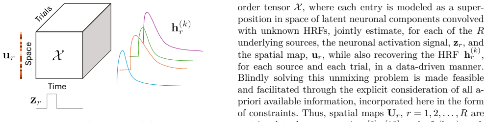
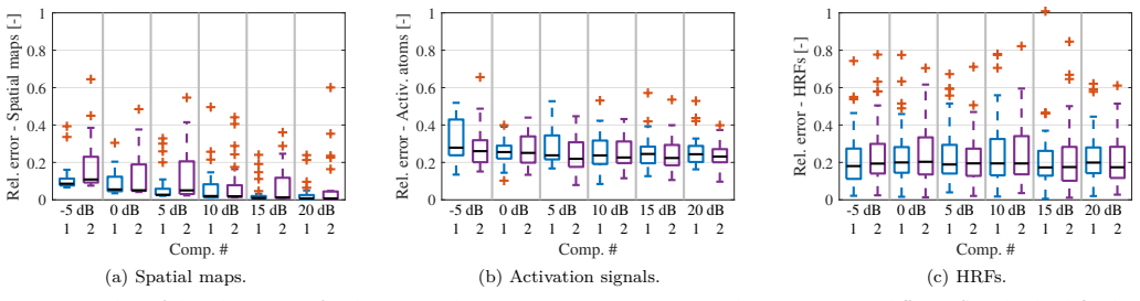
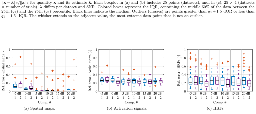
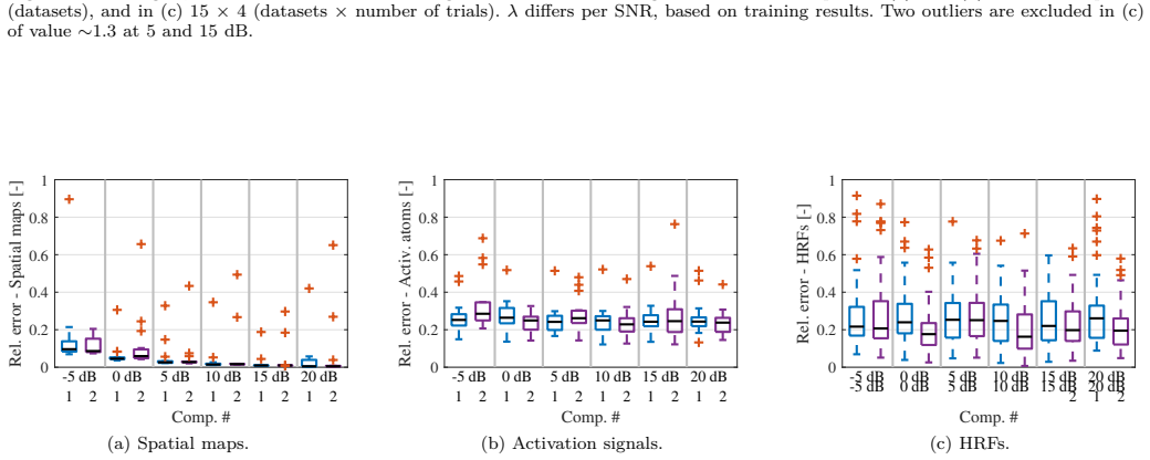
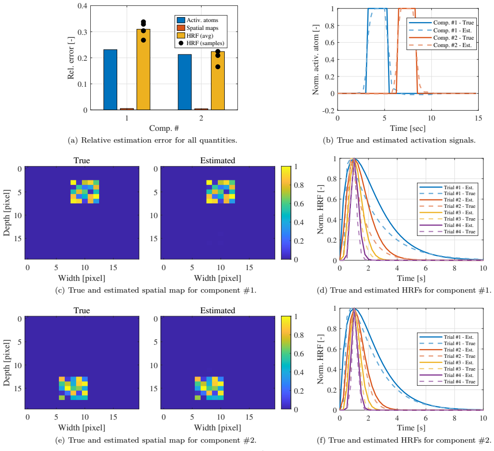
We propose a data-driven convolutive block-term tensor decomposition-based model for multi-trial fUS measurements, where each source has a spatiotemporal representation comprising a low-rank spatial map and a piecewise-constant neuronal activation signal convolved with a trial- and source-dependent hemodynamic response function (HRF) with a physiologically plausible shape. We propose a constrained optimization framework for the model computation, which consists of alternating projected gradient descent iterations. Simulation results demonstrate accurate recovery of spatial maps and reliable estimation of activation temporal profiles across various noise levels.
What carries the argument
The convolutive block-term tensor decomposition that models each source's activity as the convolution of a low-rank spatial map with a piecewise-constant neuronal signal and a source- and trial-dependent HRF.
Load-bearing premise
Each source's neuronal activation is piecewise-constant and the trial- and source-dependent HRF has a physiologically plausible shape that can be estimated jointly with the other factors.
What would settle it
If simulations with known ground-truth activations that are not piecewise-constant yield spatial maps that fail to align with expected regions even at low noise levels.
Figures
read the original abstract
Functional ultrasound (fUS) has emerged as a powerful neuroimaging modality due to its high resolution in both space and time, low cost and potential portability. Nevertheless, fUS signals provide only indirect observations of neuronal activity through the neurovascular coupling, and hence require the blind separation of latent neuronal sources while also deconvolving their hemodynamic responses. In this work, we propose a data-driven convolutive block-term tensor decomposition-based model for multi-trial fUS measurements, where each source has a spatiotemporal representation comprising a low-rank spatial map and a piecewise-constant neuronal activation signal convolved with a trial- and source-dependent hemodynamic response function (HRF) with a physiologically plausible shape. We propose a constrained optimization framework for the model computation, which consists of alternating projected gradient descent iterations. Simulation results are reported that demonstrate accurate recovery of spatial maps and reliable estimation of activation temporal profiles across various noise levels, while confirming that HRF estimation remains the most challenging part of the problem.
Editorial analysis
A structured set of objections, weighed in public.
Referee Report
Summary. The paper proposes a convolutive block-term tensor decomposition model for blind unmixing of multi-trial functional ultrasound (fUS) data. Each neuronal source is represented by a low-rank spatial map and a piecewise-constant activation signal convolved with a trial- and source-dependent HRF of physiologically plausible shape. The authors introduce a constrained optimization framework solved via alternating projected gradient descent and report simulation results claiming accurate recovery of spatial maps and temporal profiles across noise levels, while identifying HRF estimation as the most challenging component.
Significance. If validated with quantitative metrics and real data, the approach could advance blind source separation in fUS by providing a data-driven tensor framework that jointly handles spatial unmixing, activation estimation, and HRF deconvolution. The explicit modeling of trial-dependent HRFs and the use of block-term structure are strengths, as is the constrained alternating optimization. However, the current reliance on simulations without reported error metrics, ablation studies, or baseline comparisons limits the assessed impact.
major comments (2)
- [Abstract / Simulation results] Abstract and simulation results section: the claims of 'accurate recovery of spatial maps' and 'reliable estimation of activation temporal profiles' are presented without any quantitative metrics (e.g., correlation, NMSE, or Dice scores), specific noise models, number of trials, or comparisons to existing methods, which is load-bearing for evaluating whether the simulations support the central claims.
- [Constrained optimization framework] Model definition and optimization section: no convergence guarantees, initialization procedure, or sensitivity analysis for the alternating projected gradient descent are provided, which is critical because the reported recoveries depend on successful optimization of the joint factors including the HRF.
minor comments (2)
- [Abstract] The abstract should be expanded to include at least one quantitative result or table reference from the simulations to substantiate the recovery claims.
- [Model formulation] Notation for the block-term decomposition and the convolutive model should be introduced with explicit tensor dimensions and rank parameters for clarity.
Simulated Author's Rebuttal
We thank the referee for the constructive feedback. We address each major comment below and will revise the manuscript to incorporate quantitative evaluations and additional optimization details where feasible.
read point-by-point responses
-
Referee: [Abstract / Simulation results] Abstract and simulation results section: the claims of 'accurate recovery of spatial maps' and 'reliable estimation of activation temporal profiles' are presented without any quantitative metrics (e.g., correlation, NMSE, or Dice scores), specific noise models, number of trials, or comparisons to existing methods, which is load-bearing for evaluating whether the simulations support the central claims.
Authors: We agree that quantitative metrics are necessary to substantiate the claims. In the revised manuscript, we will add Pearson correlation coefficients and NMSE values for the recovered spatial maps and activation profiles, specify the additive white Gaussian noise model and number of trials (e.g., 10), and include comparisons against baselines such as ICA and NMF to demonstrate relative performance across noise levels. revision: yes
-
Referee: [Constrained optimization framework] Model definition and optimization section: no convergence guarantees, initialization procedure, or sensitivity analysis for the alternating projected gradient descent are provided, which is critical because the reported recoveries depend on successful optimization of the joint factors including the HRF.
Authors: We will expand the optimization section to describe the initialization procedure (random factors with a canonical HRF template) and include a sensitivity analysis reporting success rates over multiple random initializations. Convergence guarantees are not provided because the joint optimization is non-convex; we will instead report empirical convergence via objective value plots and discuss practical reliability in the revised text. revision: partial
Circularity Check
No significant circularity identified
full rationale
The paper proposes a data-driven convolutive block-term tensor decomposition model for multi-trial fUS measurements, with each source represented by a low-rank spatial map, piecewise-constant activation convolved with a trial- and source-dependent HRF, and a constrained optimization framework solved via alternating projected gradient descent. Simulation results demonstrate recovery of spatial maps and temporal profiles. No equations, fitting procedures, or self-citations are shown that would reduce the reported recoveries or estimates to quantities defined by the same fitted parameters by construction. The model is explicitly data-driven with stated assumptions, and the derivation chain remains self-contained against external benchmarks.
Axiom & Free-Parameter Ledger
axioms (1)
- domain assumption Each source has a piecewise-constant neuronal activation signal convolved with a trial- and source-dependent HRF of physiologically plausible shape.
Reference graph
Works this paper leans on
-
[1]
Functional ultrasound imaging of the brain,
E. Macé et al., “Functional ultrasound imaging of the brain,” Nature Meth., vol. 8, pp. 662––664, 2011
2011
-
[2]
Total activation: fMRI deconvolution through spatio-temporal regularization,
F. I. Karahanoğlu et al., “Total activation: fMRI deconvolution through spatio-temporal regularization,” NeuroImage, vol. 73, 2013
2013
-
[3]
Multivariate semi-blind deconvolution of fMRI time series,
H. Cherkaoui et al., “Multivariate semi-blind deconvolution of fMRI time series,” NeuroImage, vol. 241, 2021
2021
-
[4]
Transfer functions linking neural calcium to single voxel functional ultrasound signal,
A.-K. Aydin et al., “Transfer functions linking neural calcium to single voxel functional ultrasound signal,” Nat. Commun., vol. 11, 2020
2020
-
[5]
Joint detection-estimation of brain activity in functional MRI: A multichannel deconvolution solution,
S. Makni et al., “Joint detection-estimation of brain activity in functional MRI: A multichannel deconvolution solution,” IEEE Trans. Signal Process., vol. 53, no. 9, pp. 3488–3502, 2005
2005
-
[6]
Nonpara- metric hemodynamic deconvolution of fMRI using homomorphic filtering,
K. R. Sreenivasan, M. Havlicek, and G. Deshpande, “Nonpara- metric hemodynamic deconvolution of fMRI using homomorphic filtering,” IEEE Trans. Med. Imag., vol. 34, no. 5, pp. 1155– 1163, 2014
2014
-
[7]
Augmenting interictal mapping with neurovascular coupling biomarkers by structured factorization of epileptic eeg and fmri data,
S. Van Eyndhoven, P. Dupont, S. Tousseyn, N. Vervliet, W. Van Paesschen, S. Van Huffel, and B. Hunyadi, “Augmenting interictal mapping with neurovascular coupling biomarkers by structured factorization of epileptic eeg and fmri data,” Neu- roImage, vol. 228, p. 117652, 2021
2021
-
[8]
Event-related fMRI: Characterizing differen- tial responses,
K. Friston et al., “Event-related fMRI: Characterizing differen- tial responses,” NeuroImage, vol. 7, no. 1, pp. 30–40, 1998
1998
-
[9]
Deconvolution of the functional ultrasound response in the mouse visual pathway using block-term decom- position,
A. Erol et al., “Deconvolution of the functional ultrasound response in the mouse visual pathway using block-term decom- position,” Neuroinformatics, vol. 21, pp. 247––265, Nov. 2022
2022
-
[10]
Extracting hemodynamic activity with low-rank spatial signatures in functional ultrasound using tensor decompositions,
S.-E. Kotti and B. Hunyadi, “Extracting hemodynamic activity with low-rank spatial signatures in functional ultrasound using tensor decompositions,” in Proc. EUSIPCO, Lyon, France, Aug. 2024
2024
-
[11]
Neural correlates of blood flow measured by ultrasound,
A. O. Nunez-Elizalde et al., “Neural correlates of blood flow measured by ultrasound,” Neuron, vol. 110, no. 10, pp. 1631– 1640, 2022
2022
-
[12]
Functional ultrasound imaging and neuronal activity: How accurate is the spatiotemporal match?
T. Lambert et al., “Functional ultrasound imaging and neuronal activity: How accurate is the spatiotemporal match?” Imag. Neurosci., vol. 3, Jun. 2025. 1 2 Comp. # 0 0.1 0.2 0.3 0.4Rel. error [-] Activ. atoms Spatial maps HRF (avg) HRF (samples) (a) Relative estimation error for all quantities. 0 5 10 15 Time [sec] -0.2 0 0.2 0.4 0.6 0.8 1 Norm. activ. at...
2025
-
[13]
Tensor decomposition for signal processing and machine learning,
N. D. Sidiropoulos et al., “Tensor decomposition for signal processing and machine learning,” IEEE Trans. Signal Process., vol. 65, no. 13, pp. 3551–3582, Jul. 2017
2017
-
[14]
Block term decomposition for modelling epileptic seizures,
B. Hunyadi et al., “Block term decomposition for modelling epileptic seizures,” EURASIP J. Adv. Signal Process., vol. 2014, no. 1, 2014
2014
-
[15]
Blind fMRI source unmixing via higher-order tensor decompositions,
C. Chatzichristos et al., “Blind fMRI source unmixing via higher-order tensor decompositions,” J. Neurosci. Meth., vol. 315, Mar. 2019
2019
-
[16]
Tensor decompositions and data fusion in epileptic elec- troencephalography and functional magnetic resonance imag- ing data,
B. Hunyadi, P. Dupont, W. Van Paesschen, and S. Van Huf- fel, “Tensor decompositions and data fusion in epileptic elec- troencephalography and functional magnetic resonance imag- ing data,” Wiley Interdisciplinary Reviews: Data Mining and Knowledge Discovery, vol. 7, no. 1, p. e1197, 2017
2017
-
[17]
Tensors for neuroimaging: A review on applications of tensors to unravel the mysteries of the brain,
A. Erol and B. Hunyadi, “Tensors for neuroimaging: A review on applications of tensors to unravel the mysteries of the brain,” Tensors for Data Processing, pp. 427–482, 2022
2022
-
[18]
Tensor and coupled decom- positions: Interpretable pattern discovery in multiset and mul- timodal functional neuroimaging data,
M. Mørup, E. Acar, and T. Adalı, “Tensor and coupled decom- positions: Interpretable pattern discovery in multiset and mul- timodal functional neuroimaging data,” IEEE Signal Process. Mag., vol. 42, no. 4, pp. 41–57, 2025
2025
-
[19]
Decompositions of a higher-order tensor in block terms — Part II: Definitions and uniqueness,
L. De Lathauwer, “Decompositions of a higher-order tensor in block terms — Part II: Definitions and uniqueness,” SIAM J. Matrix Anal. Appl., vol. 30, no. 3, pp. 1033–1066, 2008
2008
-
[20]
fMRI adaptation revisited,
J. Larsson, S. G. Solomon, and A. Kohn, “fMRI adaptation revisited,” Cortex, vol. 80, pp. 154–160, 2016
2016
-
[21]
Analyzing trial-to- trial variability in the mouse visual pathway using functional ultrasound,
A. Erol, P. Kruizinga, and B. Hunyadi, “Analyzing trial-to- trial variability in the mouse visual pathway using functional ultrasound,” in Proc. ISBI, Athens, Greece, May 2024
2024
-
[22]
The variability of human, bold hemodynamic responses,
G. K. Aguirre, E. Zarahn, and M. D’Esposito, “The variability of human, bold hemodynamic responses,” NeuroImage, vol. 8, no. 4, pp. 360–369, 1998
1998
-
[23]
Varia- tion of BOLD hemodynamic responses across subjects and brain regions and their effects on statistical analyses,
D. A. Handwerker, J. M. Ollinger, and M. D’Esposito, “Varia- tion of BOLD hemodynamic responses across subjects and brain regions and their effects on statistical analyses,” NeuroImage, vol. 21, no. 4, pp. 1639–1651, 2004
2004
-
[24]
Shift-invariant multilinear decomposition of neuroimaging data,
M. Mørup et al., “Shift-invariant multilinear decomposition of neuroimaging data,” NeuroImage, vol. 42, pp. 1439–1450, Oct. 2008
2008
-
[25]
Modeling latency and shape changes in trial based neuroimaging data,
M. Mørup, L. K. Hansen, and K. H. Madsen, “Modeling latency and shape changes in trial based neuroimaging data,” in Proc. ACSSC, Pacific Grove, CA, 29 Aug.–1 Sep. 2011
2011
-
[26]
Multi-subject dictionary learning to segment an atlas of brain spontaneous activity,
G. Varoquaux et al., “Multi-subject dictionary learning to segment an atlas of brain spontaneous activity,” in Proc. IPMI, Kloster Irsee, Germany, Jul. 2011
2011
-
[27]
Spatiotemporal evolution of the functional magnetic resonance imaging response to ultrashort stimuli,
Y. Hirano, B. Stefanovic, and A. C. Silva, “Spatiotemporal evolution of the functional magnetic resonance imaging response to ultrashort stimuli,” J. Neurosci., vol. 31, no. 4, pp. 1440–1447, 2011
2011
-
[28]
Block-term tensor decomposition via constrained matrix factorization,
X. Fu and K. Huang, “Block-term tensor decomposition via constrained matrix factorization,” in Proc. MLSP, Pittsburgh, PA, Oct. 2019
2019
-
[29]
Modeling nonlinear evoked hemodynamic responses in functional ultrasound,
S.-E. Kotti, A. Erol, and B. Hunyadi, “Modeling nonlinear evoked hemodynamic responses in functional ultrasound,” in Proc. ICASSP, Rhodes, Greece, Jun. 2023
2023
-
[30]
On the Goldstein-Levitin-Polyak gradient projection method,
D. P. Bertsekas, “On the Goldstein-Levitin-Polyak gradient projection method,” IEEE Trans. Autom. Control, vol. 21, no. 2, pp. 174–184, Apr. 1976
1976
-
[31]
A constrained block-term tensor decomposition framework for spectrum cartography,
X. Chen et al., “A constrained block-term tensor decomposition framework for spectrum cartography,” IEEE Signal Process. Lett., vol. 29, pp. 1699–1703, Jul. 2022
2022
-
[32]
Signal enhancement — A composite property mapping algorithm,
J. A. Cadzow, “Signal enhancement — A composite property mapping algorithm,” IEEE Trans. Acoust., Speech, Signal Pro- cess., vol. 36, no. 1, pp. 49–62, Jan. 1988
1988
-
[33]
A direct algorithm for 1-D total variation denois- ing,
L. Condat, “A direct algorithm for 1-D total variation denois- ing,” IEEE Signal Process. Lett., vol. 20, no. 11, pp. 1054–1057, 2013
2013
-
[34]
(2017) TV Condat
——. (2017) TV Condat. Accessed: Oct. 20, 2025. [Online]. A vailable: https://lcondat.github.io/download/TV_Condat_ v2.m
2017
-
[35]
Core consistency diagnostic for the tensor block term decomposition,
S.-E. Kotti and B. Hunyadi, “Core consistency diagnostic for the tensor block term decomposition,” TechRxiv, Jun. 2025
2025
-
[36]
A projected Newton-type algorithm for rank-revealing nonneg- ative block-term tensor decomposition,
E. Kofidis, P. V. Giampouras, and A. A. Rontogiannis, “A projected Newton-type algorithm for rank-revealing nonneg- ative block-term tensor decomposition,” in Proc. EUSIPCO, Belgrade, Serbia, Sep. 2022
2022
-
[37]
Block- term tensor decomposition model selection and computation: The Bayesian way,
P. V. Giampouras, A. A. Rontogiannis, and E. Kofidis, “Block- term tensor decomposition model selection and computation: The Bayesian way,” IEEE Trans. Signal Process., vol. 70, pp. 1704–1717, Mar. 2022
2022
-
[38]
Model selection for convolutive ICA with an application to spatiotemporal analysis of EEG,
M. Dyrholm, S. Makeig, and L. K. Hansen, “Model selection for convolutive ICA with an application to spatiotemporal analysis of EEG,” Neural Comput., vol. 19, no. 4, pp. 934–955, Apr. 2007
2007
-
[39]
Deriving 3D functional brain regions from multi-slice functional ultrasound data using ICA and IV A,
I. Lehmann et al., “Deriving 3D functional brain regions from multi-slice functional ultrasound data using ICA and IV A,” in Proc. ACSSC, Pacific Grove, CA, 29 Oct.–1 Nov. 2023
2023
-
[40]
Enhanced design matrix for task-related fMRI data analysis,
M. M. Moreno et al., “Enhanced design matrix for task-related fMRI data analysis,” NeuroImage, vol. 245, Dec. 2021
2021
discussion (0)
Sign in with ORCID, Apple, or X to comment. Anyone can read and Pith papers without signing in.