Dynamic dark-field FFOCT and dynamic reflection differential phase contrast for label-free functional imaging at reflective biomaterial interfaces
Pith reviewed 2026-06-26 13:40 UTC · model grok-4.3
The pith
Two techniques recover intracellular dynamics at highly reflective biomaterial interfaces where conventional coherence tomography loses contrast.
A machine-rendered reading of the paper's core claim, the machinery that carries it, and where it could break.
Core claim
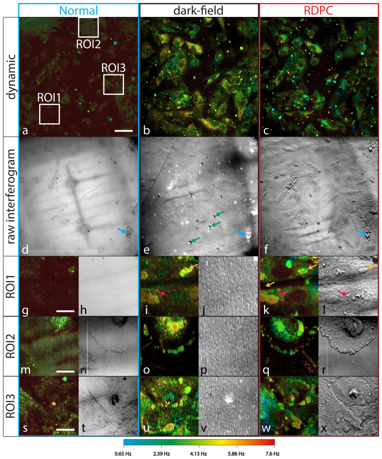
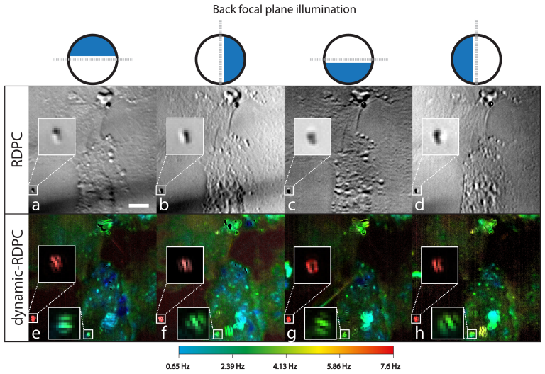
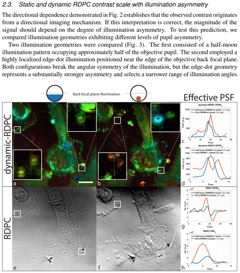
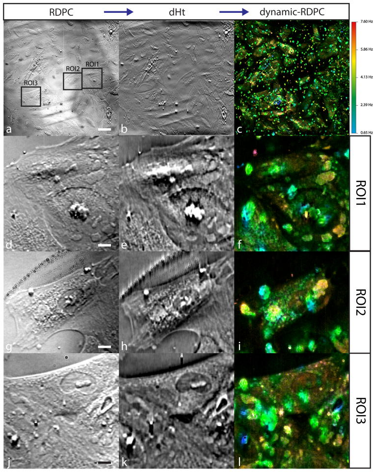
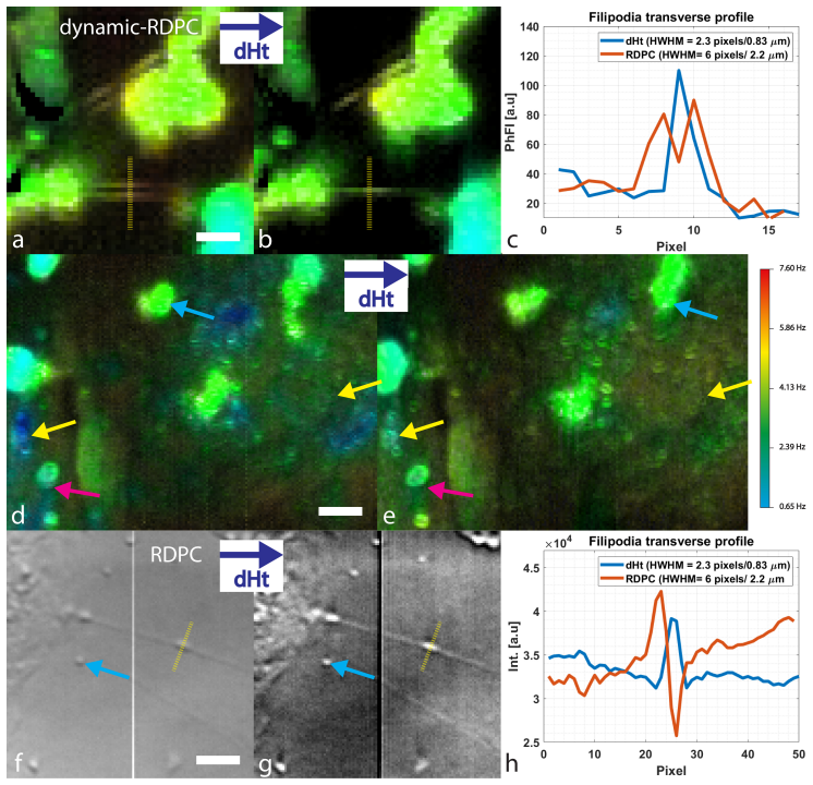
Dynamic dark-field full-field optical coherence tomography suppresses substrate reflections through selective detection of scattered light, while asymmetric illumination produces directional dynamic contrast interpreted as dynamic reflection differential phase contrast. Both recover intracellular activity at highly reflective interfaces that remains poorly visible with conventional dynamic full-field optical coherence tomography. D-RDPC exhibits contrast reversal on illumination inversion, increases with greater asymmetry, and allows spatial localization via directional Hilbert-transform reconstruction, supporting temporal fluctuation analysis.
What carries the argument
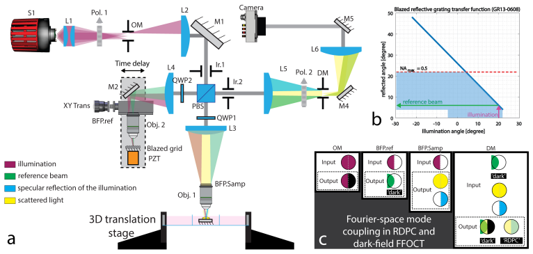
D-dFFOCT for selective scattered-light detection that suppresses substrate reflection, combined with D-RDPC that converts illumination asymmetry into directional phase-gradient contrast for dynamic imaging.
If this is right
- Label-free functional imaging becomes feasible on metallic neural electrodes and implantable devices.
- D-RDPC contrast can be reconstructed with directional Hilbert transforms to restore spatial localization.
- Temporal fluctuation analysis can be applied to the recovered phase-gradient signals for functional readouts.
- The two methods can be used together to cross-validate intracellular dynamics at the same interface.
Where Pith is reading between the lines
- The directional signatures of D-RDPC could be exploited to separate cell-induced signals from static surface roughness in post-processing.
- These contrast mechanisms may extend to other reflective interfaces in materials characterization where substrate scattering dominates.
- Combining the two modalities in a single instrument could provide complementary amplitude and phase information without additional hardware.
Load-bearing premise
The fluctuating signals detected by both methods arise from real intracellular activity inside living cells rather than from residual interface reflections or light-induced effects.
What would settle it
A side-by-side comparison of dynamic signals on identical reflective substrates with and without adherent living cells, or with cellular metabolism pharmacologically blocked, showing whether the contrast disappears when biological activity is absent.
Figures
read the original abstract
Strong reflections from metallic and engineered substrates severely limit label-free functional imaging of living cells at biomaterial interfaces, neural electrodes, and implantable devices. Here we introduce two complementary approaches for recovering intracellular dynamic contrast at highly reflective interfaces. Dynamic dark-field full-field optical coherence tomography (D-dFFOCT) suppresses the dominant substrate reflection and restores intracellular visibility through selective detection of scattered light. In parallel, asymmetric illumination generates a distinct directional dynamic contrast that is most consistently interpreted as dynamic reflection differential phase contrast (D-RDPC). Both approaches reveal intracellular activity that remains poorly visible with conventional dynamic full-field optical coherence tomography. D-RDPC exhibits characteristic signatures of phase-gradient imaging, including contrast reversal upon illumination inversion, enhancement with increasing illumination asymmetry, and recovery of spatial localization through directional Hilbert-transform reconstruction. Together, these results establish new strategies for functional imaging at reflective interfaces and suggest that differential phase contrast signals can support temporal fluctuation analysis.
Editorial analysis
A structured set of objections, weighed in public.
Referee Report
Summary. The manuscript introduces two complementary optical techniques—dynamic dark-field full-field optical coherence tomography (D-dFFOCT) and dynamic reflection differential phase contrast (D-RDPC)—to recover label-free intracellular dynamic contrast at highly reflective biomaterial interfaces where conventional dynamic FFOCT fails. D-dFFOCT suppresses specular substrate reflections via selective scattered-light detection, while D-RDPC uses asymmetric illumination to generate directional phase-gradient contrast. The work reports qualitative observations of intracellular activity and identifies three falsifiable signatures of D-RDPC (contrast reversal on illumination inversion, monotonic enhancement with asymmetry, and Hilbert-transform localization).
Significance. If the intracellular origin of the signals is rigorously validated, the methods would address a practical barrier to functional imaging on metallic and engineered substrates, with potential relevance to neural electrodes and implantable devices. The explicit falsifiable signatures for D-RDPC constitute a methodological strength that supports interpretability.
major comments (1)
- [Abstract and §3] Abstract and §3 (Results): the central claim that both techniques reveal intracellular activity (rather than residual interface artifacts or illumination-induced effects) rests on qualitative observations without reported quantitative validation, error bars, or explicit controls (e.g., fixed-cell or acellular substrate comparisons). This distinction is load-bearing for the functional-imaging conclusion.
minor comments (2)
- [Abstract] Notation for the two acronyms (D-dFFOCT, D-RDPC) is introduced without an explicit comparison table of their respective contrast mechanisms, which would aid readability.
- [§4 (Discussion)] The description of Hilbert-transform reconstruction for spatial localization would benefit from a brief schematic or equation reference to clarify the directional processing step.
Simulated Author's Rebuttal
We thank the referee for the constructive feedback. We address the major comment below and outline planned revisions.
read point-by-point responses
-
Referee: [Abstract and §3] Abstract and §3 (Results): the central claim that both techniques reveal intracellular activity (rather than residual interface artifacts or illumination-induced effects) rests on qualitative observations without reported quantitative validation, error bars, or explicit controls (e.g., fixed-cell or acellular substrate comparisons). This distinction is load-bearing for the functional-imaging conclusion.
Authors: We agree that distinguishing intracellular signals from potential artifacts is critical and that the current presentation relies primarily on qualitative observations. The manuscript highlights three explicit falsifiable signatures for D-RDPC (contrast reversal on illumination inversion, monotonic enhancement with asymmetry, and Hilbert-transform localization) that argue against simple residual reflections or uniform illumination effects. These signatures support interpreting the dynamic contrast as originating from intracellular phase gradients. However, we acknowledge the absence of quantitative validation, error bars, or explicit controls such as fixed-cell and acellular substrate comparisons. We will add these controls and any associated quantitative metrics in the revised manuscript. revision: yes
Circularity Check
No significant circularity; no derivations or equations present
full rationale
The manuscript introduces two experimental optical imaging techniques (D-dFFOCT and D-RDPC) for recovering intracellular contrast at reflective interfaces. The provided abstract and description contain no equations, parameter fits, predictions, or derivation chains. Claims rest on descriptions of physical mechanisms (dark-field suppression, asymmetric illumination) and three falsifiable signatures of phase-gradient contrast, none of which reduce to self-definition, fitted inputs renamed as predictions, or self-citation load-bearing steps. No mathematical structure exists that could exhibit circularity by construction; the work is an empirical methods paper whose central assertions are externally testable via the reported imaging signatures.
Axiom & Free-Parameter Ledger
Reference graph
Works this paper leans on
-
[1]
Foreign body reaction to biomaterials,
J. M. Anderson, A. Rodriguez, and D. T. Chang, “Foreign body reaction to biomaterials,”Seminars in Immunology, vol. 20, no. 2, pp. 86–100, 2008
2008
-
[2]
On the mechanisms of biocompatibility,
D. F. Williams, “On the mechanisms of biocompatibility,”Biomaterials, vol. 29, no. 20, pp. 2941–2953, 2008
2008
-
[3]
Immuneresponsestoimplants–areviewoftheimplications for the design of immunomodulatory biomaterials,
S.Franz,S.Rammelt,D.Scharnweber,andJ.C.Simon,“Immuneresponsestoimplants–areviewoftheimplications for the design of immunomodulatory biomaterials,”Biomaterials, vol. 32, no. 28, pp. 6692–6709, 2011
2011
-
[4]
All charged up about implanted biomaterials,
D. W. Grainger, “All charged up about implanted biomaterials,”Nature Biotechnology, vol. 31, no. 6, pp. 507–509, 2013
2013
-
[5]
Biomaterial based modulation of macrophage polarization: a review and suggested design principles,
R. Sridharan, A. R. Cameron, D. J. Kelly, C. J. Kearney, and F. J. O’Brien, “Biomaterial based modulation of macrophage polarization: a review and suggested design principles,”Materials Today, vol. 18, no. 6, pp. 313–325, 2015
2015
-
[6]
Imagingbiomaterial–tissueinteractions,
Y.S.ZhangandJ.Yao,“Imagingbiomaterial–tissueinteractions,”Trends in Biotechnology,vol.36,no.4,pp.403–414, 2018
2018
-
[7]
From seeing to believing: labelling strategies for in vivo cell-tracking experiments,
F. Progatzky, M. J. Dallman, and C. Lo Celso, “From seeing to believing: labelling strategies for in vivo cell-tracking experiments,”Interface Focus, vol. 3, no. 3, 2013
2013
-
[8]
Quantitative phase imaging in biomedicine,
Y. Park, C. Depeursinge, and G. Popescu, “Quantitative phase imaging in biomedicine,”Nature Photonics, vol. 12, no. 10, pp. 578–589, 2018
2018
-
[9]
High-resolution full-field optical coherence tomography with a Linnik microscope,
A. Dubois, K. Grieve, G. Moneron, R. Lecaque, L. Vabre, and A. C. Boccara, “High-resolution full-field optical coherence tomography with a Linnik microscope,”Applied Optics, vol. 41, no. 4, pp. 805–812, 2002
2002
-
[10]
Three-dimensional cellular-level imaging using full-field optical coherence tomography,
A. Dubois, G. Moneron, K. Grieve, and A. C. Boccara, “Three-dimensional cellular-level imaging using full-field optical coherence tomography,”Physics in Medicine & Biology, vol. 49, no. 7, pp. 1227–1234, 2004
2004
-
[11]
Dynamic full field optical coherence tomography: subcellular metabolic contrast revealed in tissues by temporal analysis of interferometric signals,
C. Apelian, F. Harms, J. Thouvenin, A. C. Boccara, and L. M. Smith, “Dynamic full field optical coherence tomography: subcellular metabolic contrast revealed in tissues by temporal analysis of interferometric signals,” Biomedical Optics Express, vol. 7, no. 4, pp. 1511–1524, 2016
2016
-
[12]
Dynamic full-field optical coherence tomography for live-cell imaging,
J. Thouvenin, C. Apelian, A. C. Boccara, and L. M. Smith, “Dynamic full-field optical coherence tomography for live-cell imaging,”Optics Letters, vol. 42, no. 7, pp. 1322–1325, 2017
2017
-
[13]
Dynamic full-field optical coherence tomography: advances and perspectives,
J. Scholler, M. F. Groux, and A. C. Boccara, “Dynamic full-field optical coherence tomography: advances and perspectives,”Journal of Biomedical Optics, vol. 25, no. 7, 2020
2020
-
[14]
Proliferative and metabolic imaging of tissues through dynamic full-field optical coherence tomography,
J. Scholler, S. Sridharan, M. F. Groux, et al., “Proliferative and metabolic imaging of tissues through dynamic full-field optical coherence tomography,”Biomedical Optics Express, vol. 10, no. 8, pp. 3850–3866, 2019
2019
-
[15]
Dynamic contrast in full-field optical coherence tomography for biological imaging,
H. Defienne, J. Thouvenin, and A. C. Boccara, “Dynamic contrast in full-field optical coherence tomography for biological imaging,”Optica, vol. 7, no. 7, pp. 744–751, 2020
2020
-
[16]
Retinal pigment epithelium imaging by Dynamic Full-Field Optical Coherence Tomography reveals the functional impact of stress and aging,
K. Groux, M. Simonutti, M. Paques, J. A. Sahel, S. Leveillard, and C. Boccara, “Retinal pigment epithelium imaging by Dynamic Full-Field Optical Coherence Tomography reveals the functional impact of stress and aging,” Communications Biology, vol. 5, no. 1, p. 566, 2022
2022
-
[17]
Dynamic full-field optical coherence tomography module adapted to commercial microscopes allows longitudinal in vitro cell culture study,
T. Monfort, S. Azzollini, J. Brogard, et al., “Dynamic full-field optical coherence tomography module adapted to commercial microscopes allows longitudinal in vitro cell culture study,”Communications Biology, vol. 6, article 992, 2023
2023
-
[18]
Drexler and J
W. Drexler and J. G. Fujimoto, Eds.,Optical Coherence Tomography: Technology and Applications, Springer, Berlin, Heidelberg, 2008
2008
-
[19]
Interface self- referenced dynamic full-field optical coherence tomography,
T. Monfort, S. Azzollini, T. Ben Yacoub, I. Audo, S. Reichman, K. Grieve, and O. Thouvenin, “Interface self- referenced dynamic full-field optical coherence tomography,”Biomedical Optics Express, vol. 14, pp. 3491–3505, 2023
2023
-
[20]
T. Monfort, “Ratio-Free Detection and Partial Field Illumination Improve Time-Domain Dynamic Full-Field Optical Coherence Tomography Sensitivity for Retinal Organoid Imaging,” 2026. doi:10.64898/2026.04.18.719402
-
[21]
Modulation contrast microscope,
R. Hoffman and L. Gross, “Modulation contrast microscope,”Applied Optics, vol. 14, no. 5, pp. 1169–1176, 1975
1975
-
[22]
Quantitative phase-gradient imaging at high resolution with asymmetric illumination-based differential phase contrast,
S. B. Mehta and C. J. R. Sheppard, “Quantitative phase-gradient imaging at high resolution with asymmetric illumination-based differential phase contrast,”Optics Letters, vol. 34, no. 13, pp. 1924–1926, 2009
1924
-
[24]
Phase-gradient microscopy in thick tissue with oblique back-illumination,
T. N. Ford, K. K. Chu, and J. Mertz, “Phase-gradient microscopy in thick tissue with oblique back-illumination,” Nature Methods, vol. 9, no. 12, pp. 1195–1197, 2012
2012
-
[25]
Phase-gradient contrast in thick tissue with a scanning microscope,
J. Mertz, “Phase-gradient contrast in thick tissue with a scanning microscope,”Biomedical Optics Express, vol. 5, no. 2, pp. 595–604, 2014
2014
-
[26]
Inverse scattering for reflection intensity phase microscopy,
A. Matlock, A. Sentenac, P. C. Chaumet, J. Yi, and L. Tian, “Inverse scattering for reflection intensity phase microscopy,”Biomedical Optics Express, vol. 11, no. 2, pp. 911–926, 2020
2020
-
[27]
Quantitative oblique back-illumination microscopy for refractive-index tomography in thick scattering samples,
P. Ledwig and F. E. Robles, “Quantitative oblique back-illumination microscopy for refractive-index tomography in thick scattering samples,”Biomedical Optics Express, vol. 12, no. 9, pp. 5482–5496, 2021
2021
-
[28]
Reflectional quantitative phase-contrast microscopy,
Y. Ma, Y. Zhang, Z. Wang, et al., “Reflectional quantitative phase-contrast microscopy,”Applied Optics, vol. 61, no. 13, pp. 3641–3648, 2022
2022
-
[29]
Using the Hilbert transform for 3D visualization of differential interference contrast microscope images,
M. R. Arnison, C. J. Cogswell, N. I. Smith, P. W. Fekete, and K. G. Larkin, “Using the Hilbert transform for 3D visualization of differential interference contrast microscope images,”Journal of Microscopy, vol. 199, no. 1, pp. 79–84, 2000
2000
-
[30]
Linear phase imaging using differential interference contrast microscopy,
M. R. Arnison, K. G. Larkin, C. J. R. Sheppard, N. I. Smith, and C. J. Cogswell, “Linear phase imaging using differential interference contrast microscopy,”Journal of Microscopy, vol. 214, no. 1, pp. 7–12, 2004
2004
-
[31]
Dark-field full-field optical coherence tomography,
E. Auksorius and A. C. Boccara, “Dark-field full-field optical coherence tomography,”Optics Letters, vol. 40, no. 14, pp. 3272–3275, 2015
2015
-
[32]
Dark-field full-field optical coherence tomography using spatial frequency filtering,
T. Xie, M. F. Groux, and A. C. Boccara, “Dark-field full-field optical coherence tomography using spatial frequency filtering,”Optics Letters, vol. 46, no. 18, pp. 4560–4563, 2021
2021
-
[33]
Dark-fieldopticalcoherencetomographyforenhancedscatteringcontrastimaging,
H.Yoo,Y.Zhang,andJ.A.Izatt,“Dark-fieldopticalcoherencetomographyforenhancedscatteringcontrastimaging,” Biomedical Optics Express, vol. 11, no. 5, pp. 2458–2471, 2020
2020
-
[34]
Epi-mode quantitative phase imaging in thick scattering samples,
P. Ledwig and F. E. Robles, “Epi-mode quantitative phase imaging in thick scattering samples,”Optics Express, vol. 27, no. 24, pp. 36047–36060, 2019
2019
-
[35]
QuantitativedifferentialphasecontrastimaginginanLEDarraymicroscope,
L.TianandL.Waller,“QuantitativedifferentialphasecontrastimaginginanLEDarraymicroscope,”Optics Express, vol. 23, no. 9, pp. 11394–11403, 2015
2015
-
[36]
Optimal illumination scheme for isotropic quantitative differential phase contrast microscopy,
Z. Fan and L. Tian, “Optimal illumination scheme for isotropic quantitative differential phase contrast microscopy,” Optics Express, vol. 27, no. 10, pp. 13885–13899, 2019
2019
-
[37]
Wide-field, high-resolution Fourier ptychographic microscopy,
G. Zheng, R. Horstmeyer, and C. Yang, “Wide-field, high-resolution Fourier ptychographic microscopy,”Nature Photonics, vol. 7, no. 9, pp. 739–745, 2013
2013
-
[38]
Label-Free Optical Transmission Tomography for Biosystems: Intracellular Structures and Dynamics,
V. Mazlin, O. Thouvenin, S. Alhaddad, M. Boccara, and C. Boccara, “Label-Free Optical Transmission Tomography for Biosystems: Intracellular Structures and Dynamics,”Biomedical Optics Express, vol. 13, no. 8, pp. 4190–4203, 2022
2022
-
[39]
Holoscopy—holographic optical coherence tomography,
D. Hillmann, G. Hulsken, H. Liu, et al., “Holoscopy—holographic optical coherence tomography,”Optica, vol. 3, no. 4, pp. 366–374, 2016
2016
-
[40]
Reproducingdiabeticretinopathy features using newly developed human induced-pluripotent stem cell-derived retinal Müller glial cells,
A. Couturier, G. Blot, L. Vignaud, C. Nanteau, C. Slembrouck-Brec, V. Fradot, N. Acar, J. A. Sahel, R. Tadayoni, G.Thuret,F.Sennlaub,J.E.Roger,O.Goureau,X.Guillonneau,andS.Reichman,“Reproducingdiabeticretinopathy features using newly developed human induced-pluripotent stem cell-derived retinal Müller glial cells,”Glia, vol. 69, no. 7, pp. 1679–1693, 2021
2021
-
[41]
Optical properties of biological tissues: a review,
S. L. Jacques, “Optical properties of biological tissues: a review,”Physics in Medicine & Biology, vol. 58, no. 11, pp. R37–R61, 2013
2013
-
[42]
Optical coherence tomography (OCT): a review,
J. M. Schmitt, “Optical coherence tomography (OCT): a review,”IEEE Journal of Selected Topics in Quantum Electronics, vol. 5, no. 4, pp. 1205–1215, 1999
1999
discussion (0)
Sign in with ORCID, Apple, or X to comment. Anyone can read and Pith papers without signing in.