REVIEW 3 major objections 5 minor 11 references
Vendor-Agnostic Joint Relaxometry and Myelin Water Fraction Mapping with B1 and Motion Correction
T0 review · 3 major / 5 minor · reviewed 2026-08-11 · deepseek-v4-flash
Pith's one-line read The paper proposes a vendor-agnostic MRI sequence that jointly maps T1, T2, and myelin water fraction from one motion-corrected 4.7-minute scan, validated in simulations, phantoms, volunteers, and pediatric cases.
desk verdict Dual T2-prep QALAS is a real, well-validated sequence modification that improves short-T2 estimation, and the vendor-agnostic open-source package is a practical contribution—but the MWF numbers rest on model assumptions and a small in vivo cohort, so the paper is conditionally acceptable after revision. read the letter →
The pith
A machine-rendered reading of the paper's core claim, the machinery that carries it, and where it could break.
The reading
What carries the argument
The load-bearing object is the MWF-QALAS pulse sequence itself, a myelin-sensitive variant of the QALAS scheme: two $T_2$-preparation modules (20 ms and 80 ms), one inversion pulse, and six FLASH readouts per repetition that sample the recovery curve at effective inversion times of 110, 1010, 1910, 2810, and 3710 ms. This design is coupled to a temporal subspace reconstruction in which the signal evolution is represented by a low-dimensional basis and a motion-aware non-uniform fast Fourier transform operator applies per-repetition rigid transforms estimated from self-navigation images. Parameter maps come from Bloch-simulated dictionary matching for $T_1$ and $T_2$, and from a partial-volume dictionary for three water compartments with fixed relaxation values: myelin water, intra/extra-cellular water, and free water. The short 20 ms preparation is what gives the sequence sensitivity to the fast-decaying myelin component that the standard 100 ms preparation cannot see.
What would settle it
Acquire the proposed sequence on a multi-compartment phantom with known short-$T_2$ volume fractions and no water exchange, or on ex vivo brain tissue with histologically stained myelin; if the fitted myelin water fraction deviates from the known fraction beyond the reported agreement limits (roughly $\pm 0.05$ absolute in vivo), the three-compartment model or the sequence's short-$T_2$ sensitivity is falsified.
Extended reading notes
Core claim
The central discovery is that the myelin-water short-$T_2$ component, which decays too quickly for the standard 100 ms $T_2$-preparation module, becomes measurable when a second preparation with 20 ms echo time is inserted before the standard one, with both followed by FLASH readouts within the same repetition. The authors show this dual-preparation design keeps $T_1$ mapping accuracy essentially unchanged while substantially reducing short-$T_2$ bias, and they integrate it with dictionary-based Bloch-simulation matching and a three-compartment partial-volume model to extract myelin water fraction. They further claim that the whole workflow, including motion estimation from undersampled self-navigation volumes and retrospective motion-corrected subspace reconstruction, can be expressed in an open, hardware-independent sequence format and run on scanners from two vendors without vendor-specific sequence code.
Load-bearing premise
The entire myelin water fraction estimate assumes each voxel is a weighted sum of exactly three non-exchanging water pools with fixed relaxation values (myelin water with $T_2$ = 15 ms, intra/extra-cellular water, and free water), so if real tissue has different relaxation times or water exchange, the fraction will be biased even with perfect acquisition and reconstruction.
Editorial extensions
If this is right
- A single 4.7-minute whole-brain acquisition replaces separate long scans for $T_1$, $T_2$, and myelin water fraction mapping, at 1 mm isotropic resolution and 3T.
- Short-$T_2$ accuracy improves enough to lower mean bias in the below-30 ms range from $-3.1$ ms to $-0.1$ ms, directly benefiting myelin-relevant measurements.
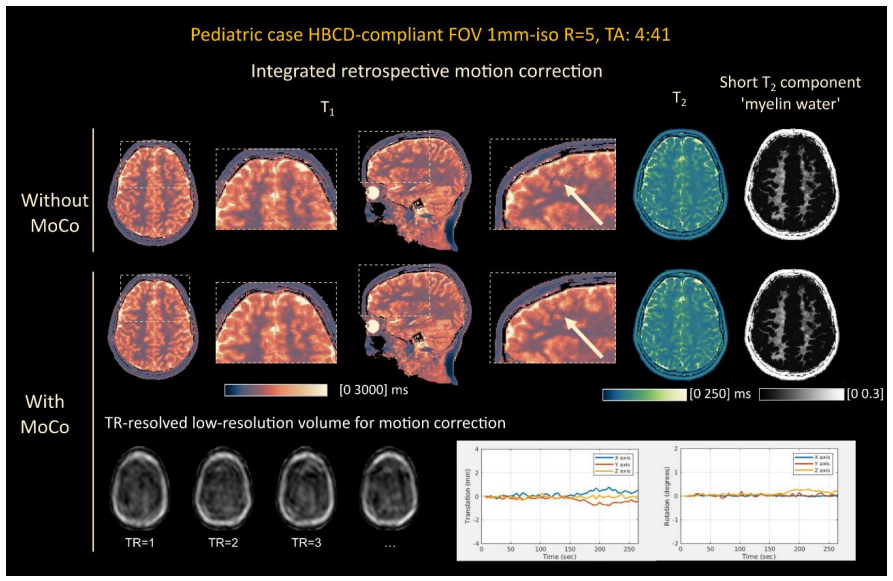
- Self-navigation motion correction removes the need for external tracking hardware; retrospective simulations reduce normalized root-mean-square error against a motion-free reference from 29.53% to 13.16% using the estimated trajectory.
- Because both sequence and reconstruction are expressed in an open, hardware-independent format, the same protocol can run on scanners from two major vendors, with cross-vendor intraclass correlations of 0.987 for $T_2$ and 0.771 for myelin water fraction.
- Open-sourced sequences and code permit other sites to run the protocol without vendor-specific re-implementation, subject only to an interpreter for the sequence format and local safety checks.
Reading between the lines
- An untested implication is that improved short-$T_2$ sensitivity may also help quantify short-$T_2$ compartments in disease, but the paper only demonstrates normal adult and pediatric brains.
- The motion-correction result at 5.4 s temporal resolution implies a practical limit: intra-repetition motion, or motion larger than about one $B_1$ voxel relative to calibration, could still bias maps because a single $B_1$ map is assumed.
- The model-derived myelin water fraction is not histologically validated; if exchange or population-specific relaxation times matter, estimates in developing or diseased tissue could diverge from true myelin content even though the acquisition is sound.
- The 13.02% mean percent difference from the reference myelin-water method suggests the two techniques measure overlapping but not identical quantities; a direct calibration study would need to decide which reference is closer to true myelin.
Signed reviews
Editorial analysis
A structured set of objections, weighed in public.
Referee Report
Summary. The paper presents MWF-QALAS, a Pulseq-based vendor-agnostic 3T quantitative MRI sequence that extends 3D-QALAS with a second short T2-preparation (TE=20 ms) and a sixth FLASH readout, enabling joint T1, T2, and myelin water fraction (MWF) mapping in about 4.7 minutes at 1 mm isotropic resolution. A center-out Cartesian spiral ordering supports self-navigated retrospective rigid motion correction in a regularized temporal subspace reconstruction. A turbo-AFI scan provides B1+ and coil sensitivity calibration, and dictionary-based Bloch simulation is used for T1/T2 and three-compartment MWF estimation. Validation includes numerical simulations, a NIST/ISMRM phantom, three healthy adult volunteers on Siemens and GE scanners, controlled motion experiments, and illustrative pediatric cases.
Significance. If the results hold, this is a practical contribution to multi-site and pediatric neuroimaging: the sequence is open-source, expressed in vendor-neutral Pulseq, includes integrated motion correction and harmonized calibration, and improves short-T2 estimation relative to standard 3D-QALAS (short-T2 simulation bias -0.1 ms vs -3.1 ms; phantom low-T2 slope 0.91 vs 0.78). The multi-layer validation and public code release are genuine strengths. The principal caveat is that MWF is a model-derived estimate under a fixed, non-exchanging three-compartment model, and the only in vivo MWF comparison is against 3D-GRASE in three adults; external validity in pediatric and pathological tissue is therefore not yet established.
major comments (3)
- [Section 2.3, 3.3, 4] The central claim of myelin water fraction mapping rests on the fixed three-compartment, no-exchange model in Section 2.3, which fixes myelin-water T2 at 15 ms and intra/extracellular T2 at 50/70 ms. The Discussion acknowledges that MWF is not a direct myelin measure and that these assumptions may vary across populations and pathologies, but the in vivo validation in Section 3.3 compares MWF-QALAS to 3D-GRASE in only three healthy adults, and 3D-GRASE is itself a model-based estimate. With ICC=0.732 and Pearson r=0.757, the agreement is moderate rather than strong. This evidence is insufficient to support unbiased MWF mapping in the pediatric and pathological populations emphasized in the Introduction. Please add a sensitivity analysis varying compartment relaxation times and/or exchange, or restrict the claims to 'short-T2 water fraction under the assumed model' throughout the title, abstract, and results.
- [Section 2.5, 3.1] The numerical simulation is a self-consistency check rather than an independent accuracy validation: the data are generated with the same Bloch signal model that builds the dictionary. The phantom experiments provide the non-circular accuracy check, but the phantom T2 regression slope is 0.86 overall and 0.91 for T2 <= 70 ms, so absolute accuracy is still imperfect. The text should describe the simulation results as demonstrating relative improvement and short-T2 sensitivity, not standalone accuracy.
- [Section 3.5] Motion robustness is a core contribution, but the quantitative motion experiments are single realizations without variance estimates, and in Experiment 6 the corrected NRMSE remains 20.47%. The Discussion acknowledges residual error, but the abstract and results should more clearly state the empirical motion range and residual errors rather than implying general motion immunity.
minor comments (5)
- [Section 2.1, 3.1] The second T2-preparation is given as TE=80 ms 'as in the original 3D-QALAS', but standard QALAS is described elsewhere as TE=100 ms; please reconcile this inconsistency.
- [Section 2.3] The dictionary grid step sizes and the wavelet regularization parameter are said to be empirically selected; please report the actual values used so that the short-T2 estimates are reproducible.
- [Section 3.4] Cross-vendor MWF agreement is moderate (ICC=0.771; pairwise Pearson 0.706-0.902); the sentence comparing these results to within-vendor MRF repeatability is not an apples-to-apples comparison and should be rephrased.
- [Section 3.3, 3.6] The pediatric cases are illustrative; the text should label them as such and not as validation of MWF accuracy in children, particularly since no reference standard was acquired in those subjects.
- [Section 2.4, abstract] The abstract mentions B1- mapping, while the methods describe coil sensitivity estimation from the ACS region; clarify the relationship between B1- and receive sensitivity.
Circularity Check
Numerical-simulation validation is a dictionary self-consistency check; independent phantom and in-vivo evidence keep the central claim non-circular.
-
self definitional
[Section 2.5 (Numerical Simulation), Section 2.3 (dictionary matching), Section 3.1 (simulation results)]
"Using the signal model of each of the imaging sequences, source contrast images were simulated. To mimic real-world experimental conditions, 1%-5% Gaussian noise was added to the simulated images... The dictionary-matching technique relied on a precomputed library of signal intensities corresponding to different combinations of T1 and T2 values (Cho et al., 2024), allowing precise estimation of these parameters from the noisy data."
The synthetic 'reference' images are generated with the same Bloch-equation signal model that is used to construct the matching dictionary. Dictionary matching therefore inverts the generative model by construction, so the reported short-T2 bias improvement (from -3.1 ms to -0.1 ms) demonstrates self-consistency of the simulator/dictionary pair rather than accuracy against independently measured tissue properties. This simulation cannot falsify the underlying signal model; the phantom and in-vivo comparisons provide the independent validation.
full rationale
The central claim is not circular: T1/T2 estimation is based on Bloch-simulated dictionary matching, a forward physics model rather than a fit to measured outputs; B1+ mapping uses the analytical AFI relation of Yarnykh; phantom validation uses inversion-recovery and spin-echo references; in-vivo MWF is compared with an external 3D-GRASE method; and motion correction is tested against motion-free references. The one genuine reduction is in the numerical simulation, where the data-generation model and the dictionary model are identical, making that validation a self-consistency check. The MWF three-compartment fixed-parameter model is an acknowledged assumption, not a circular derivation, and the 3D-GRASE comparison, while model-based and small in scale, is an independent external benchmark rather than a restatement of the paper's own inputs. Overall: one validation step reduces by construction, but the central claim retains independent phantom and in-vivo support.
Assumptions & free parameters
free parameters (5)
- Short T2-preparation echo time (TE_T2prep_short) =
20 ms
- Long T2-preparation echo time (TE_T2prep_long) =
80 ms
- Compartment relaxation times for MWF model =
myelin: T1=150 ms, T2=15 ms; ICF: T1=1100/1300 ms, T2=50/70 ms; free water: T1=4500 ms, T2=500 ms
- Dictionary grid step sizes =
T1: 10 ms to 3000 ms, 100 ms to 5000 ms; T2: 2 ms to 350 ms, 20 ms to 500 ms; B1+: 0.05 from 0.65 to 1.35
- Wavelet regularization parameter lambda and subspace dimension L =
not reported in main text
assumptions (6)
- standard math Bloch equations accurately model the implemented Pulseq sequence dynamics
- domain assumption Complete spoiling of transverse magnetization before each FLASH readout
- domain assumption Three non-exchanging compartments with fixed relaxation times represent tissue water
- domain assumption Head motion is rigid and constant within each TR (5.4 s), and the per-TR motion estimate is accurate
- domain assumption A single B1+ map is valid across the whole scan
- domain assumption Coil sensitivity maps from the tAFI ACS region are accurate
Cite this review
Pith. "Pith review of Vendor-Agnostic Joint Relaxometry and Myelin Water Fraction Mapping with B1 and Motion Correction." pith.science (2026). https://pith.science/paper/ES5IRNO5
@misc{pith2026260807687,
  author       = {Pith},
  title        = {Pith review of: Vendor-Agnostic Joint Relaxometry and Myelin Water Fraction Mapping with B1 and Motion Correction},
  year         = {2026},
  howpublished = {\url{https://pith.science/paper/ES5IRNO5}},
  note         = {Machine review of arXiv:2608.07687}
}
read the original abstract
Obtaining consistent quantitative maps of myelin content and relaxation times across different sites and vendors is essential for advancing our understanding of brain development. Herein, we present a harmonized, vendor-agnostic magnetic resonance acquisition method designed for joint T1, T2, and myelin water fraction mapping, along with a method for rapid B1+ and B1- field estimation. We used our dictionary-based fitting and multi-compartment modeling for joint mapping of T1, T2 and myelin water fraction. Self-navigation-based retrospective motion correction was integrated with subspace reconstruction to track and correct rigid head motion during scanning, operating without the need for external hardware. Simulations, phantom and in vivo experiments confirmed the sensitivity and accuracy of the method, particularly for short T2 values corresponding to myelin, and demonstrated consistent performance across multiple scanner types. Coupled with the harmonized calibration scan, the proposed package offers a practical tool for multi-site, multi-vendor neuroimaging studies in both adult and pediatric populations.
Figures


Figures from the paper (5 more)



Reference graph
Works this paper leans on
-
[1]
P., Roth, N., Roth, A., Prabhu, S
Afacan, O., Erem, B., Roby, D. P., Roth, N., Roth, A., Prabhu, S. P., & Warfield, S. K. (2016). Evaluation of motion and its effect on brain magnetic resonance image quality in children. Pediatric Radiology, 46(12), 1728–1735. A G Teixeira, R. P., Neji, R., Wood, T. C., Baburamani, A. A., Malik, S. J., & Hajnal, J. V. (2020). Controlled saturation magneti...
work page 2016
-
[95]
Wicaksono, K. P., Fushimi, Y., Nakajima, S., Sakata, A., Okuchi, S., Hinoda, T., Oshima, S., Otani, S., Tagawa, H., Urushibata, Y., & Nakamoto, Y. (2023). Accuracy, repeatability, and reproducibility of T1 and T2 relaxation times measurement by 3D magnetic resonance fingerprinting with different dictionary resolutions. European Radiology, 33(4), 2895–2904...
work page 2023
-
[962]
Saltarelli, G., Di Cerbo, G., Innocenzi, A., De Felici, C., Splendiani, A., & Di Cesare, E. (2025). Quantitative MRI in neuroimaging: A review of techniques, biomarkers, and emerging clinical applications. Brain Sciences, 15(10),
work page 2025
-
[1039]
Gómez, P. A., Cencini, M., Golbabaee, M., Schulte, R. F., Pirkl, C., Horvath, I., Fallo, G., Peretti, L., Tosetti, M., Menze, B. H., & Buonincontri, G. (2020). Rapid three- dimensional multiparametric MRI with quantitative transient-state imaging. Scientific Reports, 10(1), 13769. Griesler, T., Stebani, J., Kaplan, S., Angelov, I., Albert, P., Blaimer, M....
work page Pith review arXiv 2020
-
[1053]
Deoni, S. C. L., Rutt, B. K., Arun, T., Pierpaoli, C., & Jones, D. K. (2008). Gleaning multicomponent T1 and T2 information from steady-state imaging data: 2D Relaxometry With Steady-State Imaging. Magnetic Resonance in Medicine, 60(6), 1372–1387. Deshmane, A., McGivney, D. F., Ma, D., Jiang, Y., Badve, C., Gulani, V., Seiberlich, N., & Griswold, M. A. (2...
work page 2008
-
[1088]
I., Calamante, F., & Hu, H
Seiberlich, N., Gulani, V., Campbell-Washburn, A., Sourbron, S., Doneva, M. I., Calamante, F., & Hu, H. H. (2020). Quantitative Magnetic Resonance Imaging. Academic Press. Shrout, P. E., & Fleiss, J. L. (1979). Intraclass correlations: uses in assessing rater reliability. Psychological Bulletin, 86(2), 420–428. Somerville, L. H., Bookheimer, S. Y., Buckne...
2020
-
[1372]
National Institute on Drug Abuse. (2021, December 2). The HEALthy brain and child development study. National Institute on Drug Abuse. https://nida.nih.gov/research/nida-research-programs-activities/healthy-brain- child-development-study Nave, K.-A. (2010). Myelination and support of axonal integrity by glia. Nature, 468(7321), 244–252. Nielsen, J.-F., & ...
work page 2010
-
[1552]
Lebel, C., & Deoni, S. (2018). The development of brain white matter microstructure. NeuroImage, 182, 207–218. Lee, J., Hyun, J.-W., Lee, J., Choi, E.-J., Shin, H.-G., Min, K., Nam, Y., Kim, H. J., & Oh, S.-H. (2021). So you want to image myelin using MRI: An overview and practical guide for myelin water imaging. Journal of Magnetic Resonance Imaging, 53(...
work page 2018
Show all 11 references
-
[1682]
S., Wang, Z., Zhou, Z., Dai, E., Liberman, G., Dong, Z., Gong, T., He, H., Zhong, J., Bilgic, B., & Setsompop, K
Cao, X., Liao, C., Iyer, S. S., Wang, Z., Zhou, Z., Dai, E., Liberman, G., Dong, Z., Gong, T., He, H., Zhong, J., Bilgic, B., & Setsompop, K. (2022). Optimized multi-axis spiral projection MR fingerprinting with subspace reconstruction for rapid whole- brain high-isotropic-res...
2022
-
[2032]
Karakuzu, A., Biswas, L., Cohen-Adad, J., & Stikov, N. (2022). Vendor-neutral sequences and fully transparent workflows improve inter-vendor reproducibility of quantitative MRI. Magnetic Resonance in Medicine, 88(3), 1212–1228. Kvernby, S., Warntjes, M., Carlhäll, C.-J., Engva...
2022
-
[2486]
Veldmann, M., Ehses, P., Chow, K., Nielsen, J.-F., Zaitsev, M., & Stöcker, T. (2022). Open-source MR imaging and reconstruction workflow. Magnetic Resonance in Medicine, 88(6), 2395–2407. Wang, F., Dong, Z., Reese, T. G., Rosen, B., Wald, L. L., & Setsompop, K. (2022). 3D Echo...
2022
Reviewed August 11, 2026 · model on record in the stance chip above.
Discussion (0). Continue with ORCID to comment.