REVIEW 3 major objections 4 minor 114 references
X-ray Fokker--Planck equation for paraxial imaging
T0 review · 3 major / 4 minor · reviewed 2026-08-14 · deepseek-v4-flash
Pith's one-line read A single Fokker–Planck equation merges x-ray phase contrast with dark-field blur.
desk verdict A genuinely useful unification of phase contrast and SAXS diffusion, but the headline equation has an operator-ordering inconsistency that needs fixing before forward models should use it. read the letter →
The pith
A machine-rendered reading of the paper's core claim, the machinery that carries it, and where it could break.
The reading
What carries the argument
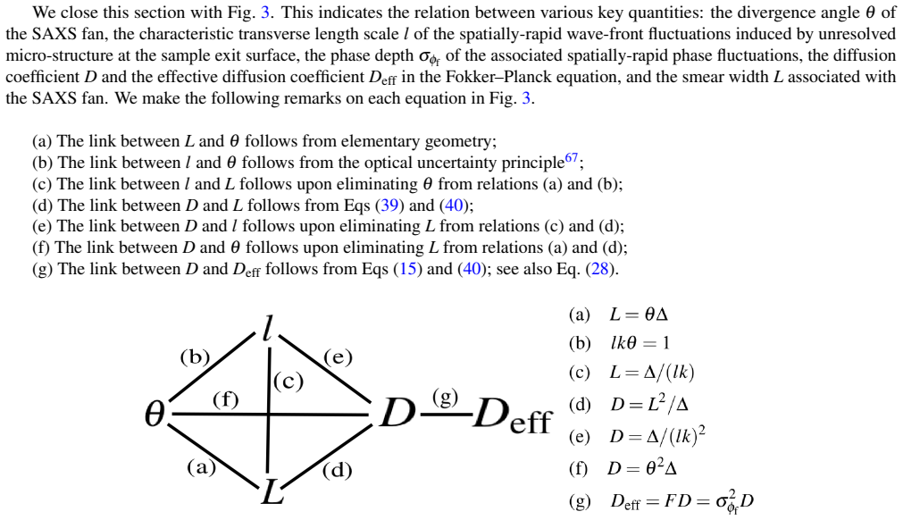
The engine of the paper is a continuity equation for transverse energy flow in which the current has two parts: a coherent Poynting-vector current $I\nabla_\perp\phi/k$ and a diffusive Fick-type current $-\nabla_\perp[DI]$ entering with a weight $F$. Combining these gives the finite-difference Fokker–Planck update of Eq. (11). The diffusion coefficient is $\Delta$-dependent, $D=L^2/\Delta$, so that the small-angle-scattering blur expands as the propagation distance itself rather than as its square root. For the extended form, the Kramers–Moyal equation replaces the scalar $D$ with a hierarchy of diffusion tensors defined as moments of the position-dependent scattering kernel, allowing elliptical and structured scattering fans to be tracked. The hierarchy also supplies a principled truncation point, since Pawula's theorem forbids stopping at any order above second short of keeping everything.
What would settle it
Take a sample whose scattering fan is known to be broad or structured and image it at three or more propagation distances; if the projected thickness recovered from each pair of distances via Eq. (61) changes with the chosen pair, or if the inferred $D_{\rm eff}/\Delta$ is not constant, the second-order diffusion truncation behind the Fokker–Planck equation has broken down.
Extended reading notes
Core claim
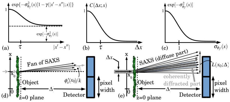
On the paper's own terms, the central claim is that the intensity of a paraxial quasi-monochromatic beam evolves as $$ \frac{\partial I}{\partial z}=-\frac{1}{k}\nabla_\perp\cdot[I\nabla_\perp\phi]+F\nabla_\$perp^{2}$[DI]. $$ The first term is the transport-of-intensity contribution from coherent refraction; the second adds a diffusive blur whose strength is the local fraction of the beam converted to small-angle scattering times a diffusion coefficient. The paper derives this equation twice: first by adding the coherent and diffusive currents in an energy-conserving way, then from first principles starting from a decomposition of the exit-surface phase into a slowly varying part and an unresolved random part. The first-principles route makes the physics explicit: the scattering fraction is $F=1-\exp(-\sigma_{\varphi_f}^2)$, the diffusion coefficient is $D=L^2/\Delta$, and the blur width $L$ grows linearly with propagation distance. When the scattering fan is anisotropic or highly structured, the same construction produces the Kramers–Moyal equation, whose successive terms are moments of the local scattering kernel.
Load-bearing premise
The load-bearing premise is that each local small-angle scattering fan is narrow, smooth, and almost forward-peaked, so that the blur it produces can be captured by a second-order Taylor expansion in spatial derivatives over the propagation distances used.
Editorial extensions
If this is right
- A near-field image of a thin sample with unresolved microstructure can be computed by one finite-difference Fokker–Planck update from the exit-surface intensity, phase, and diffusion coefficient, without resolving the sample's internal speckle.
- Two near-field images at different propagation distances can be combined, through Eqs. (60)–(61), to recover the projected thickness of a single-material sample independently of its dark-field scattering; the dark-field coefficient then follows algebraically.
- The same equations apply to paraxial imaging with visible light, electrons, and neutrons, because they depend only on the paraxial scalar wave equation and energy conservation.
- The geometric-flow speckle-tracking equation gains a diffusive term, so a single augmented equation can in principle reconstruct both phase and effective diffusion coefficient from speckle images.
- Anisotropic or structured scattering fans can be represented by the Kramers–Moyal tensors, which can be measured by raster-scanning a focused beam over the sample.
Reading between the lines
- If the two-distance inversion is stable, it suggests a practical protocol: collect two defocus distances and solve for thickness and dark-field simultaneously, rather than acquiring a full scattered-intensity map.
- The diffusion-tensor moments in the Kramers–Moyal equation could be inverted directly from ordinary near-field images, connecting phase retrieval with tensor tomography without dedicated grating or raster-scan setups.
- Because the Fokker–Planck update is linear in propagation distance for fixed $D$, time-lapse imaging of a slowly evolving sample could be modelled by the same equation with time as the evolution parameter, an extension the paper does not develop.
- The same second-order expansion of the scattering kernel suggests that resolution-dependent classification of speckle as coherent versus diffuse could be used to tune detectors or binning to suppress dark-field blur in phase-contrast imaging.
Signed reviews
Editorial analysis
A structured set of objections, weighed in public.
Referee Report
Summary. The paper proposes an x-ray Fokker–Planck equation for paraxial near-field imaging of thin samples, which augments the transport-of-intensity equation (TIE) with a diffusive term that models unresolved small-angle x-ray scattering (SAXS). Two derivations are presented: a phenomenological merging of the TIE and a diffusion equation in Sec. 2.1, and a first-principles derivation in Sec. 2.2 based on Fresnel propagation, ensemble averaging over a Gaussian stationary fast phase, and a second-order narrow-fan expansion. Section 3 generalises the result to a Kramers–Moyal equation with a hierarchy of SAXS-fan moments, and Section 4 outlines forward- and inverse-problem applications, including a two-distance phase-retrieval formula. The central claim is that Eq. (9) (and its one-dimensional form, Eq. (10)) simultaneously captures propagation-based phase contrast and dark-field blurring, with the Kramers–Moyal form, Eq. (50), handling anisotropic scattering fans.
Significance. If the proposed equations are valid, they provide a compact and practical formalism for modelling combined coherent and diffuse transport in x-ray imaging, with potential applications to phase retrieval, dark-field tomography, and imaging with visible light, electrons, and neutrons. The paper's strengths include a transparent microscopic derivation that gives independent physical meaning to the scattering fraction F, the phase depth σ_φf, the blur width L, and the diffusion coefficient D (Eqs. (28), (39), and (40)), and a Kramers–Moyal hierarchy that connects the Fokker–Planck equation to a rigorous moment expansion of the SAXS fan. The discussion of detector-resolution dependence of what is classified as SAXS is also valuable. However, the headline equation Eq. (9) has an operator-ordering inconsistency with the microscopic derivations that needs to be resolved before the paper can be accepted as is.
major comments (3)
- [§2.1.3, Eq. (9)] The diffusion term F(x,y)∇²⊥[D(x,y;Δ)I] places the diffusion coefficient inside the Laplacian, but the first-principles derivation in §2.2 (Eq. (38)) and the Kramers–Moyal expansion in §3 (Eq. (50)) both place D outside the derivatives, giving FD∇²⊥I (in the isotropic case). For spatially varying D the two forms differ by 2F∇D·∇I + FI∇²D. These are not higher-order corrections in Δ; they survive at fixed small Δ. Concretely, for I_s(x)=1 with constant F and varying D(x), Eq. (9) predicts I(z=Δ)−I_s ≈ ΔFD''(x), whereas the kernel model Eq. (42) leaves a uniform intensity unchanged and Eq. (13) gives zero. Eq. (9) is stated unconditionally and is highlighted in the abstract, so this is a load-bearing inconsistency that should be corrected by replacing ∇²⊥(DI) with D∇²⊥I (or with ∇⊥·(D∇⊥I)) whenever D varies, or by explicitly restricting Eq. (9) to ∇D≈0.
- [§2.1.2, Eq. (7)] The phenomenological diffusive current J(2)_⊥ = −∇⊥(DI) in Eq. (7) is nonstandard: combined with continuity it produces ∇²(DI), which is not the usual Fick-law form ∇·(D∇I). The uncritical use of this flux in the merging argument is the origin of the ordering discrepancy with the microscopic derivation. The authors should either justify why the current should contain I∇D or align the phenomenological derivation with the microscopic result by using the standard diffusive current −D∇I, which would yield F∇⊥·(D∇⊥I).
- [Abstract] The claim that the domain of applicability of the Fokker–Planck and Kramers–Moyal equations is 'at least as broad as that of the transport-of-intensity equation' is overstated. The diffusive term relies on the second-order narrow-fan truncation (Eqs. (34)–(35) and Eq. (44)), which requires each local SAXS fan to be narrow, smooth, and forward-peaked and the propagation distance Δ to be small enough for the Taylor expansion to be accurate. The TIE itself does not require these additional conditions. The abstract and conclusion should be qualified, for example by saying 'as broad as the TIE in the near-field limit of narrow SAXS fans.'
minor comments (4)
- [Introduction, p. 2] There is a typographical repetition: 'the the Fokker–Planck equation' should read 'the Fokker–Planck equation'.
- [Eq. (28)] The arrow notation 'weak SAXS−−−−−−→' is unusual and could be replaced with a clearer statement such as 'in the weak-SAXS limit σ²_φf ≪ 1, F(x) ≈ σ²_φf(x)'.
- [Eq. (33)] The notation kx=φ′s(x)−kx/Δ in the definition of d(x;x0) is confusing because kx appears on both sides with different meanings; using a separate symbol for the Fourier variable would improve readability.
- [§2.2, Eq. (34)] The argument that Q=1 follows solely from energy conservation is terse; a sentence clarifying that the kernel integrates to unity (cf. Eq. (45)) would make the step easier to follow.
Circularity Check
No significant circularity: the Fokker–Planck equation is derived from Fresnel propagation plus a controlled small-angle expansion, with self-citations only to standard non-load-bearing results.
full rationale
The central derivation is not circular. Section 2.2 begins from Fresnel diffraction (Eq. 16), ensemble-averaged Gaussian fast-phase correlations (Eqs. 21–25), and a second-order Taylor expansion of the narrow SAXS blur kernel (Eq. 34). This yields the propagated intensity in Eq. (38), whose diffusive increment contains σ²_φf L² d²I/dx²; the subsequent identifications D = L²/∆ and F ≈ σ²_φf give D and F microphysical content rather than fitting them to the target equation. The phenomenological derivation in Sec. 2.1 is an explicit model-building step (adding the TIE and diffusion currents), not a disguised prediction. Self-citations to Paganin's textbook, to Paganin et al. (2002) for phase retrieval, and to the authors' speckle-tracking work are to standard, externally supported results and are not load-bearing for the new Fokker–Planck/Kramers–Moyal claims; the statistical-optics steps cite Nesterets, Yashiro, Goodman, and Voronovich. The operator-ordering discrepancy between Eq. (9)'s F∇²[DI] and the D-outside form of Eq. (38) is a genuine correctness or qualification issue for position-dependent D, but it is not circularity: Eq. (38) is an independent approximation, and the paper only asserts equivalence to the simpler D-commuting form under a stated slow-D assumption. No step reduces to its own input by construction.
Assumptions & free parameters
free parameters (5)
- D(x,y;Δ) diffusion coefficient
- F(x,y) scattering fraction
- σ_φf(x) phase depth
- L(x;Δ) blur width
- Higher-order SAXS-fan moments D^(M)_m,M-m(x,y;Δ)
assumptions (6)
- domain assumption Paraxial scalar Fresnel propagation
- domain assumption Projection approximation for a thin sample
- domain assumption Gaussian, stationary statistics for the fast phase φ_f
- domain assumption Slow variation of I_s and φ_s
- domain assumption Narrow-fan second-order expansion for the blur kernel
- standard math Pawula's theorem on Kramers-Moyal truncation
Cite this review
Pith. "Pith review of X-ray Fokker--Planck equation for paraxial imaging." pith.science (2026). https://pith.science/paper/SV6SZYRT
@misc{pith2026190801473,
  author       = {Pith},
  title        = {Pith review of: X-ray Fokker--Planck equation for paraxial imaging},
  year         = {2026},
  howpublished = {\url{https://pith.science/paper/SV6SZYRT}},
  note         = {Machine review of arXiv:1908.01473}
}
read the original abstract
The Fokker--Planck Equation can be used in a partially-coherent imaging context to model the evolution of the intensity of a paraxial x-ray wave field with propagation. This forms a natural generalisation of the transport-of-intensity equation. The x-ray Fokker--Planck equation can simultaneously account for both propagation-based phase contrast, and the diffusive effects of sample-induced small-angle x-ray scattering, when forming an x-ray image of a thin sample. Two derivations are given for the Fokker--Planck equation associated with x-ray imaging, together with a Kramers--Moyal generalisation thereof. Both equations are underpinned by the concept of unresolved speckle due to unresolved sample micro-structure. These equations may be applied to the forward problem of modelling image formation in the presence of both coherent and diffusive energy transport. They may also be used to formulate associated inverse problems of retrieving the phase shifts due to a sample placed in an x-ray beam, together with the diffusive properties of the sample. The domain of applicability for the Fokker--Planck and Kramers--Moyal equations for paraxial imaging is at least as broad as that of the transport-of-intensity equation which they generalise, hence the technique is also expected to be useful for paraxial imaging using visible light, electrons and neutrons.
Figures

Reference graph
Works this paper leans on
-
[1]
(ed.) Handbook of X-Ray Imaging: Physics and Technology (CRC Press, Boca Raton, 2018)
Russo, P. (ed.) Handbook of X-Ray Imaging: Physics and Technology (CRC Press, Boca Raton, 2018)
2018
-
[2]
E., Logan, C
Martz, H. E., Logan, C. M., Schneberk, D. J. & Shull, P. J. X-Ray Imaging: Fundamentals, Industrial Techniques and Applications (CRC Press, Boca Raton, 2009)
2009
-
[3]
& Sakdinawat, A
Attwood, D. & Sakdinawat, A. X-Rays and Extreme Ultraviolet Radiation: Principles and Applications (Cambridge University Press, Cambridge, 2016), 2 edn
2016
-
[4]
Paganin, D. M. Coherent X-Ray Optics (Oxford University Press, Oxford, 2006)
2006
-
[5]
Teague, M. R. Deterministic phase retrieval: a Green’s function solution. J. Opt. Soc. Am. 73, 1434–1441 (1983)
1983
-
[6]
Paganin, D. M. & Pelliccia, D. Tutorials on x-ray phase contrast imaging: Some fundamentals and some conjectures on future developments. arXiv:1902.00364 (2019)
work page Pith review arXiv 2019
-
[7]
C., Gureyev, T
Paganin, D., Mayo, S. C., Gureyev, T. E., Miller, P. R. & Wilkins, S. W. Simultaneous phase and amplitude extraction from a single defocused image of a homogeneous object. J. Microsc. 206, 33–40 (2002)
2002
-
[8]
Green, H. S. & Wolf, E. A scalar representation of electromagnetic fields. Proc. Phys. Soc. 66, 1129–1137 (1953)
1953
Show all 114 references
-
[9]
Scattering and Diffraction in Physical Optics (John Wiley and Sons, New York, 1991)
Nieto-Vesperinas, M. Scattering and Diffraction in Physical Optics (John Wiley and Sons, New York, 1991)
1991
-
[10]
Berry, M. V . Optical currents.J. Opt. A: Pure Appl. Opt. 11, 094001 (2009)
2009
-
[11]
Morrison, G. R. & Browne, M. T. Dark-field imaging with the scanning transmission x-ray microscope. Rev. Sci. Instrum. 63, 611–614 (1992)
1992
-
[12]
& Glatter, O
Kratky, O. & Glatter, O. (eds.) Small Angle X-Ray Scattering (Academic Press, London, 1982)
1982
-
[13]
The Fokker–Planck Equation: Methods of Solution and Applications (Springer Verlag, Berlin, 1989), 2 edn
Risken, H. The Fokker–Planck Equation: Methods of Solution and Applications (Springer Verlag, Berlin, 1989), 2 edn. 15/19
1989
-
[14]
Wang, M. C. & Uhlenbeck, G. E. On the theory of the Brownian motion II. Rev. Mod. Phys. 17, 323–342 (1945)
1945
-
[15]
Singh, S. K. & Ansumali, S. Fokker–Planck model of hydrodynamics. Phys. Rev. E 91, 033303 (2015)
2015
-
[16]
& Frank, M
Olbrant, E. & Frank, M. Generalized Fokker–Planck theory for electron and photon transport in biological tissues: application to radiotherapy. Comput. Math. Methods Med. 11, 313–339 (2010)
2010
-
[17]
N., Judkewitz, B
Osnabrugge, G., Horstmeyer, R., Papadopoulos, I. N., Judkewitz, B. & Vellekoop, I. M. Generalized optical memory effect. Optica 4, 886–892 (2017)
2017
-
[18]
Akcasu, A. Z. & Larsen, E. W. Fokker–Planck description of electron and photon transport in homogeneous media. Phys. Rev. E 55, 6753–6764 (1997)
1997
-
[19]
& Tommei, G
Ferrando, R., Spadacini, R. & Tommei, G. E. Fokker–Planck dynamics at premelting surfaces. Phys. Rev. B 45, 444–447 (1992)
1992
-
[20]
K., Thantanapally, C
Singh, S. K., Thantanapally, C. & Ansumali, S. Gaseous microflow modeling using the Fokker–Planck equation. Phys. Rev. E 94, 063307 (2016)
2016
-
[21]
& Barkema, G
Kuipers, J. & Barkema, G. T. Limitations of a Fokker–Planck description of nucleation. Phys. Rev. E 82, 011128 (2010)
2010
-
[22]
& Ansumali, S
Singh, S., Subramanian, G. & Ansumali, S. Lattice Fokker Planck for dilute polymer dynamics. Phys. Rev. E 88, 013301 (2013)
2013
-
[23]
Compton Fokker–Planck equation for hot plasmas
Cooper, G. Compton Fokker–Planck equation for hot plasmas. Phys. Rev. D 3, 2312–2316 (1971)
1971
-
[24]
& Sellers, S
Kröger, M. & Sellers, S. Fokker–Planck calculations of the viscosities of biaxial fluids. Phys. Rev. E 56, 1804–1807 (1997)
1997
-
[25]
& Peinke, J
Naert, A., Friedrich, R. & Peinke, J. Fokker–Planck equation for the energy cascade in turbulence. Phys. Rev. E 56, 6719–6722 (1997)
1997
-
[26]
Selikhov, A. V . & Gyulassy, M. QCD Fokker–Planck equations with color diffusion.Phys. Rev. C 49, 1726–1729 (1994)
1994
-
[27]
Davis, T. J. Imperfect crystals and dynamical X-ray diffraction in the complex reflectance plane. Aust. J. Phys. 44, 693–704 (1991)
1991
-
[28]
Davis, T. J. Dynamical X-ray diffraction from imperfect crystals: a solution based on the Fokker–Planck equation. Acta Cryst. A 50, 224–231 (1994)
1994
-
[29]
Röntgen, W. C. On a new kind of rays. Nature 53, 274–276 (1896)
-
[30]
E., Nesterets, Ya
Gureyev, T. E., Nesterets, Ya. I., Paganin, D. M. & Wilkins, S. W. Effects of incident illumination on in-line phase-contrast imaging. J. Opt. Soc. Am. A 23, 34–42 (2006)
2006
-
[31]
Saleh, B. E. A. & Teich, M. C. Fundamentals of Photonics (Wiley, New York, 2007), 2 edn
2007
-
[32]
T., Roy, D
Gullberg, G. T., Roy, D. G., Zeng, G. L., Alexander, A. L. & Parker, D. L. Tensor tomography. IEEE Trans. Nucl. Sci. 46, 991–1000 (1999)
1999
-
[33]
Malecki, A. et al. X-ray tensor tomography. EPL (Europhys. Lett.) 105, 38002 (2014)
2014
-
[34]
Bayer, F. L.et al. Reconstruction of scalar and vectorial components in x-ray dark-field tomography. Proc. Natl. Acad. Sci. 111, 12699–12704 (2014)
2014
-
[35]
Schaff, F. et al. Six-dimensional real and reciprocal space small-angle x-ray scattering tomography. Nature 527, 353–356 (2015)
2015
-
[36]
Liebi, M. et al. Nanostructure surveys of macroscopic specimens by small-angle scattering tensor tomography. Nature 527, 349–352 (2015)
2015
-
[37]
& Lasser, T
Wieczorek, M., Schaff, F., Pfeiffer, F. & Lasser, T. Anisotropic x-ray dark-field tomography: A continuous model and its discretization. Phys. Rev. Lett. 117, 158101 (2016)
2016
-
[38]
Classical Mechanics (Addison-Wesley Publishing Company, Reading, Massachusetts, 1980), 2 edn
Goldstein, H. Classical Mechanics (Addison-Wesley Publishing Company, Reading, Massachusetts, 1980), 2 edn
1980
-
[39]
Pagot, E. et al. A method to extract quantitative information in analyzer-based x-ray phase contrast imaging. Appl. Phys. Lett. 82, 3421–3423 (2003)
2003
-
[40]
Wernick, M. N. et al. Multiple-image radiography. Phys. Medicine Biol. 48, 3875–3895 (2003)
2003
-
[41]
The Mathematics of Diffusion (Oxford University Press, Oxford, 1975), 2 edn
Crank, J. The Mathematics of Diffusion (Oxford University Press, Oxford, 1975), 2 edn
1975
-
[42]
General solution for quantitative dark-field contrast imaging with grating interferometers
Strobl, M. General solution for quantitative dark-field contrast imaging with grating interferometers. Sci. Rep. 4, 7243 (2014). 16/19
2014
-
[43]
& Uchida, F
Suzuki, Y . & Uchida, F. Dark-field imaging in hard x-ray scanning microscopy.Rev. Sci. Instrum. 66, 1468–1470 (1995)
1995
-
[44]
Nesterets, Ya. I. On the origins of decoherence and extinction contrast in phase-contrast imaging. Opt. Commun. 281, 533–542 (2008)
2008
-
[45]
& Spizzichino, A
Beckmann, P. & Spizzichino, A. The Scattering of Electromagnetic Waves from Rough Surfaces (Pergamon, Oxford, 1963)
1963
-
[46]
G.Wave Scattering from Rough Surfaces(Springer, Berlin, 1999), 2 edn
V oronovich, A. G.Wave Scattering from Rough Surfaces(Springer, Berlin, 1999), 2 edn
1999
-
[47]
& Momose, A
Yashiro, W., Terui, Y ., Kawabata, K. & Momose, A. On the origin of visibility contrast in x-ray Talbot interferometry. Opt. Express 18, 16890–16901 (2010)
2010
-
[48]
Distribution of unresolvable anisotropic microstructures revealed in visibility-contrast images using x-ray Talbot interferometry
Yashiro, W.et al. Distribution of unresolvable anisotropic microstructures revealed in visibility-contrast images using x-ray Talbot interferometry. Phys. Rev. B 84, 094106 (2011)
2011
-
[49]
Pedersen, H. M. Theory of speckle dependence on surface roughness. J. Opt. Soc. Am. 66, 1204–1210 (1976)
1976
-
[50]
Goodman, J. W. Speckle Phenomena in Optics (Roberts and Company, Englewood Colorado, 2007)
2007
-
[51]
Vartanyants, I. A. & Robinson, I. K. Origins of decoherence in coherent x-ray diffraction experiments. Opt. Commun. 222, 29–50 (2003)
2003
-
[52]
Goodman, J. W. Statistical Optics (John Wiley & Sons, New York, 1985)
1985
-
[53]
Lynch, S. K. et al. Interpretation of dark-field contrast and particle-size selectivity in grating interferometers. Appl. Opt. 50, 4310–4319 (2011)
2011
-
[54]
& Pfeiffer, F
Prade, F., Yaroshenko, A., Herzen, J. & Pfeiffer, F. Short-range order in mesoscale systems probed by x-ray grating interferometry. EPL (Europhys. Lett.) 112, 68002 (2016)
2016
-
[55]
Scattering of light by rough surfaces
Beckmann, P. Scattering of light by rough surfaces. Prog. Opt. 6, 53–69 (1967)
1967
-
[56]
K., Sirota, E
Sinha, S. K., Sirota, E. B., Garoff, S. & Stanley, H. B. X-ray and neutron scattering from rough surfaces. Phys. Rev. B 38, 2297–2311 (1988)
1988
-
[57]
Statistical dynamical theory of crystal diffraction
Kato, N. Statistical dynamical theory of crystal diffraction. I. General formulation. Acta Cryst. A 36, 763–769 (1980)
1980
-
[58]
Statistical dynamical theory of crystal diffraction
Kato, N. Statistical dynamical theory of crystal diffraction. II. Intensity distribution and integrated intensity in the Laue cases. Acta Cryst. A 36, 770–778 (1980)
1980
-
[59]
Pedersen, H. M. The roughness dependence of partially developed, monochromatic speckle patterns. Opt. Commun. 12, 156–159 (1974)
1974
-
[60]
Diffraction of partially coherent x-rays and the crystallographic phase problem
Szöke, A. Diffraction of partially coherent x-rays and the crystallographic phase problem. Acta Cryst. A 57, 586–603 (2001)
2001
-
[61]
& Gronkowski, J
Borowski, J. & Gronkowski, J. X-ray section topographs under various coherence properties of the primary beam. J. Phys. D: Appl. Phys. 34, 3496–3499 (2001)
2001
-
[62]
He, B. B. Two-Dimensional X-ray Diffraction (John Wiley and Sons, Hoboken NJ, 2009)
2009
-
[63]
Phase-sensitive x-ray imaging
Fitzgerald, R. Phase-sensitive x-ray imaging. Phys. Today 53, 23–26 (July 2000)
2000
-
[64]
& Wolf, E
Born, M. & Wolf, E. Principles of Optics (Cambridge University Press, Cambridge, 1999), 7th edn
1999
-
[65]
& Schreiber, B
Harding, G. & Schreiber, B. Coherent X-ray scatter imaging and its applications in biomedical science and industry. Radiat. Phys. Chem. 45, 229–245 (1999)
1999
-
[66]
E., Stevenson, A
Gureyev, T. E., Stevenson, A. W., Nesterets, Ya. I. & Wilkins, S. W. Image deblurring by means of defocus.Opt. Commun. 240, 81–88 (2004)
2004
-
[67]
Bracewell, R. N. The Fourier Transform and its Applications(McGraw-Hill Book Company, New York, 1986), 2nd edn
1986
-
[68]
Martin, B. R. & Shaw, G. Particle Physics (John Wiley & Sons, Chichester, 1997), 2 edn
1997
-
[69]
Sibillano, T. et al. An optimized table-top small-angle X-ray scattering set-up for the nanoscale structural analysis of soft matter. Sci. Rep. 4, 6985 (2014)
2014
-
[70]
Pawula, R. F. Approximation of the linear Boltzmann equation by the Fokker–Planck equation. Phys. Rev. 162, 186–188 (1967)
1967
-
[71]
F., Rinnerthaler, S., Roschger, P
Fratzl, P., Jakob, H. F., Rinnerthaler, S., Roschger, P. & Klaushofer, K. Position-resolved small-angle X-ray scattering of complex biological materials. J. Appl. Crystallogr. 30, 765–769 (1997). 17/19
1997
-
[72]
& Ziegler, E
David, C., Nöhammer, B., Solak, H. & Ziegler, E. Differential x-ray phase contrast imaging using a shearing interferometer. Appl. Phys. Lett. 81, 3287–3289 (2002)
2002
-
[73]
Momose, A. et al. Demonstration of X-ray Talbot interferometry. Jpn. J. Appl. Phys. 42, L866–L868 (2003)
2003
-
[74]
X-ray phase imaging with a grating interferometer
Weitkamp, T.et al. X-ray phase imaging with a grating interferometer. Opt. Express 13, 6296–6304 (2005)
2005
-
[75]
Hard-X-ray dark-field imaging using a grating interferometer
Pfeiffer, F.et al. Hard-X-ray dark-field imaging using a grating interferometer. Nat. Mater. 7, 134–137 (2008)
2008
-
[76]
Morgan, K. S. & Paganin, D. M. Applying the Fokker–Planck equation to x-ray grating-based phase and dark-field imaging. Submitted (2019)
2019
-
[77]
& Schelokov, I
Snigirev, A., Snigireva, I., Kohn, V ., Kuznetsov, S. & Schelokov, I. On the possibilities of x-ray phase contrast microimaging by coherent high-energy synchrotron radiation. Rev. Sci. Instrum. 66, 5486–5492 (1995)
1995
-
[78]
& Schlenker, M
Cloetens, P., Barrett, R., Baruchel, J., Guigay, J.-P. & Schlenker, M. Phase objects in synchrotron radiation hard x-ray imaging. J. Phys. D: Appl. Phys. 29, 133–146 (1996)
1996
-
[79]
W., Gureyev, T
Wilkins, S. W., Gureyev, T. E., Gao, D., Pogany, A. & Stevenson, A. W. Phase-contrast imaging using polychromatic hard X-rays. Nature 384, 335–338 (1996)
1996
-
[80]
& Peverini, L
Bérujon, S., Ziegler, E., Cerbino, R. & Peverini, L. Two-dimensional x-ray beam phase sensing. Phys. Rev. Lett. 108, 158102 (2012)
2012
-
[81]
S., Paganin, D
Morgan, K. S., Paganin, D. M. & Siu, K. K. X-ray phase imaging with a paper analyzer. Appl. Phys. Lett. 100, 124102 (2012)
2012
-
[82]
State of the art of X-ray speckle-based phase-contrast and dark-field imaging
Zdora, M.-C. State of the art of X-ray speckle-based phase-contrast and dark-field imaging. J. Imaging 4, 60 (2018)
2018
-
[83]
& Taibi, A
Paternò, G., Cardarelli, P., Contillo, A., Gambaccini, M. & Taibi, A. Geant4 implementation of inter-atomic interference effect in small-angle coherent X-ray scattering for materials of medical interest. Phys. Med. 51, 64–70 (2018)
2018
-
[84]
E., Raven, C., Snigirev, A., Snigireva, I
Gureyev, T. E., Raven, C., Snigirev, A., Snigireva, I. & Wilkins, S. W. Hard x-ray quantitative non-interferometric phase-contrast microscopy. J. Phys. D: Appl. Phys. 32, 563–567 (1999)
1999
-
[85]
& Nugent, K
Paganin, D. & Nugent, K. A. Noninterferometric phase imaging with partially coherent light. Phys. Rev. Lett. 80, 2586–2589 (1998)
1998
-
[86]
M., Labriet, H., Brun, E
Paganin, D. M., Labriet, H., Brun, E. & Berujon, S. Single-image geometric-flow x-ray speckle tracking. Phys. Rev. A 98, 053813 (2018)
2018
-
[87]
Lu, L. et al. Quantitative phase imaging camera with a weak diffuser. Front. Phys. 7, 77 (2019)
2019
-
[88]
Paganin, D. et al. X-ray omni microscopy. J. Microsc. 214, 315–327 (2004)
2004
-
[89]
& Momose, A
Yashiro, W. & Momose, A. Effects of unresolvable edges in grating-based X-ray differential phase imaging. Opt. Express 23, 9233–9251 (2015)
2015
-
[90]
The Bakerian lecture: On the theory of light and colours
Young, T. The Bakerian lecture: On the theory of light and colours. Phil. Trans. R. Soc. Lond. 92, 12–48 (1802)
-
[91]
Maggi, G. A. Sulla propagazione libera e perturbata delle onde luminose in un mezzo isotropo. Annali di Mat. II 16, 21–48 (1888)
-
[92]
Die Beugungswelle in der Kirchhoffschen Theorie der Beugungserscheinungen
Rubinowicz, A. Die Beugungswelle in der Kirchhoffschen Theorie der Beugungserscheinungen. Ann. Physik 53, 257–278 (1917)
1917
-
[93]
& Wolf, E
Miyamoto, K. & Wolf, E. Generalization of the Maggi–Rubinowicz theory of the boundary diffraction wave–Part I. J. Opt. Soc. Am. 52, 615–625 (1962)
1962
-
[94]
& Wolf, E
Miyamoto, K. & Wolf, E. Generalization of the Maggi–Rubinowicz theory of the boundary diffraction wave–Part II. J. Opt. Soc. Am. 52, 626–637 (1962)
1962
-
[95]
Keller, J. B. Geometrical theory of diffraction. J. Opt. Soc. Am. 52, 116–130 (1962)
1962
-
[96]
A., Paganin, D
Beltran, M. A., Paganin, D. M. & Pelliccia, D. Phase-and-amplitude recovery from a single phase-contrast image using partially spatially coherent x-ray radiation. J. Opt. 20, 055605 (2018)
2018
-
[97]
Easton Jr, R. L. Fourier Methods in Imaging (Wiley, West Sussex, 2010), 6 edn
2010
-
[98]
Brown, J. M. C., Gillam, J. E., Paganin, D. M. & Dimmock, M. R. Laplacian erosion: An image deblurring technique for multi-plane Gamma-cameras. IEEE Trans. Nucl. Sci. 60, 3333–3342 (2013)
2013
-
[99]
& Surya, G
Subbarao, M., Wei, T.-C. & Surya, G. Focused image recovery from two defocused images recorded with different camera settings. IEEE Trans. Image Process.4, 1613–1628 (1995). 18/19
1995
-
[100]
unreasonable
Gureyev, T. E., Nesterets, Y . I., Kozlov, A., Paganin, D. M. & Quiney, H. M. On the “unreasonable” effectiveness of transport of intensity imaging and optical deconvolution. J. Opt. Soc. Am. A 34, 2251–2260 (2017)
2017
-
[101]
Alonso, M. A. Wigner functions in optics: describing beams as ray bundles and pulses as particle ensembles. Adv. Opt. Photon. 3, 272–365 (2011)
2011
-
[102]
Stochastic problems in physics and astronomy
Chandrasekhar, S. Stochastic problems in physics and astronomy. Rev. Mod. Phys. 15, 1–89 (1943)
1943
-
[103]
Nugent, K. A. & Paganin, D. Matter-wave phase measurement: A noninterferometric approach. Phys. Rev. A 61, 063614 (2000)
2000
-
[104]
M., Petersen, T
Paganin, D. M., Petersen, T. C. & Beltran, M. A. Propagation of fully coherent and partially coherent complex scalar fields in aberration space. Phys. Rev. A 97, 023835 (2018)
2018
-
[105]
Paganin, D. M. & del Rio, M. S. Speckled cross-spectral densities and their associated correlation singularities for a modern source of partially coherent x rays. arXiv:1903.09754 (2019)
2019 arXiv
-
[106]
A., Paganin, D
Barty, A., Nugent, K. A., Paganin, D. & Roberts, A. Quantitative optical phase microscopy. Opt. Lett. 23, 817–819 (1998)
1998
-
[107]
Bajt, S. et al. Quantitative phase-sensitive imaging in a transmission electron microscope. Ultramicroscopy 83, 67–73 (2000)
2000
-
[108]
Allman, B. E. et al. Phase radiography with neutrons. Nature 408, 158–159 (2000)
2000
-
[109]
Klein, A. G. & Opat, G. I. Observation of 2π rotations by Fresnel diffraction of neutrons. Phys. Rev. Lett. 37, 238–240 (1976)
1976
-
[110]
Eimüller, T. et al. Transmission x-ray microscopy using x-ray magnetic circular dichroism. Appl. Phys. A 73, 697–701 (2001)
2001
-
[111]
Olivo, A. et al. An innovative digital imaging set-up allowing a low-dose approach to phase contrast applications in the medical field. Med. Phys. 28, 1610–1619 (2001)
2001
-
[112]
Neuhäusler, U. et al. X-ray microscopy in Zernike phase contrast mode at 4 keV photon energy with 60 nm resolution. J. Phys. D: Appl. Phys. 36, A79–A82 (2003)
2003
-
[113]
Neutron phase imaging and tomography
Pfeiffer, F.et al. Neutron phase imaging and tomography. Phys. Rev. Lett. 96, 215505 (2006)
2006
-
[114]
Diffraction Physics (North Holland, Amsterdam, 1995), 3 edn
Cowley, J. Diffraction Physics (North Holland, Amsterdam, 1995), 3 edn. Acknowledgements We acknowledge useful discussions with Mario Beltran, Jeremy Brown, Tim Davis, Carsten Detlefs, Timur Gureyev, Alexander Kozlov, Kieran Larkin, Thomas Leatham, Heyang (Thomas) Li, Andrew M...
1995
Reviewed August 14, 2026 · model on record in the stance chip above.
Discussion (0). Continue with ORCID to comment.