REVIEW 3 major objections 5 minor 15 references
A Feasibility Study on Deep Learning-Based Radiotherapy Dose Calculation
T0 review · 3 major / 5 minor · reviewed 2026-08-14 · deepseek-v4-flash
Pith's one-line read Deep learning dose engine matches clinical accuracy in ~1 second
desk verdict A credible feasibility study on using a ray-tracing dose prior plus a U-net to reproduce collapsed-cone dose, but the headline speed claim likely excludes the ray-tracing prior and needs a full-pipeline time measurement. read the letter →
The pith
A machine-rendered reading of the paper's core claim, the machinery that carries it, and where it could break.
The reading
What carries the argument
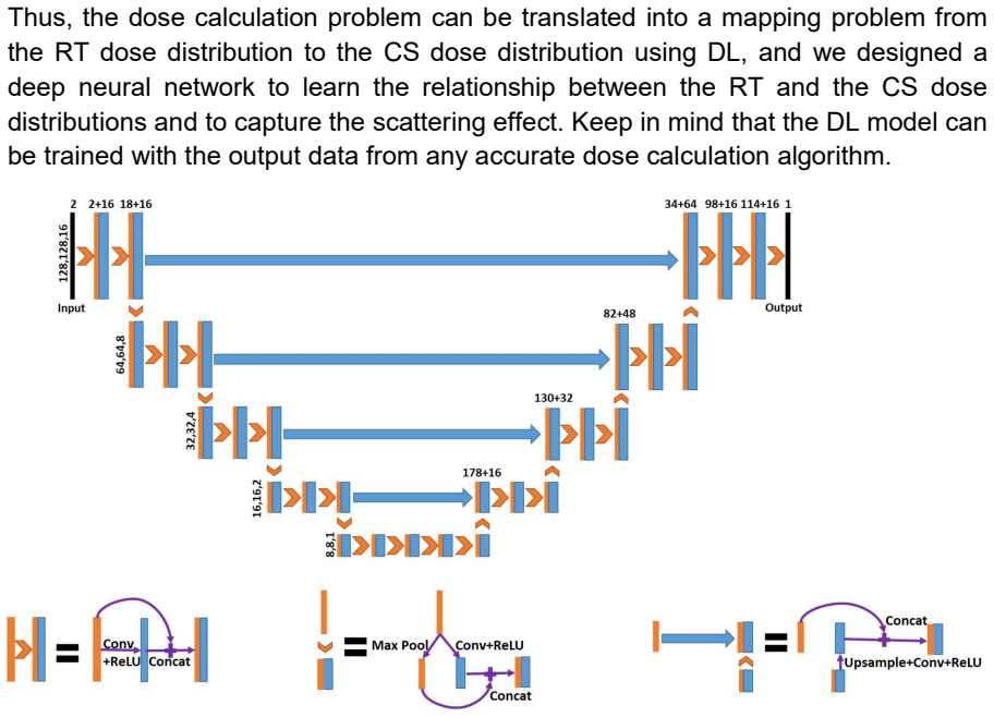
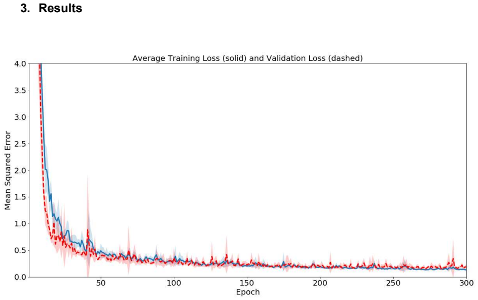
The load-bearing mechanism is the two-stage mapping. A modified ray-tracing algorithm (fluence-convolution broad-beam) generates a fast approximate 3D dose from the IMRT fluence maps and patient CT; then a Hierarchically Densely Connected U-net (HD U-net), with 3x3x3 convolutions, five downsampling levels, batch normalization, and ReLU activations, learns the residual between that ray-tracing dose and the collapsed cone convolution/superposition (CS) dose. The ray-tracing prior shrinks the domain gap between 2D fluence maps and 3D doses, so the network only has to learn the scatter correction, while the CS algorithm provides both the training labels and the clinical reference. Training uses mean squared error loss and the Adam optimizer on random 128x128x16 patches from 70 prostate patients, with five-fold cross-validation used to check stability.
What would settle it
Run the trained network on plans outside the prostate training distribution, such as lung or head-and-neck cases with air cavities, and compare its output against Monte Carlo dose: if gamma pass rates at 2%/2mm fall below the clinical threshold, or if the DL dose is no closer to Monte Carlo than the CS reference is, the central feasibility claim fails.
Extended reading notes
Core claim
The central discovery is that a first-order ray-tracing prior plus a densely connected U-net is enough to reproduce the dose of an accurate convolution/superposition algorithm. The paper recasts dose calculation as a mapping problem: project the IMRT fluence maps into a 3D dose prior with a modified ray-tracing algorithm, then train an HD U-net to correct that prior into a CS-equivalent dose, effectively learning the scatter components that the ray tracer omits. For the eight prostate test patients, the DL output is clinically identical to CS: average gamma passing rates of 98.5% at 1 mm/1% and 99.9% at 2 mm/2%, PTV D95 differences between -1.38 Gy and 0.66 Gy, and organ-at-risk volume differences mostly within ±0.4%. The network computes a full 3D dose in 1.19 ± 0.01 seconds.
Load-bearing premise
The claim rests on treating the collapsed cone dose as the true dose and on the eight prostate test patients standing in for the broader patient population; if that reference is biased or those patients are unrepresentative, the reported clinical equivalence will not generalize.
Editorial extensions
If this is right
- Dose calculation drops from minutes to about one second, removing the main computational bottleneck in real-time adaptive radiotherapy replanning.
- Because the network learns a mapping from a fast prior to a reference engine, the same scheme can be retrained on other reference algorithms, such as Monte Carlo, or on other treatment sites.
- DL-computed doses can serve as an independent secondary check of treatment-plan doses, complementing measurement-based verification.
- The reported DVH and IMRT-objective agreement means plan evaluation can be performed directly on DL doses in the tested prostate cohort.
- Fast dose evaluation makes it practical to explore many candidate fluence maps during inverse planning, potentially improving IMRT optimization.
Reading between the lines
- The method's success likely depends on the quality of the ray-tracing prior; a natural extension is to test how the network degrades when the prior is made cruder or when the anatomy (lung, head and neck) introduces strong heterogeneities the prostate cohort lacks.
- Because the network is trained to mimic CS, it will inherit any systematic errors of CS; a Monte Carlo-labeled experiment would separate true accuracy from faithful mimicry.
- The 'fast approximation plus learned correction' design could transfer to other inverse problems in physics and imaging where a cheap physical model exists but a slow accurate one defines the target.
- At roughly one second per plan on a single GPU, this could enable on-the-fly dose reconstruction during delivery, so a clinic could detect anatomical changes mid-treatment.
Signed reviews
Editorial analysis
A structured set of objections, weighed in public.
Referee Report
Summary. The paper proposes a deep learning-based dose calculation engine for radiotherapy. The method uses a two-stage pipeline: a fast ray-tracing (RT) algorithm first projects IMRT fluence maps and patient CT into an approximate 3D dose distribution, and a modified Hierarchically Densely Connected U-net (HD U-net) then maps this RT dose to a collapsed cone convolution/superposition (CS) dose distribution. The network is trained on 78 prostate IMRT plans (70 training, 8 held-out test) using five-fold cross-validation during development. Evaluation on the eight test patients reports gamma passing rates of 98.5% at 1 mm/1% and 99.9% at 2 mm/2%, small differences in IMRT optimization objectives and DVHs, and an average calculation time of 1.19 seconds for the trained model. The authors conclude that deep learning can perform dose calculation with both high accuracy and high efficiency, potentially enabling real-time adaptive radiotherapy.
Significance. If the accuracy and efficiency claims hold, this would be a valuable proof-of-concept for replacing a slow but accurate dose engine with a fast learned surrogate, with implications for real-time adaptive therapy and secondary dose verification. The paper has several strengths: the target is computed by an independent CS algorithm, providing a non-circular benchmark; the evaluation uses clinically meaningful metrics (gamma index, DVH, IMRT objectives); and the five-fold cross-validation during training addresses stability to some degree. The accuracy evidence from Tables 1 and 2 is encouraging. However, the central efficiency claim is not yet fully substantiated because the reported 1.19 s appears to refer only to the U-net forward pass, not the complete two-stage pipeline that includes the ray-tracing prior. The small, single-institution test set is a limitation but is acceptable for a feasibility study if framed cautiously.
major comments (3)
- [Section 3] The reported average calculation time of 1.19 s for the eight testing plans is described as the time for 'the trained model,' but the method defined in Section 2.1 is a two-stage pipeline: first compute a ray-tracing dose prior, then apply the HD U-net. The manuscript does not state whether the 1.19 s includes the RT prior computation, preprocessing, GPU data transfer, or any patch stitching, and it gives no timing for the RT prior. Since the abstract's claim of 'about one second' and the comparison with minutes for CS rest on this measurement, please provide an end-to-end timing measurement for the complete pipeline, with a breakdown of each stage and the hardware/software used.
- [Sections 2.1 and 2.3] The training procedure samples random patches of size 128×128×16, while the input and output volumes are 256×256×64 (or 62) for each patient. The text does not explain how full-volume inference is performed (e.g., tiling with overlap, stitching, or a single full-volume forward pass), and it is unclear whether the reported 1.19 s covers the entire volume. This ambiguity affects both the interpretation of the timing result and the reproducibility of the method, so please clarify the inference protocol explicitly.
- [Section 3, Table 1] The gamma passing rates are reported without specifying the dose threshold and normalization used (e.g., local vs. global gamma, and the percentage dose threshold such as 10% of prescription dose). These details are essential for interpreting whether the passing rates support the 'clinically identical' conclusion. Please state the gamma computation parameters and the volume or region over which gamma was evaluated.
minor comments (5)
- [Abstract and Section 3] The abstract says 'about one second,' while Section 3 reports an average of 1.19 s; please reconcile the wording for precision.
- [Section 2.1 / Figure 1] The text states that the HD U-net reduces the feature size from 128×128×16 to 8×8×1, but Section 2.2 states the patient volumes are 256×256×64; this inconsistency should be resolved, and the figure should be clarified to match the actual input dimensions.
- [Section 2.2] Please specify the treatment planning system or in-house software used to compute the CS and RT dose distributions, and confirm that the same fluence maps and beam setup were used for both algorithms.
- [Section 2.3] The description of the five-fold cross-validation could be clearer: after the five folds are used to assess stability, the final model is trained on all 70 patients, but the text does not state how the number of epochs for this final training was selected relative to the 300 epochs used in the folds.
- [References] Reference 10 (FCBB) is cited as the ray-tracing algorithm; please clarify whether the 'modified ray-tracing algorithm' used in this study is exactly the FCBB method or a different implementation, and if modified, describe the modification.
Circularity Check
No significant circularity: the DL dose network is trained by supervised regression against an independent collapsed-cone reference and tested on held-out patients.
full rationale
The paper's central claim is that an HD U-net maps a ray-tracing dose prior to a collapsed cone (CS) dose distribution, and that the resulting DL dose is clinically equivalent to the CS dose on eight held-out prostate IMRT patients. The target values are produced by the collapsed cone convolution/superposition algorithm, which is an external, independently established dose calculation method cited in references 11-13, not by the network itself or by a fitted parameter of the present study. The training data are 70 patients, the testing data are a separate set of 8 patients, and the evaluation metrics (gamma passing rates, DVH comparisons, and IMRT objective differences) compare DL output directly with CS output on those held-out cases. This is a standard supervised regression setup, so the accuracy claim does not reduce by construction to the training input. The use of a ray-tracing prior is not circular: the RT dose is an input derived from a different, lower-accuracy algorithm, and the network learns the residual mapping to CS. Self-citations to the prior HD U-net architecture (reference 9) and to the ray-tracing method (reference 10) establish provenance and technical background, but the load-bearing validation is against an external CS benchmark, so these citations do not themselves force the reported result. The efficiency claim of about 1.19 seconds may lack a complete end-to-end timing that includes the ray-tracing prior, but that is a completeness or measurement concern, not a circularity of the derivation. No equation or defined quantity in the paper is shown to be equivalent to another by construction, and no fitted parameter is renamed as a prediction. Therefore the paper exhibits no significant circularity.
Assumptions & free parameters
free parameters (5)
- learning rate =
1e-4
- number of epochs =
300
- training patch size =
128x128x16
- feature map count =
16
- dropout rate =
0
assumptions (4)
- domain assumption Collapsed cone convolution/superposition is an accurate clinical reference for photon dose.
- domain assumption Ray-tracing dose is a sufficiently informative first-order prior.
- domain assumption The 70 training patients represent the distribution of the 8 test patients and future prostate IMRT cases.
- standard math Standard neural network components (convolution, ReLU, batch normalization, Adam) behave as expected.
Cite this review
Pith. "Pith review of A Feasibility Study on Deep Learning-Based Radiotherapy Dose Calculation." pith.science (2026). https://pith.science/paper/NATHJGAT
@misc{pith2026190803159,
  author       = {Pith},
  title        = {Pith review of: A Feasibility Study on Deep Learning-Based Radiotherapy Dose Calculation},
  year         = {2026},
  howpublished = {\url{https://pith.science/paper/NATHJGAT}},
  note         = {Machine review of arXiv:1908.03159}
}
read the original abstract
Purpose: Various dose calculation algorithms are available for radiation therapy for cancer patients. However, these algorithms are faced with the tradeoff between efficiency and accuracy. The fast algorithms are generally less accurate, while the accurate dose engines are often time consuming. In this work, we try to resolve this dilemma by exploring deep learning (DL) for dose calculation. Methods: We developed a new radiotherapy dose calculation engine based on a modified Hierarchically Densely Connected U-net (HD U-net) model and tested its feasibility with prostate intensity-modulated radiation therapy (IMRT) cases. Mapping from an IMRT fluence map domain to a 3D dose domain requires a deep neural network of complicated architecture and a huge training dataset. To solve this problem, we first project the fluence maps to the dose domain using a modified ray-tracing algorithm, and then we use the HD U-net to map the ray-tracing dose distribution into an accurate dose distribution calculated using a collapsed cone convolution/superposition (CS) algorithm. Results: It takes about one second to compute a 3D dose distribution for a typical 7-field prostate IMRT plan, which can be further reduced to achieve real-time dose calculation by optimizing the network. For all eight testing patients, evaluation with Gamma Index and various clinical goals for IMRT optimization shows that the DL dose distributions are clinically identical to the CS dose distributions. Conclusions: We have shown the feasibility of using DL for calculating radiotherapy dose distribution with high accuracy and efficiency.
Figures

Reference graph
Works this paper leans on
-
[1]
Dose calculations for external photon beams in radiotherapy
Ahnesjo A, Aspradakis MM. Dose calculations for external photon beams in radiotherapy. Phys Med Biol. 1999;44(11):R99-R155
work page 1999
-
[2]
Effective approaches to attention-based neural machine translation
Luong M-T, Pham H, Manning CD. Effective approaches to attention-based neural machine translation. arXiv preprint. 2015;arXiv:1508.04025
arXiv 2015
-
[3]
Fully character-level neural machine translation without explicit segmentation
Lee J, Cho K, Hofmann T. Fully character-level neural machine translation without explicit segmentation. arXiv preprint. 2017;arXiv:1610.03017
arXiv 2017
-
[4]
Very deep convolutional networks for large-scale image recognition
Simonyan K, Zisserman A. Very deep convolutional networks for large-scale image recognition. arXiv preprint. 2014;arXiv:1409.1556
arXiv 2014
-
[5]
Deep residual learning for image recognition
He K, Zhang X, Ren S, Sun J. Deep residual learning for image recognition. arXiv preprint. 2015;arXiv:1512.03385
arXiv 2015
-
[6]
U-Net: convolutional networks for biomedical image segmentation
Ronneberger O, Fischer P, Brox T. U-Net: convolutional networks for biomedical image segmentation. Medical Image Computing and Computer-Assisted Intervention – MICCAI 2015 Lecture Notes in Computer Science. 2015;9351:234-241
work page 2015
-
[7]
Nguyen D, Long T, Jia X, et al. Dose prediction with U-net: a feasibility study for predicting dose distributions from contours using deep learning on prostate IMRT patients. arXiv preprint. 2017;arXiv:1709.09233. Scientific Reports. 2019;9(1):1076
work page Pith review arXiv 2017
-
[8]
Barragan-Montero AM, Nguyen D, Lu W, et al. Three-dimensional dose prediction for lung IMRT patients with deep neural networks: robust learning from heterogeneous beam configurations. arXiv preprint. 2018;arXiv:1812.06934
work page Pith review arXiv 2018
Show all 15 references
-
[9]
3D radiotherapy dose prediction on head and neck cancer patients with a hierarchically densely connected U-net deep learning architecture
Nguyen D, Jia X, Sher D, et al. 3D radiotherapy dose prediction on head and neck cancer patients with a hierarchically densely connected U-net deep learning architecture. arXiv preprint. 2018; arXiv:1805.10397. Phys Med Biol. 2019;64(6)
2018 arXiv
-
[10]
Fluence-convolution broad-beam (FCBB) dose calculation
Lu W, Chen M. Fluence-convolution broad-beam (FCBB) dose calculation. Phys Med Biol. 2010;55(23):7211-7229
2010
-
[11]
Collapsed cone Convolution of radiant energy for photon dose calculation in heterogeneous media
Ahnesjo A. Collapsed cone Convolution of radiant energy for photon dose calculation in heterogeneous media. Med Phys. 1989;16(4):577-592
1989
-
[12]
The inverse problem of a Gaussian convolution and its application to the finite size of the measurement chambers/detectors in photon and proton dosimetry
Ulmer W, Kaissl W. The inverse problem of a Gaussian convolution and its application to the finite size of the measurement chambers/detectors in photon and proton dosimetry. Phys Med Biol. 2003;48(6):707-727
2003
-
[13]
A 3D photon superposition/convolution algorithm and its foundation on results of Monte Carlo calculations
Ulmer W, Pyyry J, Kaissl W. A 3D photon superposition/convolution algorithm and its foundation on results of Monte Carlo calculations. Phys Med Biol. 2005;50(8):1767-1790
2005
-
[14]
Adam: A method for stochastic optimization
Kingma DP, Ba J. Adam: A method for stochastic optimization. arXiv preprint. 2014;arXiv:1412.6980
2014 arXiv
-
[15]
Tensorflow: Large-scale machine learning on heterogeneous distributed systems
Abadi M, Agarwal A, Barham P, et al. Tensorflow: Large-scale machine learning on heterogeneous distributed systems. arXiv preprint. 2016; arXiv:1603.04467
2016 arXiv
Reviewed August 14, 2026 · model on record in the stance chip above.
Discussion (0). Continue with ORCID to comment.