REVIEW 4 major objections 4 minor 64 references
Probing Tissue Microarchitecture of the Baby Brain via Spherical Mean Spectrum Imaging
T0 review · 4 major / 4 minor · reviewed 2026-08-14 · deepseek-v4-flash
Pith's one-line read Spherical mean spectrum imaging separates neurite, hindered, and free-water compartments in the developing brain from ordinary multi-shell MRI scans.
desk verdict A genuine extension of spherical-mean diffusion modeling into a spectrum framework, with a clean derivation and honest treatment of degeneracy, but the bias-removal claim depends on a heuristic that lacks independent validation. read the letter →
The pith
A machine-rendered reading of the paper's core claim, the machinery that carries it, and where it could break.
The reading
What carries the argument
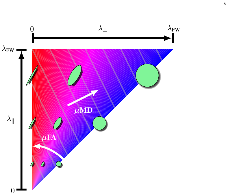
The load-bearing object is the spherical mean spectrum (SMS), the distribution $p(\lambda_\parallel,\lambda_\perp)$ over axial and radial diffusivities obtained after marginalizing over tensor orientation. Because $\bar S_b = S_0 \int p(\lambda_\parallel,\lambda_\perp)\bar h_b(\lambda_\parallel,\lambda_\perp)\,d\lambda_\parallel d\lambda_\perp$ with $\bar h_b(\lambda_\parallel,\lambda_\perp) = e^{-b\lambda_\perp}\frac{\sqrt{\pi}\,\mathrm{erf}\bigl(\sqrt{b(\lambda_\parallel-\lambda_\perp)}\bigr)}{2\sqrt{b(\lambda_\parallel-\lambda_\perp)}}$, the signal depends only on the diffusivity pairs, not on how fibers are oriented. SMSI discretizes $p$ into a dictionary of such kernels and solves a nonnegative elastic-net problem ($\nu = \arg\min_{\nu\succeq 0}\|A\nu-\bar S\|_2^2 + \gamma_1\|\mathrm{diag}(w)\nu\|_1 + \gamma_2\|\nu\|_2^2$), with an iterative reweighting step and a full-signal spectrum step that upweights atoms whose generalized fractional anisotropy falls below $0.3$. A Rician-noise debiasing transform and automatic selection of the regularization parameters complete the pipeline. All reported indices—$\mu$FA, $\mu$MD, MAI, OCI, volume fractions—are weighted summaries of this recovered spectrum.
What would settle it
On a well-characterized phantom consisting of oriented hollow fibers in a free-water bath, with known fiber volume fraction and known crossing angles, run SMSI at a signal-to-noise ratio of 20 for isotropic volume fractions from 0 to 0.9; if the recovered intra-cellular volume fraction deviates from the known fiber fraction by more than the paper's reported simulation tolerance, or if the error grows with crossing angle, the degeneracy suppression is not working.
Extended reading notes
Core claim
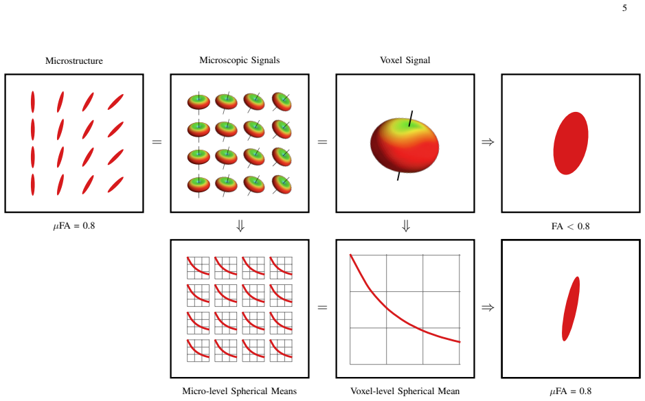
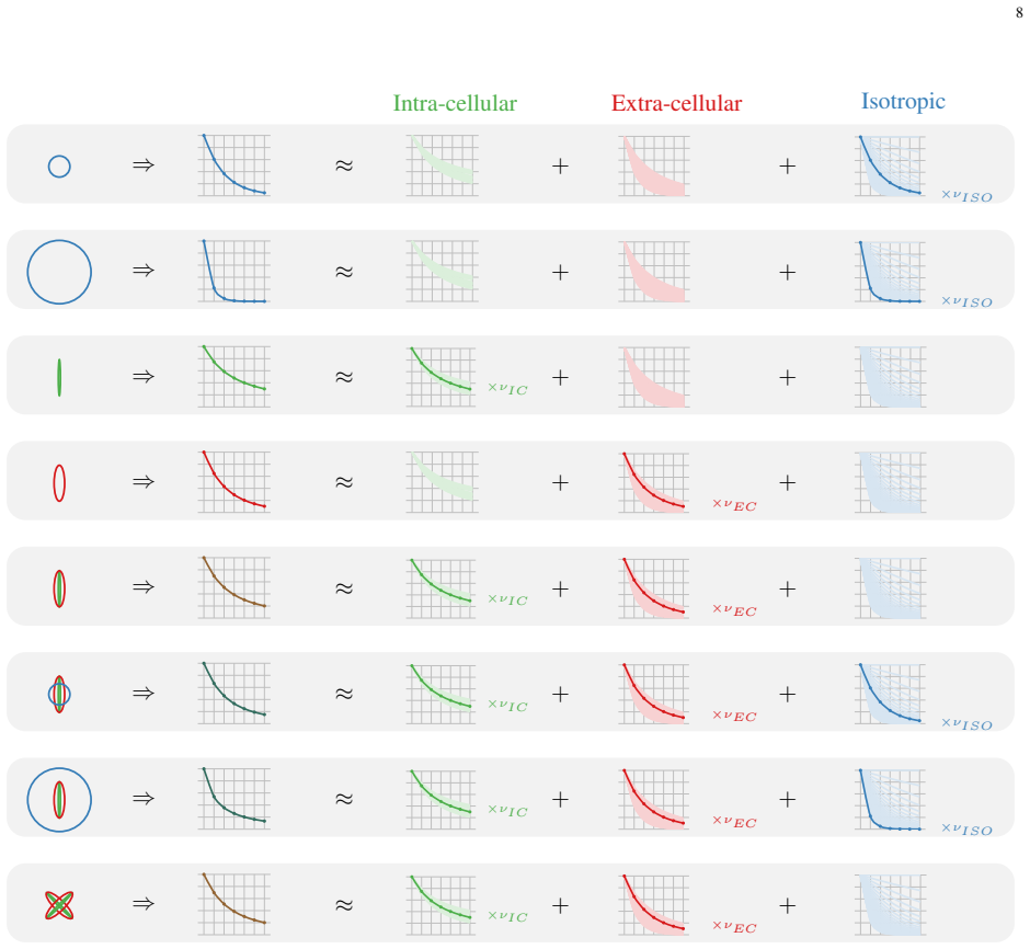
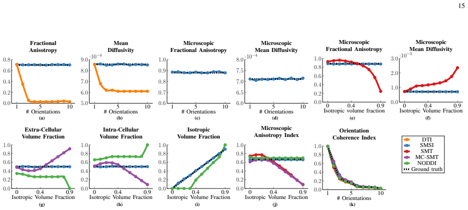
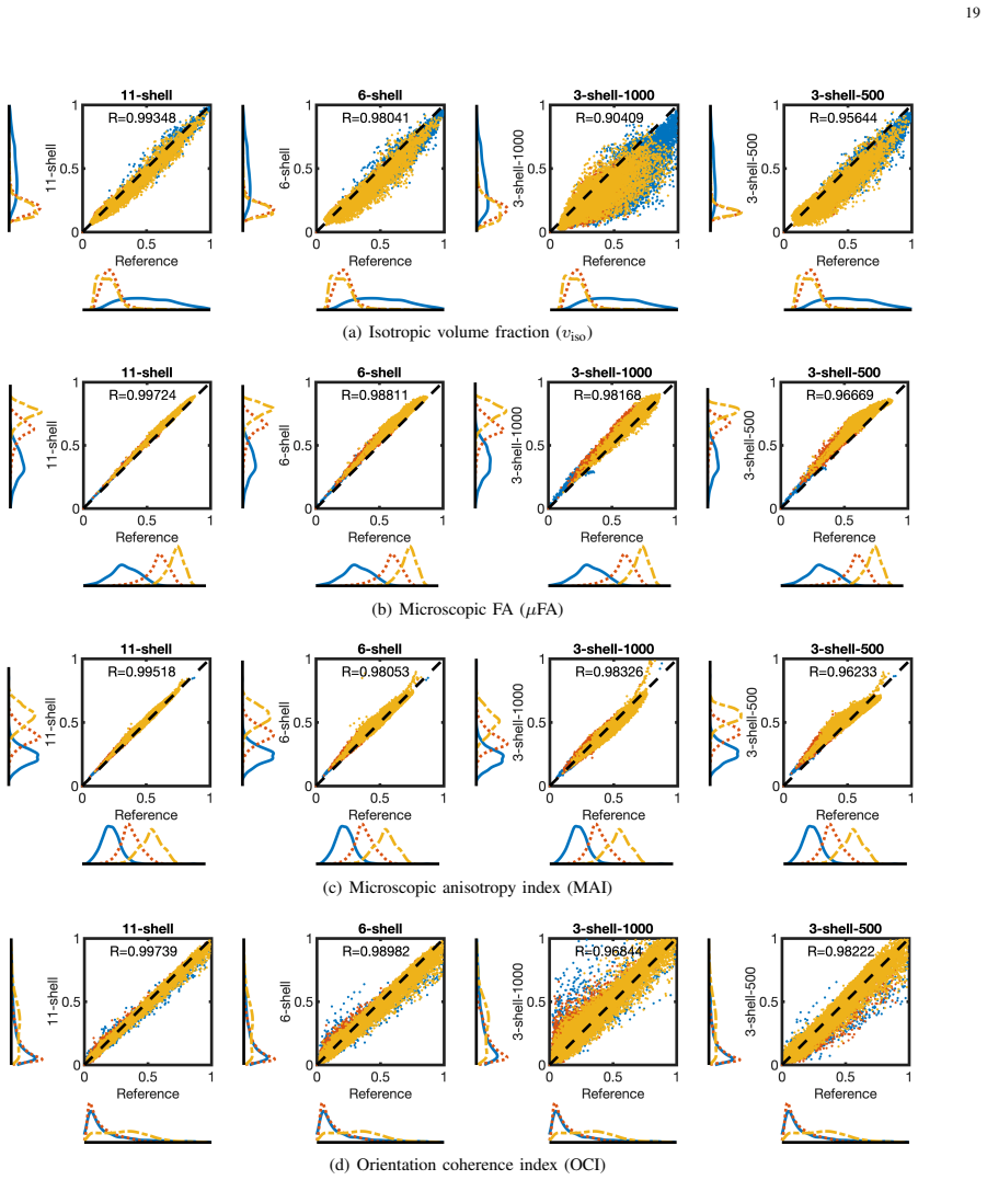
The paper's central claim is that the spherical mean of the diffusion signal—not the full direction-resolved signal—carries enough information to reconstruct a whole spectrum of tissue microenvironments, provided the degeneracy between anisotropic and isotropic compartments is handled explicitly. Starting from the spherical mean technique, SMSI represents each voxel as a nonnegative mixture of axial-symmetric tensor kernels, estimates the mixture weights by a regularized linear solve, and groups the resulting atoms into restricted (intra-cellular), hindered (extra-cellular), and isotropic compartments. On simulated data with up to ten crossing fiber orientations and isotropic volume fractions up to 0.9, the paper reports that SMSI's microscopic anisotropy, mean diffusivity, and volume fractions track the ground truth, while DTI FA and MD drop with orientation count and SMT/MC-SMT/NODDI show systematic biases in the presence of free water. The same indices on longitudinal infant scans show the expected maturation pattern: anisotropy, coherence, and intra-cellular fraction rise while isotropic and extra-cellular fractions fall. The paper further proves that the spherical-mean dictionary atoms are linearly independent, so the spectrum is identifiable in the noiseless limit; the remaining ambiguity is the anisotropic-isotropic degeneracy, which the full-signal reweighting step is designed to suppress.
Load-bearing premise
The load-bearing premise is that the extra full-signal step can identify which anisotropic-looking atoms are actually mixtures of isotropic compartments in real tissue, using a fixed anisotropy threshold ($0.3$, that is ISO $\geq 0.95$) that was validated only on synthetic mixtures; the paper's own Figure 13 shows the full signal alone does not fully resolve the ambiguity.
Editorial extensions
If this is right
- With three or more b-shells, SMSI maps whole-brain microstructure in roughly fifteen minutes, making multi-shell acquisition practical for large infant studies.
- In voxels with crossing fibers, SMSI indices such as $\mu$FA and $\mu$MD stay nearly constant as the number of simulated orientations grows from one to ten, whereas DTI FA and MD decline; this removes a major confound in cross-region comparisons.
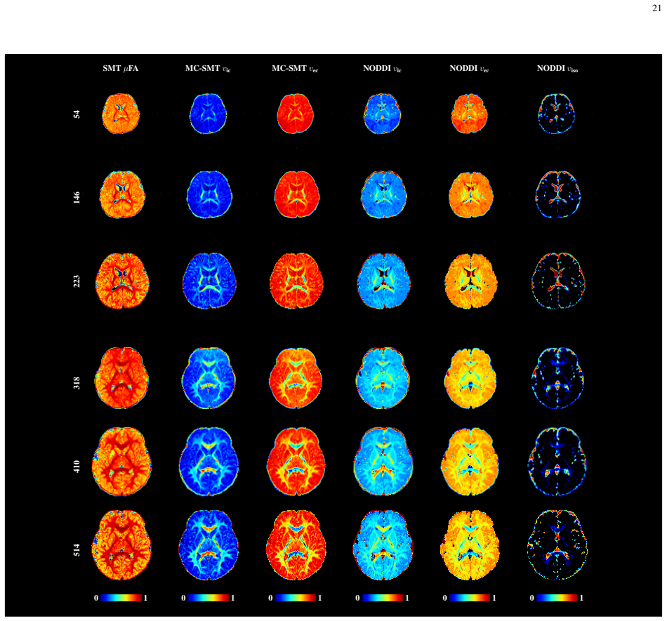
- Explicit isotropic modeling makes SMSI's intra-cellular and extra-cellular volume fractions accurate as free-water fraction rises to 0.9, while the paper reports that NODDI underestimates extra-cellular fraction and MC-SMT degrades without an isotropic compartment.
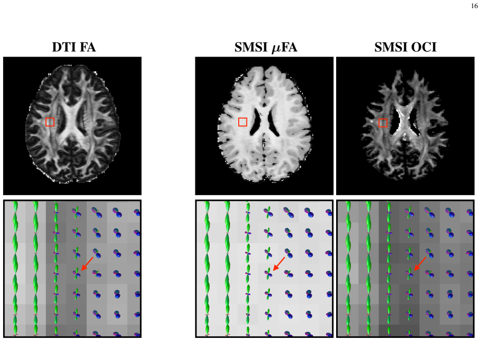
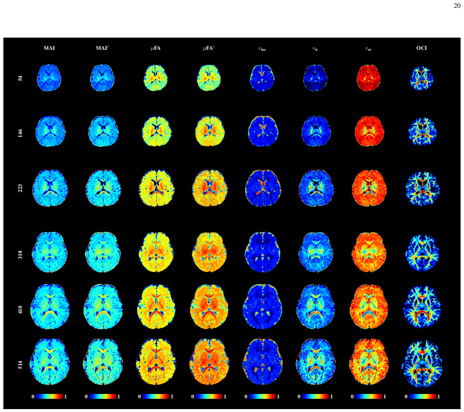
- Longitudinal infant scans show rising microscopic anisotropy, coherence, and intra-cellular fraction with age and falling isotropic and extra-cellular fractions, matching the known central-to-peripheral maturation sequence.
Reading between the lines
- Not stated in the paper, the linear-independence proof implies the spherical-mean inverse problem is identifiable from ideal noiseless data; the practical failure mode is therefore noise, b-range truncation, and the threshold heuristic, so error bounds could be derived from the dictionary's conditioning.
- A natural extension is to make the degeneracy threshold (generalized fractional anisotropy below $0.3$) adaptive to tissue type or noise level; doing so would test whether the synthetic-mixture validation transfers to pathological tissue.
- If SMSI transfers beyond the brain, the same spectrum yields two biomarkers at once: restricted fraction for cellularity and free-water fraction for edema, which would be useful in demyelinating disease where both change together.
Signed reviews
Editorial analysis
A structured set of objections, weighed in public.
Referee Report
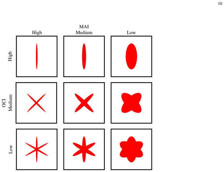
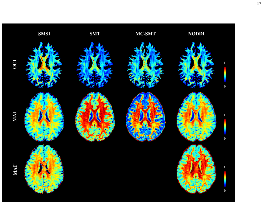
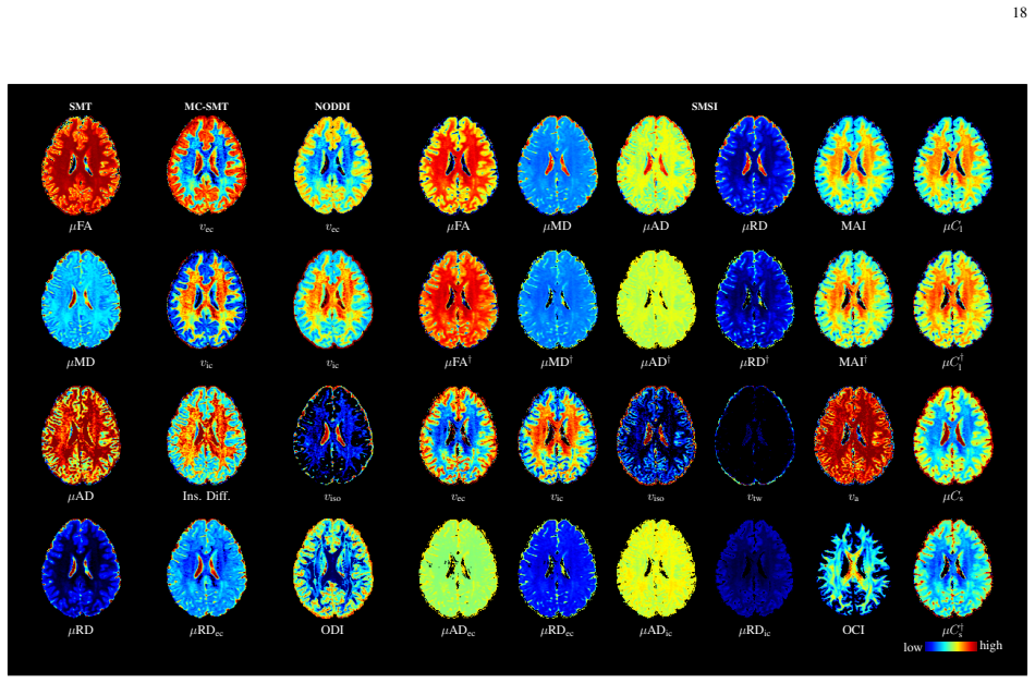
Summary. The paper proposes spherical mean spectrum imaging (SMSI), a method for estimating a spectrum of axial-symmetric tensor compartments from multi-shell diffusion MRI. The method uses the spherical mean of the diffusion signal, which is invariant to fiber orientation distribution, to decompose the signal into a dictionary of tensor compartments and estimate their volume fractions with a regularized inverse problem. From the estimated spectrum, the authors define multiple orientation-invariant indices such as microscopic FA, per-axon axial/radial diffusivity, intra-/extra-cellular volume fractions, an isotropic diffusion measure, and two new indices (MAI and OCI). They validate the method on simulated data and demonstrate maps on adult HCP and infant BCP datasets, with emphasis on longitudinal infant brain development. The central claim is that SMSI is fast, accurate, and overcomes biases of SMT, MC-SMT, and NODDI by modeling a full spectrum of diffusion scales and by explicitly handling isotropic diffusion and degeneracy.
Significance. If the claims are substantiated, SMSI would be a useful contribution to diffusion MRI microstructure imaging. The spherical mean derivation in Eqs. (10)-(14) is clean and provides a principled way to extend SMT to a multi-compartment spectrum. The proposed MAI and OCI generalize naturally to multiple compartment models. The use of convex elastic-net optimization is practical and the demonstration on longitudinal infant data addresses an important application. However, the validation is not yet sufficient to support the strong accuracy claims: the core synthetic validation uses the same tensor-compartment forward model as the estimator, and the degeneracy suppression relies on a heuristic GFA threshold with validation only on synthetic mixtures and healthy adult data. The manuscript would be strengthened by independent validation or, failing that, by substantially tempering the claims.
major comments (4)
- [§III-B and §IV-B] The synthetic validation is circular in an important sense: the simulated data in Eq. (26) are generated from the same cylinder/tensor compartment model that SMSI assumes, and the ground-truth volume fractions are exactly the compartment fractions of that model. Consequently, the accurate recovery in Fig. 5 largely confirms that the estimator can invert its own forward model, not that the model captures real tissue. An independent test using spherical tensor encoding, histology, or a different biophysical model is needed before claiming that SMSI 'can overcome the biases' of other methods.
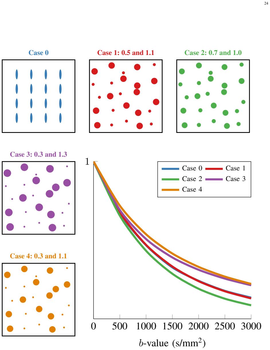
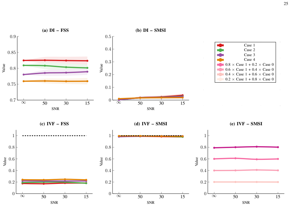
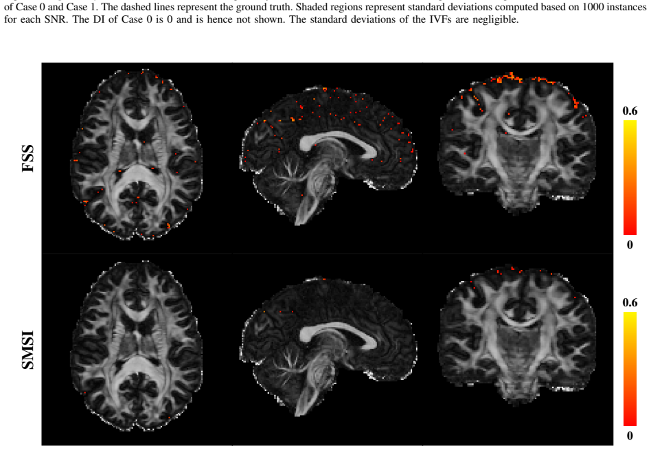
- [§II-B4 and §IV-E] The load-bearing step for resolving the spherical-mean degeneracy is the heuristic rule that anisotropic atoms with ISO ≥ 0.95 (GFA < 0.3) are 'degenerate' and should be penalized (Eqs. 27-28). Fig. 13 shows that this rule works for the synthetic Cases 1-4, but no independent validation is given for real tissue. In the fully dispersed limit, Eq. (17) shows that the full signal of uniformly oriented anisotropic tensors is exactly the spherical-mean signal, so the full signal cannot distinguish such tissue from isotropic diffusion; the threshold rule then acts as an implicit prior that could suppress genuinely anisotropic but highly dispersed white matter in the infant brain. The manuscript should either provide a validation against STE-based microscopic anisotropy, report degeneracy statistics for the BCP infant data (Table II reports only 20 HCP adults), or explicitly discuss and bound this risk.
- [§II-B4] The adaptive parameter selection procedure tunes τ, γ1, γ2, and γ3 on the same dataset used for the reported results. Specifically, τ is chosen by grid search on corpus callosum voxels using MC-SMT, and the γ parameters are chosen to minimize the difference between predicted and observed spherical mean signals in corpus callosum and ventricle voxels. This is a form of data-driven model selection without a held-out set, which can optimistically bias the reported accuracy. A cross-validation scheme or a sensitivity analysis over the selected parameters should be reported.
- [Abstract and §IV-F] The abstract claims that SMSI provides 'greater sensitivity and specificity to development related changes,' but no quantitative measure of sensitivity or specificity is presented. The longitudinal infant results in Fig. 10 are qualitative developmental trends without ground-truth microstructural measures. Either the claim should be supported by quantitative metrics (e.g., age-discrimination accuracy, effect sizes) or the wording should be weakened to 'demonstrates expected developmental trends.'
minor comments (4)
- [§II-B2] In the sentence defining the dictionary ranges, 'The ranges of λ‖[i] and λ‖[i]' should read 'λ‖[i] and λ⊥[i]'.
- [§II-B2, Eq. (15)] The text says 'γ1 and γ1 control the lasso and ridge penalty'; the second γ1 should be γ2.
- [Fig. 8] The caption contains garbled symbols such as 'µClaaaa' and 'µC†laaaa'; these should be corrected to the proper index names from Table I.
- [§III-A] The choice of axial diffusivity range (1.5-2.0 × 10^-3 mm^2/s) is stated to be 'determined using SMT,' but the details of how many voxels and subjects were used are not given; provide this information for reproducibility.
Circularity Check
Degeneracy suppression is partly self-verifying because the degeneracy index is defined from the same GFA indicator used to impose the penalty, but the core SMS decomposition is not circular.
-
self definitional
[Section II-B4 and Section V, Eqs. (24), (27)-(29), Fig. 13]
"identifying degenerate anisotropic atoms with generalized fractional anisotropy [39] (GFA) smaller than 0.3, and reapplying (24) with higher penalization of the degenerate atoms. This is implemented by doubling the corresponding elements in w′. ... Anisotropic atoms with ISO≥0.95 (GFA<0.3) are considered degenerate. This is captured by an indicator function for the i-th atom: Υ[i] = 1, ISO[i]≥0.95, 0, otherwise. ... DI = Σ_i ν[i]Υ[i]"
The same indicator Υ that flags 'degenerate' atoms (ISO≥0.95) is used both to double the penalty in Eq. (24) and to define the degeneracy index DI in Eq. (29). Penalizing exactly those atoms must reduce their fitted volume fractions ν, so the reported decrease in DI from FSS to SMSI (Fig. 13(a)-(b)) is an arithmetic consequence of the penalty, not an independent empirical finding. The paper's statement that 'SMSI suppresses the degenerate atoms and lowers the DI, resulting in accurate volume fraction estimates' therefore uses a success metric that is definitionally tied to the intervention; the volume-fraction accuracy in Fig.
full rationale
The central derivation of SMSI is not circular: Eqs. (10)-(15) define a linear mixture model in which the spherical mean signal is decomposed into dictionary atoms, and the volume fractions are estimated by elastic-net regression; no target index is defined as the fitted value of itself. The linear-independence proof in the Appendix is a genuine mathematical argument, and the synthetic validations of μFA, μMD, and compartment volume fractions are standard forward-model checks. The main circularity concern is localized to the degeneracy-resolution evidence: DI is constructed from the same GFA-based indicator that drives the suppression penalty, so showing that SMSI lowers DI is self-verifying. Additionally, the pipeline borrows calibration from the models it compares against—the anisotropic λ‖ range is set using SMT on the corpus callosum, τ is determined via MC-SMT grid search, and regularization parameters are selected using the same types of tissue regions—so the in-vivo 'overcoming biases' comparisons are not fully independent. These are calibration and validation weaknesses rather than a collapse of the derivation into its inputs, and the core spherical-mean spectrum estimation remains self-contained. Hence a moderate score of 3 is appropriate.
Assumptions & free parameters
free parameters (6)
- tau (geometric tortuosity threshold) =
approximately 2.6
- Axial diffusivity dictionary range =
1.5 to 2.0 x 10^-3 mm^2/s
- Perpendicular diffusivity lower bound =
lambda_par / lambda_perp >= 1.1
- Isotropic diffusivity grid step =
0 to 3 x 10^-3 mm^2/s, step 0.1 x 10^-3
- Regularization parameters gamma1, gamma2, gamma3 =
automatically selected from [10^-5, 1]
- GFA threshold for degenerate atoms =
GFA < 0.3 (ISO >= 0.95)
assumptions (5)
- domain assumption The diffusion signal is a noiseless spherical convolution of an antipodal fODF with an axial-symmetric diffusion-tensor kernel (Eqs. 2 and 10).
- domain assumption Membrane permeability is negligible on the diffusion timescale, so compartments do not exchange water (Section II.B3).
- standard math The spherical mean signal of a tensor kernel depends only on lambda_par and lambda_perp, not on orientation, so Eq. (12) marginalizes omega.
- standard math The dictionary atoms hbar(lambda_par[i], lambda_perp[i]) are linearly independent over b, making the inverse problem well-posed in the noiseless limit (Appendix).
- ad hoc to paper The GFA threshold with ISO >= 0.95 identifies degenerate anisotropic atoms, and reweighting the elastic net with these atoms suppressed recovers true volume fractions (Section II.B4).
Cite this review
Pith. "Pith review of Probing Tissue Microarchitecture of the Baby Brain via Spherical Mean Spectrum Imaging." pith.science (2026). https://pith.science/paper/IA7PLREI
@misc{pith2026190804483,
  author       = {Pith},
  title        = {Pith review of: Probing Tissue Microarchitecture of the Baby Brain via Spherical Mean Spectrum Imaging},
  year         = {2026},
  howpublished = {\url{https://pith.science/paper/IA7PLREI}},
  note         = {Machine review of arXiv:1908.04483}
}
abstract
During the first years of life, the human brain undergoes dynamic spatially-heterogeneous changes, involving differentiation of neuronal types, dendritic arborization, axonal ingrowth, outgrowth and retraction, synaptogenesis, and myelination. To better quantify these changes, this article presents a method for probing tissue microarchitecture by characterizing water diffusion in a spectrum of length scales, factoring out the effects of intra-voxel orientation heterogeneity. Our method is based on the spherical means of the diffusion signal, computed over gradient directions for a fixed set of diffusion weightings (i.e., b-values). We decompose the spherical mean series at each voxel into a spherical mean spectrum (SMS), which essentially encodes the fractions of spin packets undergoing fine- to coarse-scale diffusion processes, characterizing hindered and restricted diffusion stemming respectively from extra- and intra-neurite water compartments. From the SMS, multiple orientation distribution invariant indices can be computed, allowing for example the quantification of neurite density, microscopic fractional anisotropy ($\mu$FA), per-axon axial/radial diffusivity, and free/restricted isotropic diffusivity. We show maps of these indices for baby brains, demonstrating that microscopic tissue features can be extracted from the developing brain for greater sensitivity and specificity to development related changes. Also, we demonstrate that our method, called spherical mean spectrum imaging (SMSI), is fast, accurate, and can overcome the biases associated with other state-of-the-art microstructure models.
Figures
Figures from the paper (11 more)
Reference graph
Works this paper leans on
-
[1]
N. Kunz, H. Zhang, L. Vasung, K. R. O’Brien, Y . Assaf, F. Lazeyras, D. C. Alexander, and P. S. H ¨uppi, “Assessing white matter microstructure of the newborn with multi-shell diffusion MRI and biophysical compartment models.” NeuroImage, vol. 96, no. 8, pp. 288–299, 2014
work page 2014
-
[2]
B. Lampinen, F. Szczepankiewicz, J. M ˚artensson, D. van Westen, P. C. Sundgren, and M. Nilsson, “Neurite density imaging versus imaging of microscopic anisotropy in diffusion MRI: A model comparison using spherical tensor encoding,” NeuroImage, vol. 147, pp. 517–531, 2017
work page 2017
-
[3]
Composite hindered and restricted model of diffusion (CHARMED) MR imaging of the human brain,
Y . Assaf and P. J. Basser, “Composite hindered and restricted model of diffusion (CHARMED) MR imaging of the human brain,” NeuroImage, vol. 27, no. 1, pp. 48–58, 2005
work page 2005
-
[4]
AxCaliber: A method for measuring axon diameter distribution from diffusion MRI,
Y . Assaf, T. Blumenfeld-Katzir, Y . Yovel, and P. J. Basser, “AxCaliber: A method for measuring axon diameter distribution from diffusion MRI,” Magnetic Resonance in Medicine , vol. 59, no. 6, pp. 1347–1354, 2008
work page 2008
-
[5]
Orientationally invariant indices of axon diameter and density from diffusion MRI,
D. C. Alexander, P. L. Hubbard, M. G. Hall, E. A. Moore, M. Ptito, G. J. Parker, and T. B. Dyrby, “Orientationally invariant indices of axon diameter and density from diffusion MRI,” NeuroImage, vol. 52, no. 4, pp. 1374–1389, 2010
work page 2010
-
[6]
White matter characterization with diffusional kurtosis imaging,
E. Fieremans, J. H. Jensen, and J. A. Helpern, “White matter characterization with diffusional kurtosis imaging,” NeuroImage, vol. 58, no. 1, pp. 177–188, 2011
work page 2011
-
[7]
NODDI: Practical in vivo neurite orientation dispersion and density imaging of the human brain,
H. Zhang, T. Schneider, C. A. Wheeler-Kingshott, and D. C. Alexander, “NODDI: Practical in vivo neurite orientation dispersion and density imaging of the human brain,” NeuroImage, vol. 61, no. 4, pp. 1000–1016, 2012
work page 2012
-
[8]
Accelerated microstructure imaging via convex optimization (AMICO) from diffusion MRI data,
A. Daducci, E. J. Canales-Rodr ´ıguez, H. Zhang, T. B. Dyrby, D. C. Alexander, and J.-P. Thiran, “Accelerated microstructure imaging via convex optimization (AMICO) from diffusion MRI data,” NeuroImage, vol. 105, pp. 32–44, 2015
work page 2015
Show all 64 references
-
[9]
Probing tissue microstructure with restriction spectrum imaging: Histological and theoretical validation,
N. S. White, T. B. Leergaard, H. D’arceuil, J. G. Bjaalie, and A. M. Dale, “Probing tissue microstructure with restriction spectrum imaging: Histological and theoretical validation,” Human Brain Mapping , vol. 34, no. 2, pp. 327–346, 2013
2013
-
[10]
Direct estimation of the fiber orientation density function from diffusion-weighted MRI data using spherical deconvolution,
J.-D. Tournier, F. Calamante, D. G. Gadian, and A. Connelly, “Direct estimation of the fiber orientation density function from diffusion-weighted MRI data using spherical deconvolution,” NeuroImage, vol. 23, no. 3, pp. 1176–1185, 2004
2004
-
[11]
Robust determination of the fibre orientation distribution in diffusion MRI: Non-negativity constrained super- resolved spherical deconvolution,
J.-D. Tournier, F. Calamante, and A. Connelly, “Robust determination of the fibre orientation distribution in diffusion MRI: Non-negativity constrained super- resolved spherical deconvolution,” NeuroImage, vol. 35, pp. 1459–1472, 2007
2007
-
[12]
Quantitative mapping of the per-axon diffusion coefficients in brain white matter,
E. Kaden, F. Kruggel, and D. C. Alexander, “Quantitative mapping of the per-axon diffusion coefficients in brain white matter,” Magnetic Resonance in Medicine, vol. 75, no. 4, pp. 1752–1763, 2016
2016
-
[13]
Multi-compartment microscopic diffusion imaging,
E. Kaden, N. D. Kelm, R. P. Carson, M. D. Does, and D. C. Alexander, “Multi-compartment microscopic diffusion imaging,” NeuroImage, vol. 139, pp. 346–359, 2016
2016
-
[14]
Characterizing brain tissue by assessment of the distribution of anisotropic microstructural environments in diffusion-compartment imaging (DIAMOND),
B. Scherrer, A. Schwartzman, M. Taquet, M. Sahin, S. P. Prabhu, and S. K. Warfield, “Characterizing brain tissue by assessment of the distribution of anisotropic microstructural environments in diffusion-compartment imaging (DIAMOND),” Magnetic Resonance in Medicine , vol. 76, ...
2016
-
[15]
Statistical model for diffusion attenuated MR signal,
D. Yablonskiy, G. Bretthorst, and J. Ackerman, “Statistical model for diffusion attenuated MR signal,” Magnetic Resonance in Medicine , vol. 50, pp. 664–669, 2003
2003
-
[16]
Quantification of increased cellularity during inflammatory demyelination,
Y . Wang, Q. Wang, J. P. Haldar, F.-C. Yeh, M. Xie, P. Sun, T.-W. Tu, K. Trinkaus, R. S. Klein, A. H. Cross, and S.-K. Song, “Quantification of increased cellularity during inflammatory demyelination,” Brain, vol. 134, pp. 3590–3601, 2011
2011
-
[17]
Maturation of white matter in the human brain: a review of magnetic resonance studies,
T. Paus, D. Collins, A. Evans, G. Leonard, B. Pike, and A. Zijdenbos, “Maturation of white matter in the human brain: a review of magnetic resonance studies,” Brain Research Bulletin , vol. 54, no. 3, pp. 255–266, 2001
2001
-
[18]
Diffusion tensor imaging: serial quantitation of white matter tract maturity in premature newborns,
S. C. Partridge, P. Mukherjee, R. G. Henry, S. P. Miller, J. I. Berman, H. Jin, Y . Lu, O. A. Glenn, D. M. Ferriero, A. J. Barkovich, and B. V . Daniel, “Diffusion tensor imaging: serial quantitation of white matter tract maturity in premature newborns,” NeuroImage, vol. 22, n...
2004
-
[19]
Dubois, L
J. Dubois, L. Hertz-Pannier, G. Dehaene-Lambertz, Y . Cointepas, and D. Le Bihan, “Assessment of the early organization and maturation of infants’ cerebral white matter fiber bundles: a feasibility study using quantitative diffusion tensor imaging and tractography,” NeuroImage,...
2006
-
[20]
Diffusion tensor imaging with tract-based spatial statistics reveals local white matter abnormalities in preterm infants,
M. Anjari, L. Srinivasan, J. M. Allsop, J. V . Hajnal, M. A. Rutherford, A. D. Edwards, and S. J. Counsell, “Diffusion tensor imaging with tract-based spatial statistics reveals local white matter abnormalities in preterm infants,” NeuroImage, vol. 35, no. 3, pp. 1021–1027, 2007
2007
-
[21]
Microstructural brain development between 30 and 40 weeks corrected age in a longitudinal cohort of extremely preterm infants,
K. J. Kersbergen, A. Leemans, F. Groenendaal, N. E. van der Aa, M. A. Viergever, L. S. de Vries, and M. J. Benders, “Microstructural brain development between 30 and 40 weeks corrected age in a longitudinal cohort of extremely preterm infants,” NeuroImage, vol. 103, pp. 214–224, 2014
2014
-
[22]
Brain microstructural development at near-term age in very-low-birth-weight preterm infants: An atlas-based diffusion imaging study,
J. Rose, R. Vassar, K. Cahill-Rowley, X. S. Guzman, D. K. Stevenson, and N. Barnea-Goraly, “Brain microstructural development at near-term age in very-low-birth-weight preterm infants: An atlas-based diffusion imaging study,” NeuroImage, vol. 86, pp. 244–256, 2014
2014
-
[23]
Asynchrony of the early maturation of white matter bundles in healthy infants: Quantitative landmarks revealed noninvasively by diffusion tensor imaging,
J. Dubois, G. Dehaene-Lambertz, M. Perrin, J.-F. Mangin, Y . Cointepas, E. Duchesnay, D. Le Bihan, and L. Hertz-Pannier, “Asynchrony of the early maturation of white matter bundles in healthy infants: Quantitative landmarks revealed noninvasively by diffusion tensor imaging,” ...
2008
-
[24]
Diffusion tensor imaging and aging,
E. V . Sullivan and A. Pfefferbaum, “Diffusion tensor imaging and aging,” Neuroscience & Biobehavioral Reviews , vol. 30, no. 6, pp. 749–761, 2006
2006
-
[25]
One diffusion acquisition and different white matter models: How does microstructure change in human early development based on WMTI and NODDI?
I. O. Jelescu, J. Veraart, V . Adisetiyo, S. S. Milla, D. S. Novikov, and E. Fieremans, “One diffusion acquisition and different white matter models: How does microstructure change in human early development based on WMTI and NODDI?” NeuroImage, vol. 107, pp. 242–256, 2015
2015
-
[26]
Mapping an index of the myelin g-ratio in infants using magnetic resonance imaging,
D. C. Dean, J. O’muircheartaigh, H. Dirks, B. G. Travers, N. Adluru, A. L. Alexander, and S. C. Deoni, “Mapping an index of the myelin g-ratio in infants using magnetic resonance imaging,” NeuroImage, vol. 132, pp. 225–237, 2016
2016
-
[27]
D. K. Jones, Diffusion MRI. Oxford University Press, 2010. 27
2010
-
[28]
Microstructural development of human newborn cerebral white matter assessed in vivo by diffusion tensor magnetic resonance imaging,
P. S. H ¨uppi, S. E. Maier, S. Peled, G. P. Zientara, P. D. Barnes, F. A. Jolesz, and J. J. V olpe, “Microstructural development of human newborn cerebral white matter assessed in vivo by diffusion tensor magnetic resonance imaging,” Pediatric research, vol. 44, no. 4, p. 584, 1998
1998
-
[29]
The early development of brain white matter: a review of imaging studies in fetuses, newborns and infants,
J. Dubois, G. Dehaene-Lambertz, S. Kulikova, C. Poupon, P. S. H ¨uppi, and L. Hertz-Pannier, “The early development of brain white matter: a review of imaging studies in fetuses, newborns and infants,” Neuroscience, vol. 276, pp. 48–71, 2014
2014
-
[30]
Probing brain micro-architecture by orientation distribution invariant identification of diffusion compartments,
K. M. Huynh, T. Xu, Y . Wu, G. Chen, K.-H. Thung, H. Wu, W. Lin, D. Shen, P.-T. Yap, and the UNC/UMN Baby Connectome Project Consortium, “Probing brain micro-architecture by orientation distribution invariant identification of diffusion compartments,” in Medical Image Computing...
2019
-
[31]
Measurement of fiber orientation distributions using high angular resolution diffusion imaging,
A. W. Anderson, “Measurement of fiber orientation distributions using high angular resolution diffusion imaging,” Magnetic Resonance in Medicine , vol. 54, no. 5, pp. 1194–1206, 2005
2005
-
[32]
A multi-tissue global estimation framework for asymmetric fiber orientation distributions,
Y . Wu, Y . Feng, D. Shen, and P.-T. Yap, “A multi-tissue global estimation framework for asymmetric fiber orientation distributions,” inInternational Conference on Medical Image Computing and Computer-Assisted Intervention . Springer, 2018, pp. 45–52
2018
-
[33]
Regularization and variable selection via the elastic net,
H. Zou and T. Hastie, “Regularization and variable selection via the elastic net,” Journal of the Royal Statistical Society: Series B (Statistical Methodology) , vol. 67, no. 2, pp. 301–320, 2005
2005
-
[34]
Molecular diffusion, tissue microdynamics and microstructure,
D. L. Bihan, “Molecular diffusion, tissue microdynamics and microstructure,” NMR in Biomedicine , vol. 8, no. 7, pp. 375–386, 1995
1995
-
[35]
Modeling dendrite density from magnetic resonance diffusion measurements,
S. N. Jespersen, C. D. Kroenke, L. Østergaard, J. J. Ackerman, and D. A. Yablonskiy, “Modeling dendrite density from magnetic resonance diffusion measurements,” NeuroImage, vol. 34, pp. 1473–1486, 2007
2007
-
[36]
New modeling and experimental framework to characterize hindered and restricted water diffusion in brain white matter,
Y . Assaf, R. Z. Freidlin, G. K. Rohde, and P. J. Basser, “New modeling and experimental framework to characterize hindered and restricted water diffusion in brain white matter,” Magnetic Resonance in Medicine , vol. 52, no. 5, pp. 965–978, 2004
2004
-
[37]
Free water elimination and mapping from diffusion MRI,
O. Pasternak, N. Sochen, Y . Gur, N. Intrator, and Y . Assaf, “Free water elimination and mapping from diffusion MRI,” Magnetic Resonance in Medicine: An Official Journal of the International Society for Magnetic Resonance in Medicine , vol. 62, no. 3, pp. 717–730, 2009
2009
-
[38]
Quantification of microscopic diffusion anisotropy disentangles effects of orientation dispersion from microstructure: applications in healthy volunteers and in brain tumors,
F. Szczepankiewicz, S. Lasi ˇc, D. van Westen, P. C. Sundgren, E. Englund, C.-F. Westin, F. St ˚ahlberg, J. L ¨att, D. Topgaard, and M. Nilsson, “Quantification of microscopic diffusion anisotropy disentangles effects of orientation dispersion from microstructure: applications ...
2015
-
[39]
Detection of multiple pathways in the spinal cord using q-ball imaging,
J. Cohen-Adad, M. Descoteaux, S. Rossignol, R. D. Hoge, R. Deriche, and H. Benali, “Detection of multiple pathways in the spinal cord using q-ball imaging,” Neuroimage, vol. 42, no. 2, pp. 739–749, 2008
2008
-
[40]
A signal transformational framework for breaking the noise floor and its applications in MRI,
C. G. Koay, E. ¨Ozarslan, and P. J. Basser, “A signal transformational framework for breaking the noise floor and its applications in MRI,” Journal of Magnetic Resonance, vol. 197, no. 2, pp. 108–119, 2009
2009
-
[41]
The WU-Minn human connectome project: An overview,
D. C. Van Essen, S. M. Smith, D. M. Barch, T. E. Behrens, E. Yacoub, and K. Ugurbil, “The WU-Minn human connectome project: An overview,” NeuroImage, vol. 80, pp. 62–79, 2013
2013
-
[42]
The UNC/UMN Baby Connectome Project (BCP): An overview of the study design and protocol development,
B. R. Howell, M. A. Styner, W. Gao, P.-T. Yap, L. Wang, K. Baluyot, E. Yacoub, G. Chen, T. Potts, A. Salzwedel, G. Li, J. H. Gilmore, J. Piven, J. K. Smith, D. Shen, K. Ugurbil, H. Zhu, W. Lin, and J. T. Elison, “The UNC/UMN Baby Connectome Project (BCP): An overview of the st...
2018
-
[43]
Applying a free-water correction to diffusion imaging data uncovers stress-related neural pathology in depression,
M. Bergamino, O. Pasternak, M. Farmer, M. E. Shenton, and J. P. Hamilton, “Applying a free-water correction to diffusion imaging data uncovers stress-related neural pathology in depression,” NeuroImage: Clinical, vol. 10, pp. 336–342, 2016
2016
-
[44]
Normal brain maturation during childhood: developmental trends characterized with diffusion-tensor mr imaging,
P. Mukherjee, J. H. Miller, J. S. Shimony, T. E. Conturo, B. C. Lee, C. R. Almli, and R. C. McKinstry, “Normal brain maturation during childhood: developmental trends characterized with diffusion-tensor mr imaging,” Radiology, vol. 221, no. 2, pp. 349–358, 2001
2001
-
[45]
Isotropic apparent diffusion coefficient mapping of postnatal cerebral development,
K. L ¨ovblad, J. Schneider, K. Ruoss, M. Steinlin, C. Fusch, and G. Schroth, “Isotropic apparent diffusion coefficient mapping of postnatal cerebral development,” Neuroradiology, vol. 45, no. 6, pp. 400–403, 2003
2003
-
[46]
Body water distribution in newborn infants appropriate for gestational age
S. Singhi, N. Ganguli, O. Bhakoo, K. Dhall, V . Sood, and A. Kaur, “Body water distribution in newborn infants appropriate for gestational age.” The Indian journal of medical research , vol. 101, pp. 193–200, 1995
1995
-
[47]
Changes in brain water diffusion during the 1st year of life,
K. P. Forbes, J. G. Pipe, and C. R. Bird, “Changes in brain water diffusion during the 1st year of life,” Radiology, vol. 222, no. 2, pp. 405–409, 2002
2002
-
[48]
Effects of image reconstruction on fiber orientation mapping from multichannel diffusion MRI: Reducing the noise floor using SENSE,
S. N. Sotiropoulos, S. Moeller, S. Jbabdi, J. Xu, J. L. Andersson, E. J. Auerbach, E. Yacoub, D. Feinberg, K. Setsompop, L. L. Wald, T. E. J. Behrens, K. Ugurbil, and C. Lenglet, “Effects of image reconstruction on fiber orientation mapping from multichannel diffusion MRI: Redu...
2013
-
[49]
Optimizing the intrinsic parallel diffusivity in NODDI: An extensive empirical evaluation,
J. M. Guerrero, N. Adluru, B. B. Bendlin, H. H. Goldsmith, S. M. Schaefer, R. J. Davidson, S. R. Kecskemeti, H. Zhang, and A. L. Alexander, “Optimizing the intrinsic parallel diffusivity in NODDI: An extensive empirical evaluation,” PloS one, vol. 14, no. 9, 2019
2019
-
[50]
M. L. Barr, The human nervous system: An anatomical viewpoint . Harper & Row, 1974
1974
-
[51]
Blumenfeld, Neuroanatomy through clinical cases , 2nd ed
H. Blumenfeld, Neuroanatomy through clinical cases , 2nd ed. Oxford University Press, 2010
2010
-
[52]
SANDI: a compartment-based model for non-invasive apparent soma and neurite imaging by diffusion MRI,
M. Palombo, A. Ianus, D. Nunes, M. Guerreri, D. C. Alexander, N. Shemesh, and H. Zhang, “SANDI: a compartment-based model for non-invasive apparent soma and neurite imaging by diffusion MRI,” arXiv preprint arXiv:1907.02832 , 2019
1907 arXiv
-
[53]
Characterizing intra-soma diffusion with spherical mean spectrum imaging,
K. M. Huynh, Y . Wu, K.-H. Thung, S. Ahmad, H. P. Taylor IV, D. Shen, and P.-T. Yap, “Characterizing intra-soma diffusion with spherical mean spectrum imaging,” in Medical Image Computing and Computer-Assisted Intervention (MICCAI) , 2020
2020
-
[54]
Adult and neonatal human brain: diffusional anisotropy and myelination with diffusion-weighted MR imaging
H. Sakuma, Y . Nomura, K. Takeda, T. Tagami, T. Nakagawa, Y . Tamagawa, Y . Ishii, and T. Tsukamoto, “Adult and neonatal human brain: diffusional anisotropy and myelination with diffusion-weighted MR imaging.” Radiology, vol. 180, no. 1, pp. 229–233, 1991
1991
-
[55]
Identification of
D. M. Wimberger, T. P. Roberts, A. J. Barkovich, L. M. Prayer, M. E. Moseley, and J. Kucharczyk, “Identification of” premyelination” by diffusion-weighted 28 MRI.” Journal of computer assisted tomography , vol. 19, no. 1, pp. 28–33, 1995
1995
-
[56]
A review of diffusion MRI of typical white matter development from early childhood to young adulthood,
C. Lebel, S. Treit, and C. Beaulieu, “A review of diffusion MRI of typical white matter development from early childhood to young adulthood,” NMR in Biomedicine, p. e3778, 2017
2017
-
[57]
White and gray matter development in human fetal, newborn and pediatric brains,
H. Huang, J. Zhang, S. Wakana, W. Zhang, T. Ren, L. J. Richards, P. Yarowsky, P. Donohue, E. Graham, P. C. van Zijl, and S. Mori, “White and gray matter development in human fetal, newborn and pediatric brains,” Neuroimage, vol. 33, no. 1, pp. 27–38, 2006
2006
-
[58]
Accurate fibre dispersion from multiple diffusion encoding,
M. Cottaar, F. Szczepankiewicz, M. Bastiani, M. Hernandez-Fernandez, S. N. Sotiropoulos, M. Nilsson, and S. Jbabdi, “Accurate fibre dispersion from multiple diffusion encoding,” arXiv preprint arXiv:1901.05820 , 2019
1901 arXiv
-
[59]
Joint modelling of diffusion MRI and microscopy,
A. F. Howard, J. Mollink, M. Kleinnijenhuis, M. Pallebage-Gamarallage, M. Bastiani, M. Cottaar, K. L. Miller, and S. Jbabdi, “Joint modelling of diffusion MRI and microscopy,” bioRxiv, p. 563809, 2019
2019
-
[60]
Imaging diffusional variance by MRI: The role of tensor-valued diffusion encoding and tissue heterogeneity,
F. Szczepankiewicz, “Imaging diffusional variance by MRI: The role of tensor-valued diffusion encoding and tissue heterogeneity,” Ph.D. dissertation, Lund University, 2016
2016
-
[61]
Measurement tensors in diffusion MRI: generalizing the concept of diffusion encoding,
C.-F. Westin, F. Szczepankiewicz, O. Pasternak, E. ¨Ozarslan, D. Topgaard, H. Knutsson, and M. Nilsson, “Measurement tensors in diffusion MRI: generalizing the concept of diffusion encoding,” in International conference on medical image computing and computer-assisted interven...
2014
-
[62]
Tissue microstructure estimation using a deep network inspired by a dictionary-based framework,
C. Ye, “Tissue microstructure estimation using a deep network inspired by a dictionary-based framework,” Medical image analysis, vol. 42, pp. 288–299, 2017
2017
-
[63]
Multivariate quantification of brain development during the first two years of life with mahalanobis distance based on microstructure features,
K. M. Huynh, Y . Wu, K.-H. Thung, S. Ahmad, H. P. Taylor IV, W. Lin, P.-T. Yap, and the UNC/UMN Baby Connectome Project Consortium, “Multivariate quantification of brain development during the first two years of life with mahalanobis distance based on microstructure features,” i...
2020
-
[64]
Densely mapping cortical micro-architecture properties of the developing brain,
K. M. Huynh, Y . Wu, K.-H. Thung, S. Ahmad, Z. Wu, G. Li, W. Lin, P.-T. Yap, and the UNC/UMN Baby Connectome Project Consortium, “Densely mapping cortical micro-architecture properties of the developing brain,” in Proceedings of the Annual Meeting of the Organization for Human...
2020
Reviewed August 14, 2026 · model on record in the stance chip above.
Discussion (0). Continue with ORCID to comment.