REVIEW 3 major objections 5 minor 40 references
A Research Framework for Virtual Reality Neurosurgery Based on Open-Source Tools
T0 review · 3 major / 5 minor · reviewed 2026-08-14 · deepseek-v4-flash
Pith's one-line read The paper claims that an open-source VR framework can turn routine clinical MRI and CT scans into interactive 3D models for neurosurgical planning, and that this can give experienced neurosurgeons useful additional information before…
desk verdict A working open-source VR neurosurgery planning framework with an honest preliminary demo; the missing segmentation validation is the real load-bearing gap. read the letter →
The pith
A machine-rendered reading of the paper's core claim, the machinery that carries it, and where it could break.
The reading
What carries the argument
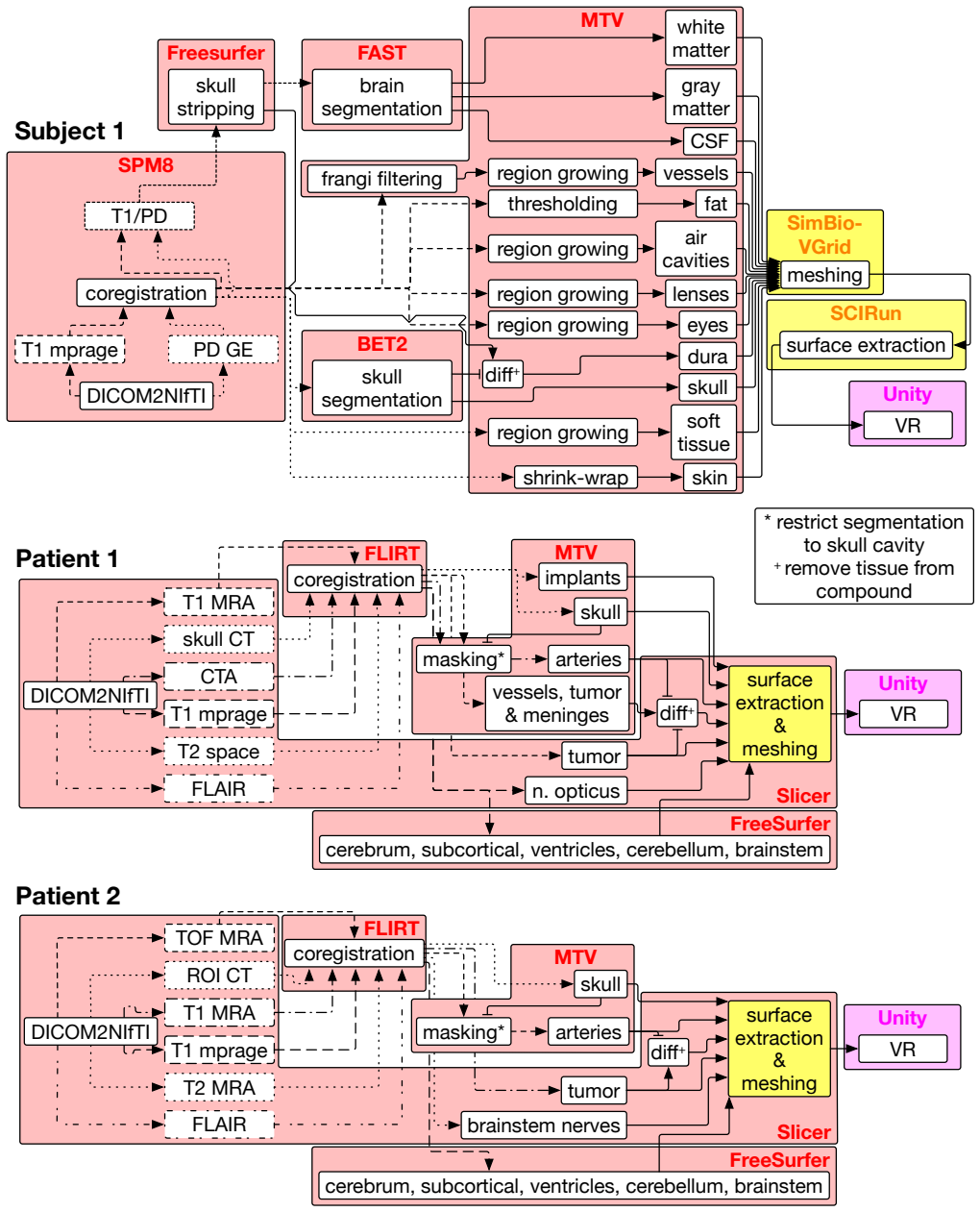
The load-bearing mechanism is a modular imaging-to-VR pipeline. Clinical image files are converted to a common format, co-registered and resliced to a reference scan, then segmented with a mix of partly automatic region growing for skull and vessels, automatic brain tissue parcellation, and manual tracing for pathology such as tumors and cranial nerves. The segmented volumes are meshed, given smoothed surfaces with correct inside/outside orientation, and imported into a VR environment built on a standard game engine, where two controller-based interaction modes let the surgeon move, rotate, scale, toggle layers, and cut cross-sectional planes through the model. Adapting each patient's pipeline to the available sequences is what makes routine clinical data usable; the renderer's stable frame rate is what makes the result usable in practice.
What would settle it
Have independent raters or an automated method re-segment the same two patients' scans and measure how much the tumor, vessel, nerve, and optic-tract boundaries overlap with the paper's models; large disagreement, or a mismatch with intraoperative findings or postoperative imaging, would show that the VR models cannot be trusted for surgical planning.
Extended reading notes
Core claim
The central claim is that the existing open-source segmentation pipeline, originally designed for research-grade images of healthy brains, can be adapted to the lower-quality, heterogeneous imaging data acquired in routine clinical care, and the resulting models can be rendered smoothly and inspected interactively in fully immersive VR. The paper demonstrates this on two tumor patients: from routine MRI and CT volumes, the pipeline produced segmented models of skull, vessels, tumor, optic tract, brainstem nerves, eyes, gray and white matter, ventricles, and other structures. Two certified senior neurosurgeons viewed the models postoperatively before seeing the 2D images, reported being surprised by their accuracy, and were able to verify their VR-based spatial understanding against the conventional 2D slices. Frame rates stayed at or above 30 frames per second on a consumer graphics card across all tested model configurations. The authors present this as first evidence that VR presentations of pre-neurosurgical data could provide additional information and potentially ease preparation of surgery.
Load-bearing premise
The framework's usefulness rests on the assumption that the segmented 3D models faithfully match the real anatomy; the manual segmentations of tumors and nerves are treated as ground truth without independent validation, so if they misrepresent the patient, the VR view could mislead rather than assist.
Editorial extensions
If this is right
- Routine clinical scans, not just high-resolution research images, can feed a VR planning environment, so the approach could be applied to normal hospital data.
- Neurosurgeons can form a 3D mental model in VR and then check it against 2D slices, suggesting VR could reduce the cognitive load of mental reconstruction.
- Because the framework uses open-source tools and consumer hardware, other research groups can reproduce, extend, and customize it without commercial licensing.
- Replacing the manual and per-patient-adapted segmentation steps with a fully automated deep-learning pipeline would turn the prototype into a scalable clinical tool, a step the paper explicitly plans.
- The positive surgeon feedback motivates a quantitative evaluation of whether VR planning changes surgical decisions or outcomes, which the authors state they are preparing.
Reading between the lines
- The decisive open question is segmentation fidelity: if the tumor, vessel, and nerve boundaries are not accurate, the immersive display could mislead rather than help, so an independent accuracy benchmark is the logical next test.
- A testable extension would be a randomized or crossover study measuring planning time, plan quality, and spatial understanding with VR versus conventional 2D review.
- The same pipeline could be pointed at surgical training, letting junior surgeons rehearse approaches on patient-specific anatomy, though the paper only hints at this direction.
- Because the two interaction modes were partly suggested by the evaluating surgeons, clinical acceptance may depend on iterative refinement with surgeons rather than generic usability heuristics.
Signed reviews
Editorial analysis
A structured set of objections, weighed in public.
Referee Report
Summary. The paper proposes a research framework for virtual reality (VR) neurosurgery built exclusively from open-source tools (3D Slicer, FreeSurfer, FSL, Blender, Unity3D) and consumer hardware (Oculus Rift, GTX 1080 Ti). The framework converts routine clinical MRI and CT data from two tumor patients and one healthy subject into segmented 3D models of brain structures, vessels, tumors, and other relevant anatomy, and presents them in an immersive VR environment with interactive controls. Preliminary evaluation consists of frame-rate measurements (always above 30 FPS) and qualitative feedback from two senior neurosurgeons who reported that the models appeared accurate and useful for surgical preparation. The paper concludes that the framework could provide experienced neurosurgeons with additional information and potentially ease surgical preparation.
Significance. If the framework's anatomical models are accurate, the work would be a valuable open-source, low-cost research platform for VR neurosurgical planning, extending prior work by using routine clinical rather than research-grade imaging data. The demonstration of smooth real-time interaction on consumer hardware is a credible strength, and the paper explicitly positions itself as a preliminary foundation. However, the absence of any quantitative validation of the segmentation accuracy, which the VR models depend on, leaves the clinical usefulness claim unsupported. A structured, blinded evaluation with a clear anatomical ground truth would substantially raise the significance of the work.
major comments (3)
- [Section 3.2] The manual segmentation of tumors, optic tract, brainstem nerves, and eyes is treated as ground truth without any validation. No Dice scores, surface-distance errors, inter-rater agreement, or comparison against intraoperative findings are reported. Because the VR models inherit the accuracy of these segmentations, the concluding claim in Section 6 that the framework 'could provide experienced neurosurgeons with additional information' is not supported for the critical anatomical details. The authors should add at least a basic validation, such as a comparison of tumor boundaries against an independent expert segmentation or against surgical observations.
- [Section 4.4] The physician evaluation is an unstructured, qualitative assessment by two senior neurosurgeons who had previously suggested interface features (Section 3.4) and who inspected the VR models postoperatively rather than in a blinded planning context. This feedback indicates perceived usefulness but cannot establish the anatomical accuracy of the models, which is the load-bearing condition for the central claim. A structured evaluation (e.g., task-based comparison with 2D planning, independent surgeon ratings, or a quantitative localization task) is needed to support the conclusion.
- [Section 3.2, Fig. 3] The segmentation pipeline is adapted individually for each patient, so the two patient cases are not replications of a fixed workflow. The paper acknowledges this as work in progress, but as a consequence the results do not yet demonstrate that the framework generalizes to new clinical datasets with different imaging protocols. The conclusion should explicitly state this limitation and avoid implying that the framework is ready as a general pipeline.
minor comments (5)
- [Section 1] In the third paragraph, 'we have started to addressed' should be 'we have started to address'.
- [Section 4.4] 'preliminarly' should be 'preliminarily', and 'A NVIDIA GTX 1080 Ti' should be 'An NVIDIA GTX 1080 Ti'.
- [Figure 4 caption] The caption contains a typo: 'asses' should be 'assess'.
- [Section 2] The claim of being 'the first dedicated, research software immersive VR framework' is asserted without a systematic literature review; consider softening to 'to the best of our knowledge'.
- [General] The paper provides supplementary videos but does not make the framework source code publicly available; for a paper whose central contribution is an open-source framework, a public repository would considerably strengthen reproducibility.
Circularity Check
No significant circularity; the paper makes no fitted predictions and its only self-citation is an independent component used as a building block.
full rationale
The paper does not derive any quantity from fitted parameters, and it makes no quantitative prediction that reduces to its inputs. It describes a software pipeline that combines open-source tools for segmentation, meshing, and VR rendering; the reported results are a frame-rate measurement and qualitative feedback from two neurosurgeons. The manual segmentations are not independently validated, which is a correctness/evidence limitation, not a circularity. The only self-referential element is the citation of the authors' prior segmentation pipeline [13], but that pipeline is a peer-reviewed, independently published component used as a starting point, not as the target conclusion. No equation is shown to equal another by construction, and no self-citation is invoked to forbid alternatives. Accordingly, the paper is self-contained with respect to its stated claims, and the circularity score is 0.
Assumptions & free parameters
assumptions (3)
- domain assumption Off-the-shelf open-source tools (FSL, FreeSurfer, 3D Slicer, Blender, Unity3D) perform their documented functions correctly and are suitable for clinical imaging data.
- domain assumption Manual segmentations of tumors, optic tract, brainstem nerves, and eyes are anatomically accurate enough for surgical planning.
- domain assumption The qualitative feedback of two senior neurosurgeons is a valid measure of the framework's usefulness.
Cite this review
Pith. "Pith review of A Research Framework for Virtual Reality Neurosurgery Based on Open-Source Tools." pith.science (2026). https://pith.science/paper/5QB6FDGH
@misc{pith2026190805188,
  author       = {Pith},
  title        = {Pith review of: A Research Framework for Virtual Reality Neurosurgery Based on Open-Source Tools},
  year         = {2026},
  howpublished = {\url{https://pith.science/paper/5QB6FDGH}},
  note         = {Machine review of arXiv:1908.05188}
}
read the original abstract
Fully immersive virtual reality (VR) has the potential to improve neurosurgical planning. For example, it may offer 3D visualizations of relevant anatomical structures with complex shapes, such as blood vessels and tumors. However, there is a lack of research tools specifically tailored for this area. We present a research framework for VR neurosurgery based on open-source tools and preliminary evaluation results. We showcase the potential of such a framework using clinical data of two patients and research data of one subject. As a first step toward practical evaluations, two certified senior neurosurgeons positively assessed the usefulness of the VR visualizations using head-mounted displays. The methods and findings described in our study thus provide a foundation for research and development aiming at versatile and user-friendly VR tools for improving neurosurgical planning and training.
Figures


Figures from the paper (4 more)


Reference graph
Works this paper leans on
- [1]
-
[2]
A. Alaraj, F. T. Charbel, D. Birk, M. Tobin, C. Luciano, P. P. Banerjee, S. Rizzi, J. Sorenson, K. Foley, K. Slavin, et al. Role of cranial and spinal virtual and augmented reality simulation using immersive touch modules in neurosurgical training. Neurosurgery, 72(suppl 1):A115– A123, 2013
work page 2013
- [3]
-
[4]
F. E. Alotaibi, G. A. AlZhrani, M. A. Mullah, A. J. Sabbagh, H. Azarnoush, A. Winkler-Schwartz, and R. F. Del Maestro. Assessing bimanual performance in brain tumor resection with neurotouch, a virtual reality simulator. Operative Neurosurgery, 11(1):89–98, 2015
work page 2015
-
[5]
G. AlZhrani, F. Alotaibi, H. Azarnoush, A. Winkler-Schwartz, A. Sab- bagh, K. Bajunaid, S. P. Lajoie, and R. F. Del Maestro. Proficiency performance benchmarks for removal of simulated brain tumors using a virtual reality simulator neurotouch. Journal of surgical education, 72(4):685–696, 2015
work page 2015
-
[6]
V . S. Arikatla, M. Tyagi, A. Enquobahrie, T. Nguyen, G. H. Blakey, R. White, and B. Paniagua. High fidelity virtual reality orthognathic surgery simulator. In Medical Imaging 2018: Image-Guided Proce- dures, Robotic Interventions, and Modeling , vol. 10576, p. 1057612. International Society for Optics and Photonics, 2018
work page 2018
-
[7]
H. Azarnoush, G. Alzhrani, A. Winkler-Schwartz, F. Alotaibi, N. Gelinas-Phaneuf, V . Pazos, N. Choudhury, J. Fares, R. DiRaddo, and R. F. Del Maestro. Neurosurgical virtual reality simulation metrics to assess psychomotor skills during brain tumor resection. International Figure 5: Model overview of P1. The upper row contains screenshots showing a frontal...
work page 2015
-
[8]
S. Chan, F. Conti, K. Salisbury, and N. H. Blevins. Virtual reality simulation in neurosurgery: technologies and evolution. Neurosurgery, 72(suppl 1):A154–A164, 2013
work page 2013
Show all 40 references
-
[9]
B. Chen, J. Moreland, and J. Zhang. Human brain functional mri and dti visualization with virtual reality. In ASME 2011 World Confer- ence on Innovative Virtual Reality, pp. 343–349. American Society of Mechanical Engineers, 2011
2011
-
[10]
A. M. Dale, B. Fischl, and M. I. Sereno. Cortical surface-based analysis: I. segmentation and surface reconstruction. Neuroimage, 9(2):179–194, 1999
1999
-
[11]
Delorme, D
S. Delorme, D. Laroche, R. DiRaddo, and R. F. Del Maestro. Neuro- touch: a physics-based virtual simulator for cranial microneurosurgery training. Operative Neurosurgery, 71(suppl 1):ons32–ons42, 2012
2012
-
[12]
Fedorov, R
A. Fedorov, R. Beichel, J. Kalpathy-Cramer, J. Finet, J.-C. Fillion- Robin, S. Pujol, C. Bauer, D. Jennings, F. Fennessy, M. Sonka, J. Buatti, S. Aylward, J. V . Miller, S. Pieper, and R. Kikinis. 3d Slicer as an image computing platform for the Quantitative Imaging Network. M...
2012 doi
-
[13]
L. D. J. Fiederer, J. V orwerk, F. Lucka, M. Dannhauer, S. Yang, M. D¨umpelmann, A. Schulze-Bonhage, A. Aertsen, O. Speck, C. H. Wolters, et al. The role of blood vessels in high-resolution volume conductor head modeling of eeg. NeuroImage, 128:193–208, 2016
2016
-
[14]
Fischl, A
B. Fischl, A. Van Der Kouwe, C. Destrieux, E. Halgren, F. S´egonne, D. H. Salat, E. Busa, L. J. Seidman, J. Goldstein, D. Kennedy, et al. Automatically parcellating the human cerebral cortex. Cerebral cortex, 14(1):11–22, 2004
2004
-
[15]
D. N. Greve and B. Fischl. Accurate and robust brain image alignment using boundary-based registration. NeuroImage, 48(1):63–72, Oct
-
[16]
H ¨anel, P
C. H ¨anel, P. Pieperhoff, B. Hentschel, K. Amunts, and T. Kuhlen. Interactive 3d visualization of structural changes in the brain of a person with corticobasal syndrome. Frontiers in neuroinformatics , 8:42, 2014
2014
-
[17]
B. K. Hendricks, J. Hartman, and A. A. Cohen-Gadol. Cerebrovascular operative anatomy: An immersive 3d and virtual reality description. Operative Neurosurgery, 15(6):613–623, 2018
2018
-
[18]
B. K. Hendricks, A. J. Patel, J. Hartman, M. F. Seifert, and A. Cohen- Gadol. Operative anatomy of the human skull: a virtual reality expedi- tion. Operative Neurosurgery, 15(4):368–377, 2018
2018
-
[19]
Jenkinson, P
M. Jenkinson, P. Bannister, M. Brady, and S. Smith. Improved Opti- mization for the Robust and Accurate Linear Registration and Motion Correction of Brain Images. NeuroImage, 17(2):825–841, Oct. 2002. doi: 10.1006/nimg.2002.1132
2002
-
[20]
Jenkinson and S
M. Jenkinson and S. Smith. A global optimisation method for robust affine registration of brain images. Medical Image Analysis, 5(2):143– 156, June 2001. doi: 10.1016/S1361-8415(01)00036-6
2001 doi
-
[21]
Kikinis, S
R. Kikinis, S. D. Pieper, and K. G. V osburgh. 3d Slicer: A Platform for Subject-Specific Image Analysis, Visualization, and Clinical Support. In F. A. Jolesz, ed.,Intraoperative Imaging and Image-Guided Therapy, pp. 277–289. Springer New York, New York, NY , 2014. doi: 10.1007...
2014
-
[22]
T. Kin, H. Oyama, K. Kamada, S. Aoki, K. Ohtomo, and N. Saito. Prediction of surgical view of neurovascular decompression using interactive computer graphics. Neurosurgery, 65(1):121–129, 2009
2009
-
[23]
R. A. Kockro and P. Y . Hwang. Virtual temporal bone: An interac- tive 3-dimensional learning aid for cranial base surgery. Operative Neurosurgery, 64(suppl 5):ons216–ons230, 2009
2009
-
[24]
Litjens, T
G. Litjens, T. Kooi, B. E. Bejnordi, A. A. A. Setio, F. Ciompi, M. Ghafoorian, J. A. Van Der Laak, B. Van Ginneken, and C. I. S´anchez. A survey on deep learning in medical image analysis. Medical image analysis, 42:60–88, 2017
2017
-
[25]
D. Low, C. K. Lee, L. L. T. Dip, W. H. Ng, B. T. Ang, and I. Ng. Augmented reality neurosurgical planning and navigation for surgical excision of parasagittal, falcine and convexity meningiomas. British journal of neurosurgery, 24(1):69–74, 2010
2010
-
[26]
Luciano, P
C. Luciano, P. Banerjee, G. M. Lemole, and F. Charbel. Second generation haptic ventriculostomy simulator using the immersivetouch system. Studies in health technology and informatics , 119:343, 2005
2005
-
[27]
Maier, B
O. Maier, B. H. Menze, J. von der Gablentz, L. H¨ani, M. P. Heinrich, M. Liebrand, S. Winzeck, A. Basit, P. Bentley, L. Chen, et al. Isles 2015- a public evaluation benchmark for ischemic stroke lesion segmentation from multispectral mri. Medical image analysis, 35:250–269, 2017
2015
-
[28]
A. M. Mendrik, K. L. Vincken, H. J. Kuijf, M. Breeuwer, W. H. Bouvy, J. De Bresser, A. Alansary, M. De Bruijne, A. Carass, A. El-Baz, et al. Mrbrains challenge: online evaluation framework for brain image segmentation in 3t mri scans. Computational intelligence and neuroscienc...
2015
-
[29]
B. H. Menze, A. Jakab, S. Bauer, J. Kalpathy-Cramer, K. Farahani, J. Kirby, Y . Burren, N. Porz, J. Slotboom, R. Wiest, et al. The mul- timodal brain tumor image segmentation benchmark (brats). IEEE transactions on medical imaging, 34(10):1993, 2015
1993
-
[30]
I. Ng, P. Y . Hwang, D. Kumar, C. K. Lee, R. A. Kockro, and Y . Sitoh. Surgical planning for microsurgical excision of cerebral arterio-venous malformations using virtual reality technology. Acta neurochirurgica, 151(5):453, 2009
2009
-
[31]
P. E. Pelargos, D. T. Nagasawa, C. Lagman, S. Tenn, J. V . Demos, S. J. Lee, T. T. Bui, N. E. Barnette, N. S. Bhatt, N. Ung, et al. Utilizing virtual and augmented reality for educational and clinical enhancements in neurosurgery. Journal of Clinical Neuroscience, 35:1–4, 2017
2017
-
[32]
T.-m. Qiu, Y . Zhang, J.-S. Wu, W.-J. Tang, Y . Zhao, Z.-G. Pan, Y . Mao, and L.-F. Zhou. Virtual reality presurgical planning for cerebral gliomas adjacent to motor pathways in an integrated 3-d stereoscopic visual- ization of structural mri and dti tractography. Acta neuroch...
2010
-
[33]
R. A. Robison, C. Y . Liu, and M. L. Apuzzo. Man, mind, and machine: the past and future of virtual reality simulation in neurologic surgery. World neurosurgery, 76(5):419–430, 2011
2011
-
[34]
C. M. Schirmer, J. B. Elder, B. Roitberg, and D. A. Lobel. Virtual reality–based simulation training for ventriculostomy: an evidence- based approach. Neurosurgery, 73(suppl 1):S66–S73, 2013
2013
-
[35]
C. M. Schirmer, J. Mocco, and J. B. Elder. Evolving virtual reality simulation in neurosurgery. Neurosurgery, 73(suppl 1):S127–S137, 2013
2013
-
[36]
D. Shen, G. Wu, and H.-I. Suk. Deep learning in medical image Figure 7: Preliminary Performance Evaluation of the VR Visualization. Performance is stable across interactions but decreases with increasing number of vertices passed to the GPU. Vertices of multiple models are acc...
2017
-
[37]
G. K. Wong, C. X. Zhu, A. T. Ahuja, and W. Poon. Stereoscopic virtual reality simulation for microsurgical excision of cerebral arteriovenous malformation: case illustrations. Surgical neurology, 72(1):69–72, 2009
2009
-
[38]
G. K. Wong, C. X. Zhu, A. T. Ahuja, and W. S. Poon. Craniotomy and clipping of intracranial aneurysm in a stereoscopic virtual reality environment. Neurosurgery, 61(3):564–569, 2007
2007
-
[39]
D. L. Yang, Q. W. Xu, X. M. Che, J. S. Wu, and B. Sun. Clinical evaluation and follow-up outcome of presurgical plan by dextroscope: a prospective controlled study in patients with skull base tumors.Surgical neurology, 72(6):682–689, 2009
2009
-
[2009]
doi: 10.1016/j.neuroimage.2009.06.060
2009 doi
Reviewed August 14, 2026 · model on record in the stance chip above.
Discussion (0). Continue with ORCID to comment.