REVIEW 2 major objections 4 minor 30 references
On Using Signal Magnitude in Diffusion Magnetic Resonance Measurements of Restricted Motion
T0 review · 2 major / 4 minor · reviewed 2026-08-14 · deepseek-v4-flash
Pith's one-line read Using the magnitude of the diffusion MR signal forces the reconstructed displacement distribution to be symmetric, erasing asymmetric microstructure.
desk verdict A mathematically correct but textbook-level cautionary note: magnitude processing in diffusion MRI forces symmetry, and the simulations illustrate it well, but the practical inference claim is overreached. read the letter →
The pith
A machine-rendered reading of the paper's core claim, the machinery that carries it, and where it could break.
The reading
What carries the argument
The load-bearing object is the displacement integral $W^d_i$, the difference between the time integrals of a magnetic moment's path during the two pulsed gradients of a PGSE sequence; its distribution $P^{nmr}_{cfd}$ is real-valued and is the physical quantity of interest. Equations (7) and (8) express the noiseless complex signal as the Fourier transform of this distribution, so Hermitian symmetry is the mechanism: since $P^{nmr}_{cfd}$ is real, $S^{nmr}_{cfd}$ is Hermitian, and taking $|S^{nmr}_{cfd}|$ before the inverse transform collapses the phase and symmetrizes the recovered distribution.
What would settle it
Take any real, asymmetric displacement-integral distribution $P$, compute $F^{-1}(|F(P)|)$, and compare with $P$; the paper's central identity says they differ whenever $P$ is asymmetric, so an example where they are equal would falsify the mathematical claim, while a biological phantom with known asymmetric microstructure whose magnitude-based and complex-based inferences agree would falsify the practical-impact claim.
Extended reading notes
Core claim
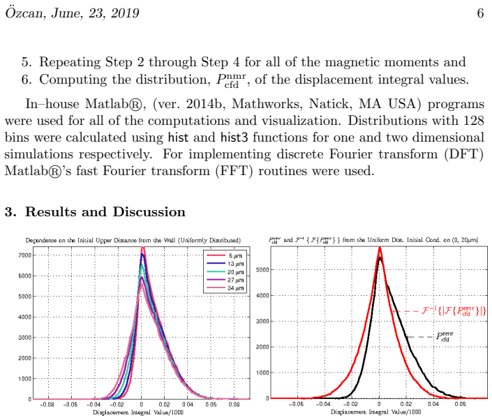
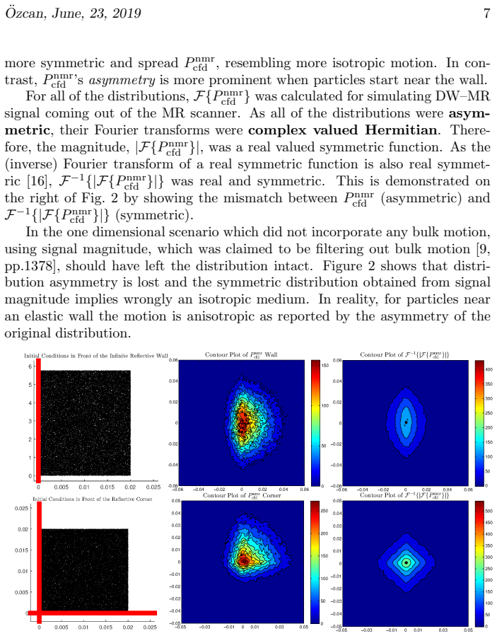
The central claim is the inequality in Eq. (12): $F^{-1}(|S^{nmr}_{cfd}|) \neq P^{nmr}_{cfd} = F^{-1}(S^{nmr}_{cfd})$, where $S^{nmr}_{cfd}$ is the complex diffusion MR signal and $P^{nmr}_{cfd}$ is the distribution of displacement integrals. Because $P^{nmr}_{cfd}$ is real, its Fourier transform is Hermitian, so its magnitude is real and symmetric; the inverse Fourier transform of that magnitude is therefore real and symmetric and cannot reproduce an asymmetric distribution. The paper shows this concretely in simulations: a wall appears as a tube, a corner appears as a box, and a spurious spike at zero displacement integral suggests stationary spins that are not there. The conclusion is that magnitude processing, often used to remove bulk motion, removes asymmetry as well and should be replaced by complex-valued processing with phase correction.
Load-bearing premise
The asymmetric displacement distributions produced by a single reflective wall and an orthogonal corner are representative of the asymmetry that matters in biological tissue, so that the demonstrated distortion actually impedes microstructure inference in practice.
Editorial extensions
If this is right
- Magnitude-based diffusion MR models cannot recover asymmetric displacement distributions, so they bias microstructure inference in any geometry where motion is asymmetric.
- The spurious zero-displacement peak introduced by magnitude processing can be misinterpreted as stationary or trapped water, inflating estimates of restricted compartments.
- The distortion is present in ideal noiseless simulations, so it is intrinsic to the magnitude operation rather than a noise artifact.
- Complex-valued processing that restores Hermitian symmetry, such as phase correction, avoids this distortion and also removes the Rician noise assumption.
- The paper's recommendation is to let data analysis reveal symmetry or its absence instead of imposing symmetry through signal magnitude or symmetric basis expansions.
Reading between the lines
- By the same Fourier argument, any real asymmetric displacement distribution will be symmetrized by magnitude processing, so the wall and corner results are instances of a general mathematical fact rather than geometry-specific accidents.
- A testable extension would be to quantify how much the inferred microstructure is distorted as a function of asymmetry strength and signal-to-noise ratio; the paper establishes the noiseless floor but not the practical error size.
- If biological tissue contains asymmetric restrictions such as curved axons, branching, or boundaries at multiple scales, magnitude-based estimates of axon diameter and density could be systematically biased in those regions.
- The linear-phase signature of bulk motion, which magnitude processing was meant to remove, could instead be estimated and corrected from the complex signal; the paper leaves that phase-correction algorithm as open future work.
Signed reviews
Editorial analysis
A structured set of objections, weighed in public.
Referee Report
Summary. This paper addresses the common practice in diffusion MR of using the magnitude of the complex MR signal in q-space and related analyses. The author shows that for a real-valued displacement-integral distribution P, the noiseless NMR signal S is its Fourier transform and is Hermitian. Taking |S| before inverse Fourier transformation yields a real, symmetric function F^{-1}(|S|), which generally differs from the true asymmetric P. The paper supports this with random-walk simulations of particles near a reflecting wall and an orthogonal corner, comparing the true P with F^{-1}(|S|). The visual results show that magnitude processing produces symmetric reconstructions that resemble a tube instead of a wall and a box instead of a corner, and it adds an artificial spike at the origin. The author recommends using complex-valued processing as in the CFD-MRI framework.
Significance. If correct, the paper provides a clear, elementary demonstration that magnitude-based q-space analysis cannot recover asymmetric displacement-integral distributions. The core identity, Eq. (12), is a direct consequence of Fourier transform properties and is correctly implemented and visualized in the simulations. The paper is honest about the idealized nature of the simulations and explicitly notes that the chosen geometries are not representative of biological tissue. Its practical significance for diffusion MRI inference, however, is not yet established: the simulations show distribution mismatch but do not quantify its effect on any parameter-estimation task, and the extrapolation to tissue is not supported. The paper would be a useful cautionary note and a motivation for complex-valued methods, provided the inference claims are strengthened.
major comments (2)
- [Section 4, Eq. (12)] The central claim that magnitude usage 'impedes DW-MR from accurately inferring microstructural information' is not supported by the evidence presented. Eq. (12) is a correct Fourier-theoretic statement, and Figures 2 and 3 visually confirm the distribution mismatch, but no inference or parameter-estimation is performed. The statements that a wall 'is inferred as a vertically oriented tube' and a corner 'appears as a box' are qualitative visual labels, not results of any model-fitting procedure. To substantiate the inference claim, the authors should run a downstream estimation task (e.g., fitting wall position, tube radius, or compartment size) on the simulated complex signal and on its magnitude, and report bias or error for both. As written, the paper establishes a distribution-reconstruction mismatch, not an inference failure.
- [Sections 1 and 4] The generalization from the two open-boundary geometries to biological tissue is acknowledged by the authors as unsupported: Section 1 states that these geometries 'are not necessarily representatives of biological tissue's microstructure.' Nevertheless, Section 4 recommends abandoning magnitude processing in future in and ex vivo diffusion imaging research. This extrapolation requires either additional simulations in tissue-like geometries with closed boundaries (e.g., cylinders or spheres) or an explicit argument for why the open-boundary asymmetry persists after orientation averaging in a voxel. Without such support, the practical relevance of the demonstrated distortion to typical diffusion MRI inference remains an open question.
minor comments (4)
- [Section 2.2] The simulations use 120000 particles but no random seeds or repeated runs are reported; please provide reproducibility details (seed or code) and, if possible, error bars on the displayed distributions to rule out single-realization artifacts.
- [Section 3] The statement that 'taking the magnitude transfers the imaginary portion to the real axis, thereby adding a positive constant to the signal' is imprecise: |S| - Re(S) is nonnegative pointwise but is not a constant. The spike at the origin is the zero-frequency consequence of the integral of |S|, rather than of adding a constant to the signal; please rephrase.
- [Section 2.1] The phrase 'the signal magnitude becomes a symmetric' is missing a noun; it should read 'a symmetric function.'
- [Eq. (2)] The exponential phase term appears malformed in the submitted manuscript; please check the typesetting of the term -iγΩ_i.
Circularity Check
No circularity: Eq. (12) follows from standard Fourier theory and independent simulations; the CFD-MRI self-citation is not load-bearing.
full rationale
I walked the derivation chain. The central claim, Eq. (12), is F^{-1}(|S^{nmr}_{cfd}|) != P^{nmr}_{cfd} = F^{-1}(S^{nmr}_{cfd}). This is obtained by applying standard Fourier reciprocity to the simulated displacement-integral distributions. P^{nmr}_{cfd} is computed from random-walk trajectories; S^{nmr}_{cfd} is computed as the Fourier transform of P^{nmr}_{cfd}; no parameter is fitted to a subset of the data and then used to predict a closely related quantity. The inequality is not assumed: it follows because an asymmetric real P has a complex Hermitian Fourier transform, whose magnitude is real and symmetric, so its inverse Fourier transform is real and symmetric and cannot coincide with the asymmetric P. This mathematical content is independent of the author's CFD-MRI framework, which is cited [4] for notation and recommended as a solution, but Eq. (12) does not depend on that citation. The generalization from wall/corner geometries to practical microstructural inference is an extrapolation, not a circular step; that concern belongs to correctness risk rather than circularity. Therefore no circular step is present.
Assumptions & free parameters
assumptions (4)
- standard math The Fourier transform of a real-valued function is Hermitian, and the inverse Fourier transform of a real, even function is real and even.
- domain assumption The DW-MR signal from a PGSE sequence is the Fourier transform of the distribution of displacement integrals, as defined in Eqs. (4)-(7).
- domain assumption Brownian motion of non-interacting particles with elastic collisions at reflecting walls accurately simulates diffusion near boundaries.
- ad hoc to paper Initial positions uniformly distributed in a strip or square near the wall or corner produce a representative displacement integral distribution for the geometry.
Cite this review
Pith. "Pith review of On Using Signal Magnitude in Diffusion Magnetic Resonance Measurements of Restricted Motion." pith.science (2026). https://pith.science/paper/CKV2JLOH
@misc{pith2026190806749,
  author       = {Pith},
  title        = {Pith review of: On Using Signal Magnitude in Diffusion Magnetic Resonance Measurements of Restricted Motion},
  year         = {2026},
  howpublished = {\url{https://pith.science/paper/CKV2JLOH}},
  note         = {Machine review of arXiv:1908.06749}
}
read the original abstract
Tissue microstructure has significance as a biomarker, however its accurate inference with diffusion magnetic resonance (MR) is still an open problem. With few exceptions, diffusion weighted (DW) MR models either process diffusion MR data using signal magnitude, whereby microstructural information is forcefully confined to symmetry due to Fourier transform properties, or directly use symmetric basis expansions. Herein, information loss from magnitude utilization is demonstrated by numerically simulating particles undergoing diffusion near a fully reflective infinite wall and an orthogonal corner. Simulation results show that the loss of the Hermitian property when using signal magnitude impedes DW--MR from accurately inferring microstructural information in both of the geometries.
Figures

Reference graph
Works this paper leans on
-
[1]
A. ¨Ozcan, K. H. Wong, L. Larson-Prior, Z.-H. Cho, S. K. Mun, Back- ground and mathematical analysis of diffusion MRI methods, Intern a- tional Journal of Imaging Systems and Technology 22 (1) (2012) 4 4–52. doi:10.1002/ima.22001. URL http://dx.doi.org/10.1002/ima.22001
-
[2]
E. ¨Ozarslan, C. G. Koay, T. M. Shepherd, M. E. Komlosh, M. O. ˙Irfano¯ glu, C. Pierpaoli, P. J. Basser, Mean apparent propagator (MAP) MRI: A novel diffusion imaging method for mapping tissue microstructure, NeuroI mage 78 (0) (2013) 16 – 32. doi:10.1016/j.neuroimage.2013.04.016. URL http://www.sciencedirect.com/science/article/pii/S1053811913003431 ¨Ozca...
-
[3]
C. Liu, R. Bammer, B. Acar, M. E. Moseley, Characterizing non–g aussian diffusion by using generalized diffusion tensors, Magnetic Resonance in Medicine 51 (5) (2004) 924–937
work page 2004
-
[4]
A. ¨Ozcan, Complete fourier direct magnetic resonance imaging (CFD– MRI) for diffusion MRI, Frontiers in Integrative Neuroscience 7 (18 ) (2013). doi:10.3389/fnint.2013.00018. URL http://www.frontiersin.org/integrative neuroscience/10.3389/fnint.2013.00018/abstract
arXiv 2013
- [5]
-
[6]
J. Mattiello, P. Basser, D. Lebihan, Analytical expressions for t he b matrix in NMR diffusion imaging and spectroscopy, Journal of Magnet ic Resonance, Series A 108 (2) (1994) 131 – 141. doi:10.1006/jmra.19 94.1103. URL http://www.sciencedirect.com/science/article/pii/S106418588471103X
-
[7]
L. R. Frank, Anisotropy in high angular resolution diffusion–weight ed MRI, Magnetic Resonance in Medicine 45 (6) (2001) 935–1141
work page 2001
-
[8]
P. T. Callaghan, Translational Dynamics and Magnetic Resonance : Princi- ples of Pulsed Gradient Spin Echo NMR, Oxford University Press, Oxf ord [England] New York, 2011
work page 2011
Show all 30 references
-
[9]
V. J. Wedeen, P. Hagmann, W.-Y. I. Tseng, T. G. Reese, R. M. We isskoff, Mapping complex tissue architecture with diffusion spectrum magnet ic res- onance imaging, Magnetic Resonance in Medicine 54 (6) (2005) 1377– 1385
2005
-
[10]
D. S. Tuch, Q–ball imaging, Magnetic Resonance in Medicine 52 (6) (2004) 1358–1372
2004
-
[11]
R. C. Wayne, R. M. Cotts, Nuclear–magnetic–resonance stud y of self–diffusion in a bounded medium, Phys. Rev. 151 (1966) 264–272. doi:10.1103/PhysRev.151.264. URL https://link.aps.org/doi/10.1103/PhysRev.151.264
1966 doi
-
[12]
Robertson, Spin–echo decay of spins diffusing in a bounded re gion, Phys
B. Robertson, Spin–echo decay of spins diffusing in a bounded re gion, Phys. Rev. 151 (1966) 273–277. doi:10.1103/PhysRev.151.273. URL https://link.aps.org/doi/10.1103/PhysRev.151.273
1966 doi
-
[13]
Barzykin, Theory of spin echo in restricted geometries unde r a step–wise gradient pulse sequence, Journal of Magnetic Resonance 139 (2) (1999) 342 – 353
A. Barzykin, Theory of spin echo in restricted geometries unde r a step–wise gradient pulse sequence, Journal of Magnetic Resonance 139 (2) (1999) 342 – 353. doi:https://doi.org/10.1006/jmre.1999.1778. URL http://www.sciencedirect.com/science/article/pii/S1090780799917780
1999
-
[14]
Brown, Y
R. Brown, Y. Cheng, E. Haacke, M. Thompson, R. Venkatesan , Magnetic Resonance Imaging: Physical Principles and Sequence Design, John Wiley and Sons, New York, 2014. ¨Ozcan, June, 23, 2019 11
2014
-
[15]
A. ¨Ozcan, Comparison of the Complete Fourier Direct MRI with existing diffusion weighted MRI methods, in: Proceedings of the 2011 IEEE In ter- national Symposium on Biomedical Imaging, Chicago, Illinois, USA, 201 1, pp. 931–934
2011
-
[16]
R. N. Bracewell, The Fourier transform and its applications, 3rd Edition, McGraw-Hill series in electrical and computer engineering. Circuits a nd systems, McGraw Hill, Boston, 2000, (DLC) 99021139 Ronald N. Bra cewell. ill. ; 25 cm. Includes bibliographical references and index
2000
-
[17]
D. L. Ermak, A computer simulation of charged particles in solutio n. I. Technique and equilibrium properties, The Journal of Chemical Phys ics 62 (10) (1975) 4189–4196. doi:10.1063/1.430300. URL https://doi.org/10.1063/1.430300
1975 doi
-
[18]
¨Ozcan, J
A. ¨Ozcan, J. Quirk, Y. Wang, Q. Wang, P. Sun, W. Spees, S.-K. Song, The validation of complete fourier direct MR method for diffusion MRI via biological and numerical phantoms, in: Engineering in Medicine and Biology Society,EMBC, 2011 Annual International Conference of t he I...
2011
-
[19]
¨Ozarslan, C
E. ¨Ozarslan, C. G. Koay, P. J. Basser, Remarks on q–space MR propagator in partially restricted, axially–symmetric, and isotropic environments, Magnetic Resonance Imaging 27 (6) (2009) 834 – 84 4. doi:10.1016/j.mri.2009.01.005. URL http://www.sciencedirect.com/science/article...
2009 doi
-
[20]
¨Ozarslan, U
E. ¨Ozarslan, U. Nevo, P. J. Basser, Anisotropy induced by macroscopic boundaries: Surface–normal mapping using diffusion– weighted imaging, Biophysical Journal 94 (7) (2008) 2809 – 2818. doi:10.1529/biophysj.107.124081. URL http://www.sciencedirect.com/science/article/pii/S000...
2008 doi
-
[21]
D. C. Alexander, P. L. Hubbard, M. G. Hall, E. A. Moore, M. Ptito , G. J. Parker, T. B. Dyrby, Orientationally invariant indices of axon diamet er and density from diffusion MRI, NeuroImage 52 (4) (2010) 1374 – 13 89. doi:https://doi.org/10.1016/j.neuroimage.2010.05.043. URL ht...
2010 doi
-
[22]
Panagiotaki, T
E. Panagiotaki, T. Schneider, B. Siow, M. G. Hall, M. F. Lythgoe, D. C. Alexander, Compartment models of the diffusion MR signal in brain whit e matter: A taxonomy and comparison, NeuroImage 59 (3) (2012) 2 241 –
2012
-
[23]
C. D. Kroenke, G. L. Bretthorst, T. E. Inder, J. J. Neil, Mode ling water diffusion anisotropy within fixed newborn primate brain using bayesian probability theory, Magnetic Resonance in Medicine 55 (1) (2006) 18 7–197. ¨Ozcan, June, 23, 2019 12
2006
-
[24]
M. D. Budde, J. A. Frank, Neurite beading is sufficient to decrea se the apparent diffusion coefficient after ischemic stroke, Proceedin gs of the National Academy of Sciences 107 (32) (2010) 14472–1447 7. doi:10.1073/pnas.1004841107. URL http://www.pnas.org/content/107/32/14472.abstract
2010 doi
-
[25]
C. D. Kroenke, Using diffusion anisotropy to study cerebral co rtical gray matter development, Journal of Magnetic Resonance 292 (2018) 106 – 116. doi:https://doi.org/10.1016/j.jmr.2018.04.011. URL http://www.sciencedirect.com/science/article/pii/S1090780718301150
2018 doi
-
[26]
Snaar, H
J. Snaar, H. Vanas, NMR self-diffusion measurements in a bounded system with loss of magnetization at the walls, Jour- nal of Magnetic Resonance, Series A 102 (3) (1993) 318 – 326. doi:https://doi.org/10.1006/jmra.1993.1110. URL http://www.sciencedirect.com/science/article/pii/...
1993
-
[27]
P. N. Sen, L. M. Schwartz, P. P. Mitra, B. I. Halperin, Surface relaxation and the long–time diffusion coefficient in porous media: Periodic geome- tries, Phys. Rev. B 49 (1994) 215–225. doi:10.1103/PhysRevB.49.2 15. URL https://link.aps.org/doi/10.1103/PhysRevB.49.215
1994 doi
-
[28]
J. E. Tanner, Transient diffusion in a system partitioned by perm e- able barriers. Application to NMR measurements with a pulsed field gradient, The Journal of Chemical Physics 69 (4) (1978) 1748–17 54. doi:10.1063/1.436751. URL https://doi.org/10.1063/1.436751
1978 doi
-
[29]
Gudbjartsson, S
H. Gudbjartsson, S. Patz, The Rician distribution of noisy MRI d ata, Mag- netic Resonance in Medicine 34 (6) (1995) 910–914
1995
-
[2254]
URL http://www.sciencedirect.com/science/article/pii/S1053811911011566
doi:http://dx.doi.org/10.1016/j.neuroimage.2011.09.081. URL http://www.sciencedirect.com/science/article/pii/S1053811911011566
2011 doi
Reviewed August 14, 2026 · model on record in the stance chip above.
Discussion (0). Continue with ORCID to comment.