REVIEW 5 major objections 4 minor 26 references
K-margin-based Residual-Convolution-Recurrent Neural Network for Atrial Fibrillation Detection
T0 review · 5 major / 4 minor · reviewed 2026-08-14 · deepseek-v4-flash
Pith's one-line read A K-margin-based residual-convolution-recurrent network detects atrial fibrillation from noisy single-lead ECGs with an F1NAOP of 0.8125, beating deep learning baselines by 6.8 percent.
desk verdict Useful engineering on AF detection, but the headline improvement is an apples-to-oranges comparison against challenge scores and is not established. read the letter →
The pith
A machine-rendered reading of the paper's core claim, the machinery that carries it, and where it could break.
The reading
What carries the argument
The load-bearing mechanism is the K-margin uncertainty selector. For every augmented segment, the network computes the difference between the top-1 and second-most-probable class probabilities; smaller differences mean the segment is confidently classified. The K segments with the smallest margin are treated as the most informative, are used in a fine-tuning cross-entropy objective, and their predicted labels vote for the record-level diagnosis. Around it sits the RCR-net: a 33-layer residual block for local beat morphology, followed by a Bi-LSTM for rhythm trend, and a skewness-driven augmentation that sets the segment stride inversely to how rare the record label is.
What would settle it
Take a random sample of test records, have experts label each augmented segment individually, and count how many records have fewer than the K used in the paper (K=3) segments matching the record-level label; if that fraction is non-negligible, the K-margin mechanism is being trained on systematically wrong labels and the reported gains must come from elsewhere.
Extended reading notes
Core claim
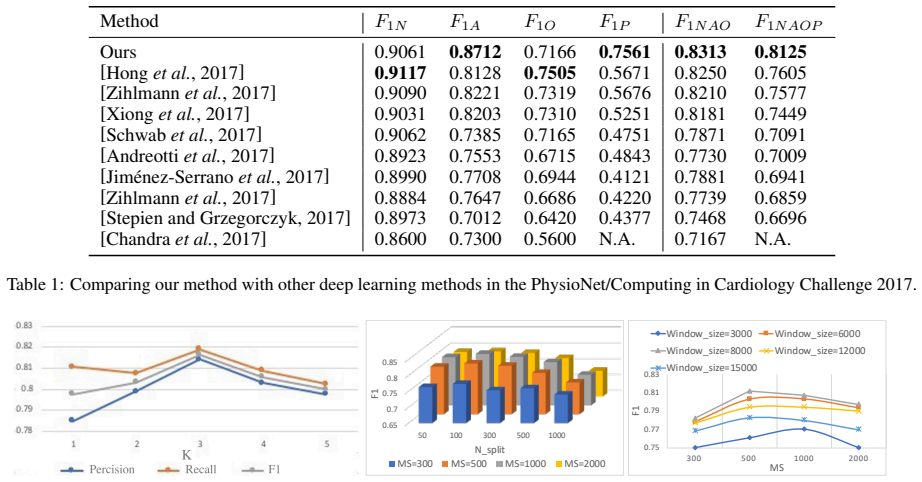
The central claim is that the K-margin-based RCR-net outperforms all compared deep learning baselines on the four-class AF detection task, achieving an F1NAOP of 0.8125 versus 0.7605 for the next-best listed method. The contribution is the specific combination of three mechanisms: dynamic augmentation whose stride is smaller for rarer classes, a multi-view network in which residual blocks extract local heartbeat-level features and a Bi-LSTM layer captures long-term rhythm-level structure, and a K-margin-based selector that identifies the top-K segments with the smallest margin between the two most probable predicted classes. These segments are used for a fine-tuning objective and then vote to label each record. The paper reports that this design improves the F1 of the 'too noisy to classify' class in particular, and that its deep features are well separated across the four classes.
Load-bearing premise
The method's training and voting scheme assumes that every record has at least K segments whose true rhythm matches the record-level label, because all segments inherit that label; if a record is mostly noise or contains multiple rhythms, every segment is mislabeled and K-margin selection cannot repair it.
Editorial extensions
If this is right
- Automated AF detection becomes practical for short, noisy single-lead ECGs, since the model learns beat-level and rhythm-level features end to end without hand-crafted cardiology features.
- The 'too noisy to classify' class benefits the most: the F1 for that class rises to 0.7561, compared with roughly 0.48–0.57 for the listed baselines, so noisy records are less likely to be misread or discarded.
- Only the top-K most confident segments of each record take part in fine-tuning and voting, so the method can be interpreted as an automatic noise-filtering layer inside a deep network.
- The same K-margin selection scheme can be attached to other segment-based deep classifiers for any multi-class time-series task, not only ECG rhythm classification.
Reading between the lines
- Because the paper does not remove the K-margin block while keeping the same architecture and augmentation, the relative contributions of augmentation versus segment selection remain untangled; a controlled ablation would make the source of the 6.8 percent gain explicit.
- The K-margin selector depends on the calibration of the network's softmax probabilities: on very noisy records the top-1 minus top-2 gap may be high for a confident but wrong class. Measuring calibration on the 'too noisy' class would show where the reliance on confidence is safe.
- Because the augmentation stride is set only by class frequency, the same idea could be extended to a noise-aware stride that also considers per-record signal quality; the current formulation has no such term.
Signed reviews
Editorial analysis
A structured set of objections, weighed in public.
Referee Report
Summary. The paper proposes a K-margin-based Residual-Convolution-Recurrent Network (K-margin-based RCR-net) for atrial fibrillation detection from noisy single-lead ECGs. The method combines skewness-driven dynamic data augmentation, a residual-convolutional recurrent architecture, and a confidence-based segment selection and voting scheme. On the PhysioNet/Computing in Cardiology Challenge 2017 dataset, the authors report an F1NAOP score of 0.8125 and claim a 6.8% improvement over all state-of-the-art deep learning baselines.
Significance. If the reported results were obtained under a controlled and reproducible protocol, the proposed method would be a useful contribution to automated ECG analysis: the idea of focusing training and inference on high-confidence segments is intuitively appealing, and the residual-convolution-recurrent architecture combines local and rhythm-level features in a principled way. However, the paper does not provide code, confidence intervals, or a clearly specified evaluation protocol, and the comparison against published baselines is uncontrolled. The central comparative claim is therefore not currently supported, despite the plausibility of the underlying architecture.
major comments (5)
- [§4.1 and §4.2] The evaluation protocol is ambiguous and the comparison underlying the headline claim is uncontrolled. Section 4.1 states that the authors 'randomly split 80% for model training, and evaluate on remaining 20% testing data' and also that they report averages 'by repeatedly running 20 times using 5-fold cross validation', without specifying which protocol produced the 0.8125 F1NAOP score. Table 1 then lists scores from published Challenge 2017 papers, which were obtained on the official hidden test set under the Challenge scoring rule, not on a random 80/20 split. The 6.8% improvement over these baselines is therefore not established; it may reflect differences in test data, tuning, or scoring rather than method quality.
- [§4.1, Precision and Recall definitions] The formulas for Precision and Recall have swapped or inconsistent denominators. Precision is written with the denominator |{i|y(i)=c}|, which is the number of true examples of class c, and Recall is written with the denominator |{i|hat y(i)=c}|, which is the number of predicted examples. With these definitions the reported F1 values, including the headline 0.8125, cannot be interpreted as standard F1 scores. The authors should either correct the formulas or clarify that a nonstandard challenge-specific metric is being used, and then recompute all reported numbers accordingly.
- [§3.4, Eq. (6)] The K-margin-based segment selection creates a self-training loop that can reinforce confident but wrong predictions. The model's own softmax output is used to decide which segments contribute to the cross-entropy loss, and for records with average confidence below 0.5 the algorithm selects the complement of the top-K confident segments, i.e., the 'hard' segments. Because all segments of a record inherit the record-level label (Section 3.2), a record that contains mostly noise or multiple rhythm types will have incorrect segment labels, and the selection procedure cannot recover the true class. No per-segment annotations or analysis of how often the required assumption (at least K segments predominantly of the true class) holds are provided.
- [Figure 7 / Section 4.2] Hyperparameters are selected on the same test set used for the final reported numbers. Figure 7 reports F1 scores for different values of K, N_split, MS, and window size, with no separate validation set described. This means the reported 0.8125 score is partly the result of test-set tuning, which inflates the apparent performance and makes the comparison with fixed baseline configurations unfair.
- [Section 4.2] No confidence intervals, standard deviations, or statistical significance tests are reported, despite the claim that runs are repeated 20 times. Without such statistics, the 6.8% margin cannot be assessed, especially since the baseline scores in Table 1 are single published values rather than distributions under the same protocol.
minor comments (4)
- [Abstract and Section 4.2] The abstract and Section 4.2 state that the method outperforms all state-of-the-art methods 'by 6.8%', but the same section also mentions a range of 6.8%–21.3% when discussing Table 1; the two statements should be reconciled.
- [Figure 2] Figure 2 contains garbled or OCR-corrupted labels such as '7UDLQLQJ' and '7HVWLQJ' and the text 'Noise diagnotor'; these should be cleaned up before any revision.
- [Section 4.2] The text mentions AlexNet and VGG in the discussion of Figure 5, but these methods are not defined or listed among the compared baselines, and no results for them are shown.
- [Section 3.4, Eqs. (2)-(4)] The notation dot-hat and double-dot-hat in Eqs. (2)-(4) is confusing; the definitions of the most and second-most probable classes should be written explicitly to avoid ambiguity between arg-min and arg-max formulations.
Circularity Check
No significant circularity: the reported predictions are not equivalent to the method's inputs by construction; the noted weaknesses are experimental-design concerns, not circular reductions.
full rationale
The paper's derivation chain is empirical and not circular. The record-level label is assigned to all augmented segments (Section 3.2), then a residual-convolution-recurrent network is trained with cross-entropy on selected segments (Eq. 7). The K-margin selection (Eqs. 2-6, Algorithm 1) uses the model's own confidence scores to choose which segments contribute to the loss, but the training objective is still evaluated against the ground-truth record labels y(i); model predictions are never substituted for labels. The final record prediction is a majority vote (Eq. 9) over selected segment predictions, which is a standard inference procedure and is not defined in terms of the reported F1 score. Self-citations to [Hong et al., 2017] and [Hong et al., 2019] appear only as related work and as a baseline in Table 1; the method does not depend on an unverified theorem from those papers. The weaknesses in the paper—test-set hyperparameter selection (Figure 7), an unclear train/test protocol (Section 4.1), and comparison against published challenge scores under a different protocol (Table 1)—are methodological correctness risks, not circular reductions. No equation in the paper reduces to its own input by construction.
Assumptions & free parameters
free parameters (6)
- window size w =
6000
- maximum stride threshold MS =
500
- K (number of top segments) =
3
- N_split (number of fragments for RNN) =
300
- alpha threshold for segment selection =
0.5
- train/test split ratio =
80/20
assumptions (4)
- domain assumption Record-level labels in the PhysioNet/CinC 2017 dataset are correct ground truth.
- ad hoc to paper Each ECG record contains at least K segments that are predominantly of the true class.
- standard math Standard neural network training (backpropagation, softmax, cross-entropy, Bi-LSTM) behaves as expected.
- domain assumption The random 80/20 split is representative of the official challenge test distribution.
Cite this review
Pith. "Pith review of K-margin-based Residual-Convolution-Recurrent Neural Network for Atrial Fibrillation Detection." pith.science (2026). https://pith.science/paper/XQEUOOLO
@misc{pith2026190806857,
  author       = {Pith},
  title        = {Pith review of: K-margin-based Residual-Convolution-Recurrent Neural Network for Atrial Fibrillation Detection},
  year         = {2026},
  howpublished = {\url{https://pith.science/paper/XQEUOOLO}},
  note         = {Machine review of arXiv:1908.06857}
}
read the original abstract
Atrial Fibrillation (AF) is an abnormal heart rhythm which can trigger cardiac arrest and sudden death. Nevertheless, its interpretation is mostly done by medical experts due to high error rates of computerized interpretation. One study found that only about 66% of AF were correctly recognized from noisy ECGs. This is in part due to insufficient training data, class skewness, as well as semantical ambiguities caused by noisy segments in an ECG record. In this paper, we propose a K-margin-based Residual-Convolution-Recurrent neural network (K-margin-based RCR-net) for AF detection from noisy ECGs. In detail, a skewness-driven dynamic augmentation method is employed to handle the problems of data inadequacy and class imbalance. A novel RCR-net is proposed to automatically extract both long-term rhythm-level and local heartbeat-level characters. Finally, we present a K-margin-based diagnosis model to automatically focus on the most important parts of an ECG record and handle noise by naturally exploiting expected consistency among the segments associated for each record. The experimental results demonstrate that the proposed method with 0.8125 F1NAOP score outperforms all state-of-the-art deep learning methods for AF detection task by 6.8%.
Figures


Figures from the paper (4 more)


Reference graph
Works this paper leans on
-
[1]
[Andreotti et al., 2017] Fernando Andreotti, Oliver Carr, and Marco AF Pimentel, et al. Comparing feature-based clas- sifiers and convolutional neural networks to detect arrhyth- mia from short segments of ecg. InCinC, pages 1–4. IEEE,
work page 2017
-
[4]
[Clifford et al., 2017] Gari D Clifford, Chengyu Liu, and Benjamin Moody, et al. Af classification from a short sin- gle lead ecg recording: the physionet/computing in cardi- ology challenge
work page 2017
-
[5]
Real-time ecg monitoring and arrhythmia detection using android-based mobile devices
[Gradl et al., 2012] Stefan Gradl, Patrick Kugler, and Clemens Lohm ¨uller, et al. Real-time ecg monitoring and arrhythmia detection using android-based mobile devices. In EMBC, pages 2452–2455. IEEE,
work page 2012
-
[9]
[Hong et al., 2019] Shenda Hong, Yuxi Zhou, and Meng Wu, et al. Combining deep neural networks and engineered fea- tures for cardiac arrhythmia detection from ECG record- ings. Physiological Measurement, 40(5):054009,
work page 2019
-
[10]
[Jim´enez-Serrano et al., 2017] Santiago Jim ´enez-Serrano, Jaime Yag¨ue-Mayans, and Elena Simarro-Mond´ejar, et al. Atrial fibrillation detection using feedforward neural networks and automatically extracted signal features. In CinC, pages 1–4. IEEE,
work page 2017
-
[11]
[Kamaleswaran et al., 2018] Rishikesan Kamaleswaran, Ruhi Mahajan, and Oguz Akbilgic. A robust deep convolutional neural network for the classification of abnormal cardiac rhythm using single lead electrocardio- grams of variable length. Physiological measurement , 39(3):035006,
work page 2018
-
[14]
Accurate, automated detec- tion of atrial fibrillation in ambulatory recordings
[Linker, 2016] David Linker. Accurate, automated detec- tion of atrial fibrillation in ambulatory recordings. CVET, 7(2):182–189,
work page 2016
-
[15]
Robust greedy deep dictionary learning for ecg arrhythmia classification
[Majumdar and Ward, 2017] Angshul Majumdar and Rabab Ward. Robust greedy deep dictionary learning for ecg arrhythmia classification. In IJCNN, pages 4400–4407. IEEE,
work page 2017
Show all 26 references
-
[16]
Beat by beat: Classifying cardiac ar- rhythmias with recurrent neural networks
[Schwab et al., 2017] Patrick Schwab, Gaetano C Scebba, and Jia Zhang, et al. Beat by beat: Classifying cardiac ar- rhythmias with recurrent neural networks. In CinC, pages 1–4. IEEE,
2017
-
[17]
Errors in the computerized electrocardiogram inter- pretation of cardiac rhythm
[Shah and Rubin, 2007] Atman P Shah and Stanley A Ru- bin. Errors in the computerized electrocardiogram inter- pretation of cardiac rhythm. Journal of electrocardiology, 40(5):385–390,
2007
-
[20]
Classification of ecg recordings with neu- ral networks based on specific morphological features and regularity of the signal
[Stepien and Grzegorczyk, 2017] Katarzyna Stepien and Iga Grzegorczyk. Classification of ecg recordings with neu- ral networks based on specific morphological features and regularity of the signal. In CinC, pages 1–4. IEEE,
2017
-
[21]
Automatic detection of atrial fibrillation using the coefficient of varia- tion and density histograms of rr andδrr intervals
[Tateno and Glass, 2001] K Tateno and L Glass. Automatic detection of atrial fibrillation using the coefficient of varia- tion and density histograms of rr andδrr intervals. MBEC, 39(6):664–671,
2001
-
[23]
A novel method for classification of ecg arrhythmias using deep belief networks
[Wu et al., 2016] Zhiyong Wu, Xiangqian Ding, and Guan- grui Zhang. A novel method for classification of ecg arrhythmias using deep belief networks. IJCIA, 15(04):1650021,
2016
-
[24]
Robust ecg signal classification for detec- tion of atrial fibrillation using a novel neural network
[Xiong et al., 2017] Zhaohan Xiong, Martin K Stiles, and Jichao Zhao. Robust ecg signal classification for detec- tion of atrial fibrillation using a novel neural network. In CinC, pages 1–4. IEEE,
2017
-
[25]
Arrhythmia detection and classifi- cation using morphological and dynamic features of ecg signals
[Ye et al., 2010] Can Ye, Miguel Tavares Coimbra, and BVK Vijaya Kumar. Arrhythmia detection and classifi- cation using morphological and dynamic features of ecg signals. In EMBC, pages 1918–1921. IEEE, Aug
2010
-
[2001]
Ensembling convolutional and long short-term memory networks for electrocardio- gram arrhythmia detection
[Warrick and Homsi, 2018] Philip A Warrick and Ma- sun Nabhan Homsi. Ensembling convolutional and long short-term memory networks for electrocardio- gram arrhythmia detection. Physiological measurement, 39(11):114002,
2018
-
[2005]
Real-time patient-specific ecg classifi- cation by 1-d convolutional neural networks
[Kiranyaz et al., 2015] Serkan Kiranyaz, Turker Ince, and Moncef Gabbouj. Real-time patient-specific ecg classifi- cation by 1-d convolutional neural networks. IEEE Trans- actions on Biomedical Engineering, 63(3):664–675,
2015
-
[2007]
Improving the quality of ecgs collected using mobile phones: The physionet/computing in cardiology challenge
[Silva et al., 2011] Ikaro Silva, George B Moody, and Leo Celi. Improving the quality of ecgs collected using mobile phones: The physionet/computing in cardiology challenge
2011
-
[2010]
Convolu- tional recurrent neural networks for electrocardiogram classification
[Zihlmann et al., 2017] Martin Zihlmann, Dmytro Perekrestenko, and Michael Tschannen. Convolu- tional recurrent neural networks for electrocardiogram classification. In CinC, pages 1–4. IEEE, 2017
2017
-
[2011]
A convolutional neural network for ecg annotation as the basis for classification of cardiac rhythms
[Sodmann et al., 2018] Philipp Sodmann, Marcus V ollmer, and Neetika Nath, et al. A convolutional neural network for ecg annotation as the basis for classification of cardiac rhythms. Physiological measurement , 39(10):104005,
2018
-
[2012]
Cardiologist-level ar- rhythmia detection and classification in ambulatory elec- trocardiograms using a deep neural network
[Hannun et al., 2019] Awni Y Hannun, Pranav Rajpurkar, and Masoumeh Haghpanahi, et al. Cardiologist-level ar- rhythmia detection and classification in ambulatory elec- trocardiograms using a deep neural network. Nature medicine, 25(1):65,
2019
-
[2015]
Atrial fibrillation detection using con- volutional neural networks
[Chandra et al., 2017] BS Chandra, Challa S Sastry, and Soumya Jana, et al. Atrial fibrillation detection using con- volutional neural networks. In CinC, pages 1–4. IEEE,
2017
-
[2016]
Encase: An ensemble classifier for ecg classification using expert features and deep neural networks
[Hong et al., 2017] Shenda Hong, Meng Wu, and Yuxi Zhou, et al. Encase: An ensemble classifier for ecg classification using expert features and deep neural networks. In CinC, pages 1–4. IEEE,
2017
-
[2017]
Heart rate dynamics distinguish among atrial fibrillation, normal sinus rhythm and sinus rhythm with frequent ectopy
[Carrara et al., 2015] Marta Carrara, Luca Carozzi, and Travis J Moss, et al. Heart rate dynamics distinguish among atrial fibrillation, normal sinus rhythm and sinus rhythm with frequent ectopy. Physiological measurement, 36(9):1873,
2015
-
[2018]
Basis and treatment of cardiac arrhythmias , vol- ume
[Kass and Clancy, 2005] Robert E Kass and Colleen E Clancy. Basis and treatment of cardiac arrhythmias , vol- ume
2005
-
[2019]
Deep residual learning for image recogni- tion
[He et al., 2016] Kaiming He, Xiangyu Zhang, and Shao- qing Ren, et al. Deep residual learning for image recogni- tion. In CVPR, pages 770–778. IEEE,
2016
Reviewed August 14, 2026 · model on record in the stance chip above.
Discussion (0). Continue with ORCID to comment.