REVIEW 3 major objections 5 minor 8 references
Sequential Rib Labeling and Segmentation in Chest X-Ray using Mask R-CNN
T0 review · 3 major / 5 minor · reviewed 2026-08-14 · deepseek-v4-flash
Pith's one-line read The paper reports the first approach to simultaneously segment and anatomically label ribs in chest X-rays, with mean Dice of 0.846 for bounding boxes and 0.733 for masks.
desk verdict First genuine combination of rib segmentation and labeling in chest X-ray, but the reported numbers are compromised by anchor boxes fitted to the whole dataset before cross-validation. read the letter →
The pith
A machine-rendered reading of the paper's core claim, the machinery that carries it, and where it could break.
The reading
What carries the argument
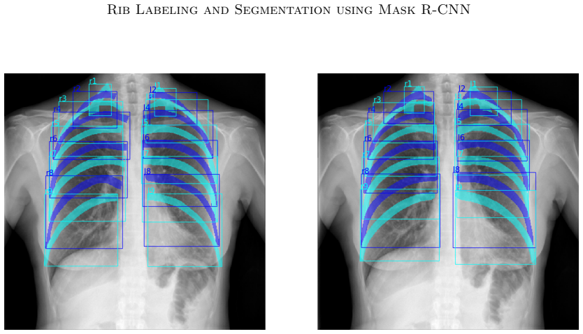
The load-bearing idea is sequential, rib-specific instance segmentation. For each label 1 through 9, a Mask R-CNN branch is trained with its own set of anchor boxes; these boxes come from Mean Shift clustering of all ground-truth rib boxes normalized to image size, producing 30 prototypical locations. For ribs 2 to 9, the predicted mask of the rib above is inserted as the third input channel, with the grayscale X-ray replicated in the other two channels, so the network can use the upper neighbor's shape and position to disambiguate the current rib. That sequential context is what gives the labels anatomical consistency rather than just separating arbitrary instances.
What would settle it
On a hold-out set of posterior-anterior chest X-rays with rib masks produced from CT bone segmentations, retrain and evaluate per-label Dice; if the sequential channel ceases to improve labeling accuracy over the non-sequential baseline, or if ribs 7–9 labels are frequently confused, the central claim is not supported.
Extended reading notes
Core claim
The central claim is that a Mask R-CNN, split into rib-specific networks and fed the segmentation of the upper neighboring rib as contextual input, is the first method to both segment ribs and assign anatomical labels in chest radiographs. The reported results—mean Dice 0.846 for bounding-box detection and 0.733 for segmentation on a five-fold cross-validation of 174 posterior-anterior chest X-rays—are said to be comparable to or better than published methods that only segment, and an improvement of over 5% and 23% over the base Mask R-CNN implementation. The authors also report higher accuracy, sensitivity, and specificity than an atlas-based rib-detection baseline. The claim matters because rib labeling and rib segmentation have previously been separate problems, and solving them together removes a preprocessing step and enables automatic rib counting for inhalation assessment.
Load-bearing premise
The accuracy of the hand-contoured rib masks in the 174 training images, and the consistent visibility of ribs 1–9, are assumed; errors there propagate into every anchor, segmentation target, and reported Dice value.
Editorial extensions
If this is right
- A single forward pass through this network yields both rib masks and rib labels, so chest X-ray pipelines could replace separate bone-suppression and rib-labeling stages.
- The large jump from the base architecture suggests that domain-specific anchors and sequential context are the main levers for instance segmentation of repetitive anatomy, not just more training data.
- The reported speed advantage over atlas-based registration opens the possibility of real-time rib counting for assessing inspiratory effort in bedside or screening settings.
Reading between the lines
- The upper-neighbor input channel is a transferable design: any elongated, serially ordered anatomy—vertebrae, teeth, bowel segments—could be labeled by feeding the previous segment's mask back into the next detection head.
- Because the reference masks cover only visible rib shadows, the reported Dice measures fidelity to visible anatomy rather than bone recovery; a CT-derived ground truth would likely be a sterner test and might reveal systematic under-segmentation.
- The normalization of anchor boxes by image size implies the same architecture should carry over to X-rays of different resolutions without retuning the proposal geometry, as long as the anatomy is similarly positioned in the frame.
Signed reviews
Editorial analysis
A structured set of objections, weighed in public.
Referee Report
Summary. The paper presents an adaptation of Mask R-CNN for simultaneous detection, anatomical labeling, and segmentation of ribs in PA chest X-ray images. The method introduces three extensions: rib-specific networks, dedicated anchor boxes computed by Mean Shift clustering from ground-truth bounding boxes, and a sequential processing scheme in which the segmentation of the upper neighbor rib is provided as an additional input channel for ribs 2-9. The authors report a five-fold cross-validation on 174 images, achieving mean Dice values of 0.846 for bounding-box detection and 0.733 for segmentation, and they claim improvements of more than 5% and 23% over the original Mask R-CNN implementation, as well as better performance than an atlas-based rib detection method.
Significance. If the reported results hold, this would be the first method to provide both rib segmentation and anatomical labeling in chest radiographs, which has practical value for rib counting, report generation, and follow-up correspondence. The paper builds on a publicly available Mask R-CNN implementation, uses a clearly described dataset, and reports quantitative results from cross-validation. The strengths are the explicit architectural extensions (rib-specific anchors and sequential neighbor-mask input) and the direct comparison to a baseline implementation. However, the central empirical claims depend on the soundness of the evaluation protocol, and the current manuscript leaves two methodological points unresolved: anchor-box information leakage across cross-validation folds, and the training/inference protocol for the sequential network input. These issues must be addressed before the reported gains can be considered reliable.
major comments (3)
- [Section 2 (Data and Method) and Section 3 (Results)] The anchor boxes are computed by Mean Shift clustering from all ground-truth bounding boxes of the entire dataset, as stated in Section 2. In the five-fold cross-validation of Section 3, the ground-truth boxes of the test fold therefore contribute to the anchor geometry used by the model evaluated on that fold. Because the modified RPN determines shifts directly from these anchors without non-maximum suppression, the anchors are not a neutral training hyperparameter; they encode test-fold rib locations and sizes. This introduces a favorable bias of unknown magnitude into the reported Dice values (0.846 detection, 0.733 segmentation) and into the claimed >5% and >23% improvements over the original implementation. The authors should recompute the anchor boxes using only each training fold's ground truth, or otherwise demonstrate that the evaluation is insensitive to this leakage.
- [Section 2, paragraph on sequential processing] The text states that for ribs 2 to 9, 'the output of the segmentation of the above rib is used for the third channel,' but it does not specify whether this output is the predicted mask from the network or the ground-truth mask during training. If ground-truth masks are used in training and predicted masks in testing, the network sees perfect upper-rib segmentations during training, creating a train-test mismatch that can inflate the reported segmentation accuracy. If predicted masks are used, the propagation order and the handling of errors in upstream ribs need to be described. This ambiguity is load-bearing because the sequential input is a central novelty of the method.
- [Section 3, comparison with Candemir et al.] The comparison with the atlas-based method of Candemir et al. reports accuracy, sensitivity, and specificity values, but the text does not define the units, the reference standard, or the spatial extent over which these metrics are computed (e.g., pixel-level, rib-level, or image-level). Without a precise definition, and without confidence intervals or a statistical significance test, the statement that the proposed algorithm 'achieves better results in all categories' is not supported. The authors should specify the evaluation protocol used for this comparison and report variability across the cross-validation folds.
minor comments (5)
- [Abstract and Section 2] The abstract describes the images as 'anterior-posterior' while Section 2 states 'posterior-anterior'; the terminology should be made consistent.
- [Section 3, Table 1] The table heading 'Detection' should clarify that the reported value is the Dice coefficient for bounding boxes, and the spacing in '0 .841' should be corrected.
- [Section 3, baseline comparison] The comparison to the 'original implementation' of Mask R-CNN would be more informative if the anchor configuration and training details of that baseline were described, since the claimed improvement depends on the baseline setup.
- [Section 4, Conclusion] The claim of 'very fast run-times that enable realtime analysis' is not supported by any timing measurement in the paper; a statement of inference time per image would be needed.
- [References] The reference for He et al. contains a formatting error: 'InComputer Vision (ICCV)' should be 'In Computer Vision (ICCV)'.
Circularity Check
Anchor boxes computed from all ground-truth boxes leak test-fold rib locations into five-fold cross-validation, inflating the reported Dice improvements.
-
fitted input called prediction
[Section 2 (Data and Method, dedicated anchor boxes) and Section 3 (Results, five-fold cross-validation)]
"In this experiment, the anchor boxes have been estimated using the Mean Shift algorithm (Comaniciu and Meer, 2002). Therefore, 30 cluster boxes were computed from all ground truth bounding boxes of the entire dataset for all labels. ... For an evaluation of the proposed architecture, a five-fold cross-validation was performed."
The anchor boxes are a fitted parameter, and they are fitted to all ground-truth bounding boxes, including those of every fold's validation images, before the five-fold cross-validation is run. Because the modified RPN 'determines shifts on the basis of anchor boxes' without non-maximum suppression, the proposal locations and scales for a test image are derived from that image's own ground-truth boxes via the cluster centers. The reported detection Dice (0.846) and segmentation Dice (0.733), and the claimed more-than-5% and 23% improvements over the original implementation, therefore are not independent predictions: a load-bearing part of the bounding-box result is built from the very labels used for evaluation.
full rationale
The paper's contributions are empirical; there is no equation-level derivation to reduce. The sequential rib-labeling scheme is a legitimate architectural contribution, and the comparison with Candemir et al. uses external metrics. The central quantitative claim, however, rests on a five-fold cross-validation in which the anchor boxes, an active component of the modified RPN, are computed from all ground-truth bounding boxes of the entire dataset, including each validation fold. This is a concrete case of a fitted input being used to 'predict' closely related quantities (bounding boxes and, indirectly, masks), and it is load-bearing because the claimed improvements over the original Mask R-CNN are measured against this setup. The hand-contoured dataset is not itself circular: it is external annotation from prior work and is used as ground truth, not as the source of the derived claim. No self-citation chain forces the central result. The score is 6 rather than higher because the mask predictions still require the network to produce segmentation output and because the anchors are only one component of the architecture; the reported numbers are partially, not entirely, determined by construction.
Assumptions & free parameters
free parameters (1)
- Anchor box clusters (mean shift) =
30
assumptions (5)
- domain assumption Hand-contoured rib masks in the von Berg et al. (2016) dataset are accurate and reflect all visible rib shadows.
- domain assumption Ribs 1 through 9 are consistently visible in all 174 PA chest X-rays and can be assigned stable anatomical labels.
- domain assumption Mask R-CNN with ResNet50 and FPN, pretrained on natural images, transfers to grey-value chest X-ray anatomy.
- domain assumption Feeding the segmentation of the upper neighbor rib as an additional input channel does not accumulate errors across the rib sequence.
- domain assumption Mean shift clustering of ground truth boxes yields representative anchor priors for all rib locations and image sizes.
Cite this review
Pith. "Pith review of Sequential Rib Labeling and Segmentation in Chest X-Ray using Mask R-CNN." pith.science (2026). https://pith.science/paper/T2SV2Z7Z
@misc{pith2026190808329,
  author       = {Pith},
  title        = {Pith review of: Sequential Rib Labeling and Segmentation in Chest X-Ray using Mask R-CNN},
  year         = {2026},
  howpublished = {\url{https://pith.science/paper/T2SV2Z7Z}},
  note         = {Machine review of arXiv:1908.08329}
}
read the original abstract
Mask R-CNN is a state-of-the-art network architecture for the detection and segmentation of object instances in the computer vision domain. In this contribution, it is used to localize, label and segment individual ribs in anterior-posterior chest X-ray images. For this purpose, several extensions have been made to the original architecture, in order to address the specific challenges of this application. This includes the use of rib specific networks, facilitating dedicated anchor boxes sampled from a training set, as well as a sequential processing of all ribs. Here, the segmentation result of the upper neighbor rib is used as additional input to the network. This approach is the first addressing both rib segmentation and anatomical labeling in chest radiographs. The results are comparable or even better than existing methods aiming only at segmentation.
Figures
Reference graph
Works this paper leans on
-
[1]
Atlas-based rib-bone detection in chest X -rays
Sema Candemir, Stefan Jaeger, Sameer Antani, Ulas Bagci, Les R Folio, Ziyue Xu, and George Thoma. Atlas-based rib-bone detection in chest X -rays. Computerized Medical Imaging and Graphics, 51: 0 32--39, 2016
work page 2016
-
[2]
Mean shift: A robust approach toward feature space analysis
Dorin Comaniciu and Peter Meer. Mean shift: A robust approach toward feature space analysis. IEEE Transactions on Pattern Analysis and Machine Intelligence, 24 0 (5): 0 603--619, 2002
2002
-
[3]
Deep residual learning for image recognition
Kaiming He, Xiangyu Zhang, Shaoqing Ren, and Jian Sun. Deep residual learning for image recognition. In Proceedings of the IEEE Conference on Computer Vision and Pattern Recognition, pages 770--778, 2016
2016
-
[4]
Kaiming He, Georgia Gkioxari, Piotr Doll \'a r, and Ross Girshick. Mask R-CNN . In Computer Vision (ICCV), 2017 IEEE International Conference on, pages 2980--2988. IEEE, 2017
work page 2017
-
[5]
Iterative fully convolutional neural networks for automatic vertebra segmentation and identification
Nikolas Lessmann, Bram van Ginneken, Pim A de Jong, and Ivana I s gum. Iterative fully convolutional neural networks for automatic vertebra segmentation and identification. Medical Image Analysis, 53: 0 142--155, 2019
work page 2019
-
[6]
Feature pyramid networks for object detection
Tsung-Yi Lin, Piotr Doll \'a r, Ross B Girshick, Kaiming He, Bharath Hariharan, and Serge J Belongie. Feature pyramid networks for object detection. In CVPR, volume 1, page 4, 2017
work page 2017
-
[7]
Alexander Oliver Mader, Jens von Berg, Alexander Fabritz, Cristian Lorenz, and Carsten Meyer. Localization and labeling of posterior ribs in chest radiographs using a CRF -regularized FCN with local refinement. In International Conference on Medical Image Computing and Computer-Assisted Intervention, pages 562--570. Springer, 2018
work page 2018
-
[8]
A novel bone suppression method that improves lung nodule detection
Jens von Berg, Stewart Young, Heike Carolus, Robin Wolz, Axel Saalbach, Alberto Hidalgo, Ana Gim \'e nez, and Tom \'a s Franquet. A novel bone suppression method that improves lung nodule detection. International Journal of Computer Assisted Radiology and Surgery, 11 0 (4): 0 641--655, 2016
work page 2016
Reviewed August 14, 2026 · model on record in the stance chip above.
Discussion (0). Continue with ORCID to comment.