REVIEW 2 major objections 4 minor 1 cited by
Artifacts in Photoacoustic Imaging: Origins and Mitigations
T0 review · 2 major / 4 minor · reviewed 2026-08-16 · deepseek-v4-flash
Pith's one-line read The paper argues that every photoacoustic imaging artifact arises from insufficient data or incorrect assumptions in reconstruction, organized into five artifact sources with simulation and clinical examples.
desk verdict Useful educational taxonomy of PAI artifacts, honestly scoped in the discussion but overbroad in title and abstract; worth publishing after a scope-claim fix. read the letter →
The pith
A machine-rendered reading of the paper's core claim, the machinery that carries it, and where it could break.
The reading
What carries the argument
The organizing device is the two-cause taxonomy and the assumption list behind it. The physical backbone is the initial-pressure equation $p_0(\mathbf{x}) = \Gamma(\mathbf{x})\mu_a(\mathbf{x})\Phi(\mathbf{x})$ for light absorption and the linear acoustic wave equation for propagation, and each artifact corresponds to a violated assumption in that chain. The taxonomy maps every artifact source to a cause, such as fluence decay, spectral coloring, sound-speed mismatch, or limited view, and to a visual effect, such as dislocation, splitting, blurring, clutter, or signal loss. Because the simulations isolate one violated assumption at a time, the machinery also provides a controlled way to learn what each artifact looks like.
What would settle it
A single clinical or simulated artifact that persists when the data are complete, noise-free, and broadband and the reconstruction uses an exact physical model would falsify the two-cause claim. A more practical check is to simulate two artifacts together, such as fluence decay plus sound-speed error, and see whether their isolated signatures still predict the combined image; if they mask or transform each other, the single-cause illustrations cannot be extended directly to clinical images.
Extended reading notes
Core claim
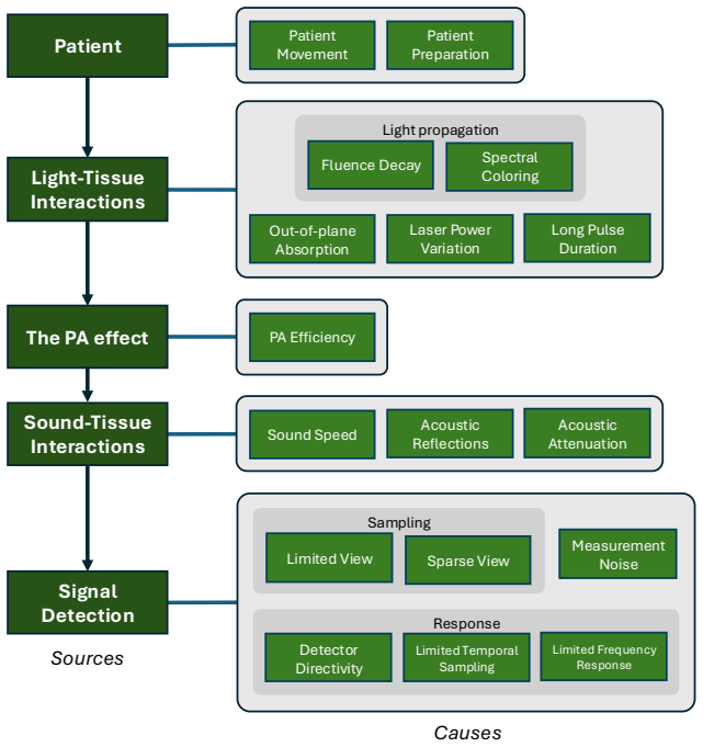
On the paper's own terms, the central claim is that the confusing variety of photoacoustic artifacts is not arbitrary. Every artifact is produced either because the measured data are incomplete—limited detector coverage, sparse elements, finite bandwidth, noise—or because the reconstruction model assumes something false, such as uniform sound speed, no acoustic attenuation, omnidirectional detectors, or spatially constant light fluence. The authors organize the resulting phenomena into five sources (patient, light-tissue interactions, the photoacoustic effect, sound-tissue interactions, and signal detection) and connect each to concrete assumptions in the image-formation chain $p_0(\mathbf{x}) = \Gamma(\mathbf{x})\mu_a(\mathbf{x})\Phi(\mathbf{x})$, followed by linear acoustic propagation to the detectors. Simulated phantoms that deliberately break one assumption at a time, together with in vivo examples, demonstrate the characteristic signature of each artifact.
Load-bearing premise
The whole didactic apparatus depends on whether an artifact's isolated signature in an ideal simulation still appears recognizably the same in a real clinical image, where many assumptions fail at once and the reconstruction algorithm is different.
Editorial extensions
If this is right
- Clinicians using multispectral PAI can learn to recognize spectral coloring and out-of-plane absorption as causes of false or missing features in oxygenation maps, reducing the risk of misdiagnosis.
- Knowing that sound-speed mismatch causes dislocation, splitting, and blurring, scanner designers can incorporate measured sound-speed maps to recover vessels that are invisible under constant-sound-speed reconstruction.
- For hardware, the taxonomy makes trade-offs explicit: more detector coverage or denser elements reduces limited- and sparse-view clutter, while detector directivity and bandwidth set a floor on resolution that deconvolution can only partially recover.
- Artifacts can sometimes carry diagnostic information, so identifying an artifact correctly may assist diagnosis rather than merely confound it, as with the comet-tail artifact in ultrasound.
- Because mitigation splits into improving the model versus supplementing the data, any reconstruction algorithm, including deep learning, will still produce artifacts whenever its implicit physics model is wrong or the data are incomplete.
Reading between the lines
- Extension: If the two-cause framing is right, artifact identification becomes a diagnostic inverse problem: the violated assumption could be inferred from the image itself, pointing toward automated artifact annotation tools for clinical workflows.
- Extension: The same two-cause dichotomy likely transfers to other hybrid modalities that reconstruct from physical models, so the taxonomy may apply to ultrasound-guided or optoacoustic tomography with re-labelled sources.
- Extension: The paper leaves reconstruction-induced artifacts out of scope, so a natural extension of the classification would add a sixth source for algorithm-specific artifacts, including deep-learning hallucinations.
- Extension: A testable extension of the isolation protocol is to break two assumptions at once; if the resulting signatures interact nonlinearly, clinical users would need signatures learned from combined artifact conditions rather than from single-cause examples.
Signed reviews
Editorial analysis
A structured set of objections, weighed in public.
Referee Report
Summary. This review paper provides a systematic account of image artifacts in photoacoustic imaging (PAI). It proposes that the artifacts considered arise from two fundamental causes, insufficient data and incorrect assumptions embedded in the reconstruction model, and organizes them into five sources: patient, light-tissue interactions, the photoacoustic effect, sound-tissue interactions, and signal detection. For each source, the paper explains the underlying physics, illustrates the artifact with simulations that break exactly one idealizing assumption, and provides experimental images from the literature. It closes with a discussion of advanced mitigation strategies, including improving the physics model, supplementing the data, deep learning, and deconvolution. The simulation code is provided through SIMPA, and the figures are generally clear.
Significance. If the scope issue is resolved, this will be a timely and valuable educational resource for clinical users and a useful reference for system developers. The simulation protocol is transparent, uses no fitted parameters, and is reproducible via the linked code. The simulated artifact signatures are corroborated by experimental examples from the literature, including in vivo images. The proposed taxonomy provides a helpful mental model even though, as discussed below, its claimed completeness needs to be scoped.
major comments (2)
- [Section 2 and Section 11] The paper frames the artifact taxonomy as covering PAI artifacts generally, with Section 2 stating that there are two corresponding classes of underlying causes (insufficient data and incorrect assumptions in the image reconstruction algorithm). Section 11, however, explicitly limits the scope to artifacts that originate outside the reconstruction step and acknowledges that reconstruction algorithms can introduce new artifacts, such as backprojection streaks, negative-value artifacts, and deep-learning hallucinations. These reconstruction-induced artifacts are not classified in the five-source taxonomy, so the classification is not exhaustive as presented. The title, abstract, and Section 2 should either incorporate reconstruction-induced artifacts or state prominently that the taxonomy covers only artifacts arising before the reconstruction step; otherwise the reader is left without an entry point for a whole class of artifacts.
- [Section 4] The isolation methodology in Section 4 (ideal settings except for the one difference giving rise to the artifact) is clear pedagogically, but the paper does not address how artifact signatures interact when several assumptions are violated simultaneously, which is the clinical situation. Because the stated purpose is to train clinical users to identify artifacts, the Discussion should warn explicitly that combined artifacts may not have the same visual signature as the isolated demonstrations and, ideally, include at least one simulation with multiple simultaneous violations to illustrate the interaction. The experimental examples partially mitigate this concern, but they are not a substitute for a stated caveat.
minor comments (4)
- [Abstract] The phrase 'assess whether their impact' is incomplete; the sentence needs an object, e.g., 'assess whether their impact is clinically significant'.
- [Section 9.1] The text 'For a typical resolution of 100 µs' should read '100 µm'; the current wording mixes time and length units in the motion estimate.
- [Table 1] Assumption H7 is listed as 'Detectors are perfectly directional,' but Section 8.2.1 states the ideal assumption is omnidirectionality; the table wording should be corrected to avoid contradiction.
- [Section 4] The simulation volume is described with '0.25 mm/pixel'; for a 3D volume this should be '0.25 mm/voxel'.
Circularity Check
No circularity: the paper is a self-contained educational taxonomy with no derived predictions or fitted parameters.
full rationale
This manuscript is an expository review that organizes known photoacoustic imaging artifacts into a taxonomy by source and cause. It introduces no fitted parameters, no self-derived constants, no inversion formula, and no quantitative prediction whose output is defined as its input. The central classification (Section 2) groups artifacts by their physical origin, while Section 4 explicitly states that the simulations isolate each artifact by deliberately breaking exactly one reconstruction assumption, which is a didactic construction rather than a claimed derivation. The paper's two-cause framing (insufficient data and incorrect reconstruction assumptions) is a high-level conceptual statement, not a mathematical result that could reduce to itself. Self-citations such as Dantuma et al. 2023 (Ref. 33) and Vogt et al. 2019 (Ref. 29) are used only as illustrative experimental examples of artifacts already demonstrated by the authors' own simulations; the taxonomy does not depend on any uniqueness theorem or load-bearing self-cited result. Section 11's explicit limitation that reconstruction-induced artifacts are outside scope is an honest scoping statement, not a circular step, and it does not undermine the internal consistency of the taxonomy as presented for its stated scope. No equation in the paper is equivalent by construction to any other equation, and no fitted value is renamed as a prediction. The paper is therefore self-contained against external benchmarks and exhibits no significant circularity.
Assumptions & free parameters
assumptions (4)
- standard math Photoacoustic pressure is given by p0 = Γ(x) μa(x) Φ(x), where Γ is the Gruneisen parameter.
- standard math The radiative transfer equation (Eq. 1) describes light transport in tissue.
- domain assumption Acoustic propagation obeys the linear wave equation (Eq. 5) with homogeneous initial conditions (Eq. 6).
- domain assumption Each artifact can be isolated by breaking exactly one assumption in an otherwise ideal simulation.
Cite this review
Pith. "Pith review of Artifacts in Photoacoustic Imaging: Origins and Mitigations." pith.science (2026). https://pith.science/paper/225L3ROJ
@misc{pith2026250412772,
  author       = {Pith},
  title        = {Pith review of: Artifacts in Photoacoustic Imaging: Origins and Mitigations},
  year         = {2026},
  howpublished = {\url{https://pith.science/paper/225L3ROJ}},
  note         = {Machine review of arXiv:2504.12772}
}
read the original abstract
Photoacoustic imaging (PAI) is rapidly moving from the laboratory to the clinic, increasing the need to understand confounders which might adversely affect patient care. Over the past five years, landmark studies have shown the clinical utility of PAI, leading to regulatory approval of several devices. In this article, we describe the various causes of artifacts in PAI, providing schematic overviews and practical examples, simulated as well as experimental. This work serves two purposes: (1) educating clinical users to identify artifacts, understand their causes, and assess whether their impact, and (2) providing a reference of the limitations of current systems for those working to improve them. We explain how two aspects of PAI systems lead to artifacts: their inability to measure complete data sets, and embedded assumptions during reconstruction. We describe the physics underlying PAI, and propose a classification of the artifacts. The paper concludes by discussing possible advanced mitigation strategies.
Figures


Figures from the paper (16 more)


Forward citations
Cited by 1 Pith paper
-
Digital twins enable full-reference quality assessment of photoacoustic image reconstructions
Digital twins provide a simulated reference that enables full-reference quality comparison of photoacoustic image reconstructions on experimental data.
Reference graph
Works this paper leans on
-
[1]
Suetens, Fundamentals of medical imaging, Cambridge university press, 2017
P. Suetens, Fundamentals of medical imaging, Cambridge university press, 2017
2017
-
[2]
Sureshbabu, O
W. Sureshbabu, O. Mawlawi, Pet/ct imaging artifacts, Journal of nuclear medicine technology 33 (3) (2005) 156–161
2005
-
[3]
E. M. Bellon, E. M. Haacke, P. E. Coleman, D. C. Sacco, D. A. Steiger, R. E. Gangarosa, Mr artifacts: a review, American Journal of Roentgenology 147 (6) (1986) 1271–1281
1986
-
[4]
D. A. Boas, D. H. Brooks, E. L. Miller, C. A. DiMarzio, M. Kilmer, R. J. Gaudette, Q. Zhang, Imaging the body with diffuse optical tomography, IEEE signal processing magazine 18 (6) (2001) 57–75
2001
-
[5]
L. V. Wang, H.-i. Wu, Biomedical optics: principles and imaging, John Wiley & Sons, 2007
2007
-
[6]
L. V. Wang, S. Hu, Photoacoustic tomography: in vivo imaging from organelles to organs, science 335 (6075) (2012) 1458–1462
2012
-
[7]
T. Chen, L. Liu, X. Ma, Y. Zhang, H. Liu, R. Zheng, J. Ren, H. Zhou, Y. Ren, R. Gao, et al., Dedicated photoacoustic imaging instrument for human periphery blood vessels: a new paradigm for understanding the vascular health, IEEE Transactions on Biomedical Engineering 69 (3) (2021) 1093–1100
2021
-
[8]
E. L. Brown, T. L. Lefebvre, P. W. Sweeney, B. J. Stolz, J. Gröhl, L. Hacker, Z. Huang, D.-L. Couturier, H. A. Harrington, H. M. Byrne, et al., Quantification of vascular networks in photoacoustic mesoscopy, Photoacoustics 26 (2022) 100357
2022
Show all 168 references
-
[9]
A. B. E. Attia, G. Balasundaram, M. Moothanchery, U. Dinish, R. Bi, V. Ntziachristos, M. Olivo, A review of clinical photoacoustic imaging: Current and future trends, Photoacoustics 16 (2019) 100144
2019
-
[10]
J. Jo, G. Xu, M. Cao, A. Marquardt, S. Francis, G. Gandikota, X. Wang, A functional study of human inflammatory arthritis using photoacoustic imaging, Scientific reports 7 (1) (2017) 15026
2017
-
[11]
Knieling, C
F. Knieling, C. Neufert, A. Hartmann, J. Claussen, A. Urich, C. Egger, M. Vetter, S. Fischer, L. Pfeifer, A. Hagel, et al., Multispectral optoacoustic tomography for assessment of crohn’s disease activity, New England Journal of Medicine 376 (13) (2017) 1292–1294
2017
-
[12]
A. P. Regensburger, E. Brown, G. Krönke, M. J. Waldner, F. Knieling, Optoacoustic imaging in inflammation, Biomedicines 9 (5) (2021) 483
2021
-
[13]
M. Li, Y. Tang, J. Yao, Photoacoustic tomography of blood oxygenation: a mini review, Photoacoustics 10 (2018) 65–73
2018
-
[14]
Gröhl, T
J. Gröhl, T. Kirchner, T. J. Adler, L. Hacker, N. Holzwarth, A. Hernández-Aguilera, M. A. Herrera, E. Santos, S. E. Bohndiek, L. Maier-Hein, Learned spectral decoloring enables photoacoustic oximetry, Scientific reports 11 (1) (2021) 6565
2021
-
[15]
W. C. Vogt, X. Zhou, R. Andriani, K. A. Wear, T. J. Pfefer, B. S. Garra, Photoacoustic oximetry imaging performance evaluation using dynamic blood flow phantoms with tunable oxygen saturation, Biomedical optics express 10 (2) (2019) 449–464
2019
-
[16]
Ntziachristos, D
V. Ntziachristos, D. Razansky, Molecular imaging by means of multispectral optoacoustic tomography (msot), Chemical reviews 110 (5) (2010) 2783–2794
2010
-
[17]
J. Yao, L. V. Wang, Recent progress in photoacoustic molecular imaging, Current opinion in chemical biology 45 (2018) 104–112
2018
-
[18]
Weber, P
J. Weber, P. C. Beard, S. E. Bohndiek, Contrast agents for molecular photoacoustic imaging, Nature methods 13 (8) (2016) 639–650
2016
-
[19]
Q. Fu, R. Zhu, J. Song, H. Yang, X. Chen, Photoacoustic imaging: contrast agents and their biomedical applications, Advanced Materials 31 (6) (2019) 1805875
2019
-
[20]
L. Lin, L. V. Wang, The emerging role of photoacoustic imaging in clinical oncology, Nature Reviews Clinical Oncology 19 (6) (2022) 365–384
2022
-
[21]
Karlas, M
A. Karlas, M. A. Pleitez, J. Aguirre, V. Ntziachristos, Optoacoustic imaging in endocrinology and metabolism, Nature Reviews Endocrinology 17 (6) (2021) 323–335
2021
-
[22]
Oraevsky, B
A. Oraevsky, B. Clingman, J. Zalev, A. Stavros, W. Yang, J. Parikh, Clinical optoacoustic imaging combined with ultrasound for coregistered functional and anatomical mapping of breast tumors, Photoacoustics 12 (2018) 30–45
2018
-
[23]
M. E. Noltes, M. Bader, M. J. Metman, J. Vonk, P. J. Steinkamp, J. Kukačka, H. E. Westerlaan, R. A. Dierckx, B. M. van Hemel, A. H. Brouwers, et al., Towards in vivo characterization of thyroid nodules suspicious for malignancy using multispectral optoacoustic tomography, Euro...
2023
-
[24]
J. L. Sandell, T. C. Zhu, A review of in-vivo optical properties of human tissues and its impact on pdt, Journal of biophotonics 4 (11-12) (2011) 773–787
2011
-
[25]
Gröhl, K
J. Gröhl, K. K. Dreher, M. Schellenberg, T. Rix, N. Holzwarth, P. Vieten, L. Ayala, S. E. Bohndiek, A. Seitel, L. Maier- Hein, Simpa: an open-source toolkit for simulation and image processing for photonics and acoustics, Journal of biomed- ical optics 27 (8) (2022) 083010–083010
2022
-
[26]
Kirchner, F
T. Kirchner, F. Sattler, J. Gröhl, L. Maier-Hein, Signed real-time delay multiply and sum beamforming for multispectral photoacoustic imaging, Journal of Imaging 4 (10) (2018).doi:10.3390/jimaging4100121. URL https://www.mdpi.com/2313-433X/4/10/121
2018 doi
-
[27]
B. T. Cox, B. E. Treeby, Artifact trapping during time reversal photoacoustic imaging for acoustically heterogeneous media, IEEE transactions on medical imaging 29 (2) (2009) 387–396
2009
-
[28]
Tarvainen, B
T. Tarvainen, B. Cox, Quantitative photoacoustic tomography: modeling and inverse problems, Journal of Biomedical Optics 29 (S1) (2024) S11509–S11509
2024
-
[29]
W. C. Vogt, X. Zhou, R. Andriani, K. A. Wear, T. J. Pfefer, B. S. Garra, Photoacoustic oximetry imaging performance evaluation using dynamic blood flow phantoms with tunable oxygen saturation, Biomed. Opt. Express 10 (2) (2019) 449–464. doi:10.1364/BOE.10.000449. URL https://o...
2019 doi
-
[30]
Gröhl, K
J. Gröhl, K. Yeung, K. Gu, T. R. Else, M. Golinska, E. V. Bunce, L. Hacker, S. E. Bohndiek, Distribution-informed and wavelength-flexible data-driven photoacoustic oximetry, Journal of Biomedical Optics 29 (S3) (2024) S33303–S33303
2024
-
[31]
M. R. Tomaszewski, M. Gehrung, J. Joseph, I. Quiros-Gonzalez, J. A. Disselhorst, S. E. Bohndiek, Oxygen-enhanced and dynamic contrast-enhanced optoacoustic tomography provide surrogate biomarkers of tumor vascular function, hypoxia, and necrosis, Cancer research 78 (20) (2018)...
2018
-
[32]
Kirchner, J
T. Kirchner, J. Gröhl, L. Maier-Hein, Context encoding enables machine learning-based quantitative photoacoustics, Journal of biomedical optics 23 (5) (2018) 056008–056008
2018
-
[34]
H. N. Y. Nguyen, W. Steenbergen, Three-dimensional view of out-of-plane artifacts in photoacoustic imaging using a laser-integrated linear-transducer-array probe, Photoacoustics 19 (2020) 100176.doi:https://doi.org/10.1016/j.pacs. 2020.100176. URL https://www.sciencedirect.com...
2020
-
[35]
S. A. Akhmanov, V. A. Vysloukh, A. S. Chirkin, Y. Atanov, Optics of femtosecond laser pulses, Springer, 1992
1992
-
[36]
Özsoy, A
Ç. Özsoy, A. Cossettini, A. Özbek, S. Vostrikov, P. Hager, X. L. Deán-Ben, L. Benini, D. Razansky, LightSpeed: A Compact, High-Speed Optical-Link-Based 3D Optoacoustic Imager, IEEE Transactions on Medical Imaging 40 (8) (2021) 2023–2029. doi:10.1109/TMI.2021.3070833
2021
-
[37]
X. Liu, S. K. Kalva, B. Lafci, D. Nozdriukhin, X. L. Deán-Ben, D. Razansky, Full-view LED-based optoacoustic tomog- raphy, Photoacoustics 31 (2023) 100521.doi:10.1016/j.pacs.2023.100521
2023
-
[38]
N. A. Rejesh, H. Pullagurla, M. Pramanik, Deconvolution-based deblurring of reconstructed images in photoacous- tic/thermoacoustic tomography, J. Opt. Soc. Am. A 30 (10) (2013) 1994–2001.doi:10.1364/JOSAA.30.001994. URL https://opg.optica.org/josaa/abstract.cfm?URI=josaa-30-10-1994
2013 doi
-
[39]
T. J. Allen, P. C. Beard, Pulsed near-infrared laser diode excitation system for biomedical photoacoustic imaging, Opt. Lett. 31 (23) (2006) 3462–3464.doi:10.1364/OL.31.003462. URL https://opg.optica.org/ol/abstract.cfm?URI=ol-31-23-3462
2006 doi
-
[40]
M. W. Sigrist, Laser generation of acoustic waves in liquids and gases, Journal of applied physics 60 (7) (1986) R83–R122
1986
-
[41]
G. Diot, S. Metz, A. Noske, E. Liapis, B. Schroeder, S. V. Ovsepian, R. Meier, E. Rummeny, V. Ntziachristos, Multi- spectral optoacoustic tomography (msot) of human breast cancer, Clinical Cancer Research 23 (22) (2017) 6912–6922
2017
-
[42]
Daoudi, P
K. Daoudi, P. Van Es, S. Manohar, W. Steenbergen, Two-dimensional spatiotemporal monitoring of temperature in photothermal therapy using hybrid photoacoustic–ultrasound transmission tomography, Journal of biomedical optics 18 (11) (2013) 116009–116009
2013
-
[43]
H. Assi, C. Yang, E. Shaswary, M. Tam, J. Tavakkoli, M. Kolios, G. Peyman, C. Kumaradas, Real-time control of nanoparticle-mediated thermal therapy using photoacoustic imaging, IEEE Transactions on Biomedical Engineering 68 (7) (2020) 2188–2194
2020
-
[44]
J. Kim, W. Choi, E.-Y. Park, Y. Kang, K. J. Lee, H. H. Kim, W. J. Kim, C. Kim, Real-time photoacoustic thermometry combined with clinical ultrasound imaging and high-intensity focused ultrasound, IEEE Transactions on Biomedical Engineering 66 (12) (2019) 3330–3338
2019
-
[45]
E. V. Petrova, H. P. Brecht, M. Motamedi, A. A. Oraevsky, S. A. Ermilov, In vivo optoacoustic temperature imaging for image-guided cryotherapy of prostate cancer, Physics in Medicine & Biology 63 (6) (2018) 064002
2018
-
[46]
Y. Zhou, M. Li, W. Liu, G. Sankin, J. Luo, P. Zhong, J. Yao, Thermal memory based photoacoustic imaging of temper- ature, Optica 6 (2) (2019) 198–205
2019
-
[47]
Fonseca, L
M. Fonseca, L. An, P. Beard, B. Cox, Sulfates as chromophores for multiwavelength photoacoustic imaging phantoms, Journal of Biomedical optics 22 (12) (2017) 125007–125007
2017
-
[48]
I. G. Calasso, W. Craig, G. J. Diebold, Photoacoustic point source, Physical Review Letters 86 (16) (2001) 3550
2001
-
[49]
Y.-S. Chen, W. Frey, S. Aglyamov, S. Emelianov, Environment-dependent generation of photoacoustic waves from plas- monic nanoparticles, Small (Weinheim an der Bergstrasse, Germany) 8 (1) (2011) 47. 28
2011
-
[50]
G. A. Pang, J. Laufer, R. Niessner, C. Haisch, Photoacoustic signal generation in gold nanospheres in aqueous solution: signal generation enhancement and particle diameter effects, The Journal of Physical Chemistry C 120 (48) (2016) 27646–27656
2016
-
[51]
Triki, M
F. Triki, M. Vauthrin, Mathematical modeling of the photoacoustic effect generated by the heating of metallic nanopar- ticles, Quarterly of Applied Mathematics 76 (4) (2018) 673–698
2018
-
[52]
URL https://www.sciencedirect.com/science/article/pii/S2213597916300714
E.Petrova, A.Liopo, A.A.Oraevsky, S.A.Ermilov, Temperature-dependentoptoacousticresponseandtransientthrough zero grüneisen parameter in optically contrasted media, Photoacoustics 7 (2017) 36–46.doi:https://doi.org/10.1016/ j.pacs.2017.06.002. URL https://www.sciencedirect.com/...
2017
-
[53]
M. A. Anastasio, J. Zhang, X. Pan, Y. Zou, G. Ku, L. V. Wang, Half-time image reconstruction in thermoacoustic tomography, IEEE transactions on medical imaging 24 (2) (2005) 199–210
2005
-
[54]
B. E. Treeby, T. K. Varslot, E. Z. Zhang, J. G. Laufer, P. C. Beard, Automatic sound speed selection in photoacoustic image reconstruction using an autofocus approach, Journal of biomedical optics 16 (9) (2011) 090501–090501
2011
-
[55]
C. Yoon, J. Kang, S. Han, Y. Yoo, T.-K. Song, J. H. Chang, Enhancement of photoacoustic image quality by sound speed correction: ex vivo evaluation, Optics express 20 (3) (2012) 3082–3090
2012
-
[56]
B. Cong, K. Kondo, T. Namita, M. Yamakawa, T. Shiina, Photoacoustic image quality enhancement by estimating mean sound speed based on optimum focusing, Japanese Journal of Applied Physics 54 (7S1) (2015) 07HC13
2015
-
[57]
Zhang, L
Y. Zhang, L. Wang, Video-rate full-ring ultrasound and photoacoustic computed tomography with real-time sound speed optimization, Biomedical Optics Express 13 (8) (2022) 4398–4413
2022
-
[58]
T. R. Else, L. Hacker, J. Gröhl, E. V. Bunce, R. Tao, S. E. Bohndiek, Effects of skin tone on photoacoustic imaging and oximetry, Journal of Biomedical Optics 29 (S1) (2024) S11506–S11506
2024
-
[59]
T. R. Else, C. Loreno, A. Groves, B. T. Cox, J. Gröhl, I. Modolell, S. E. Bohndiek, A. Roshan, The confounding effects of skin colour in photoacoustic imaging, medRxiv (2025).arXiv:https://www.medrxiv.org/content/early/2025/03/30/ 2025.03.28.25324605.full.pdf, doi:10.1101/2025...
2025 doi
-
[60]
M. K. A. Singh, W. Steenbergen, Photoacoustic-guided focused ultrasound (pafusion) for identifying reflection artifacts in photoacoustic imaging, Photoacoustics 3 (4) (2015) 123–131
2015
-
[61]
Preisser, G
S. Preisser, G. Held, H. G. Akarçay, M. Jaeger, M. Frenz, Study of clutter origin in in-vivo epi-optoacoustic imaging of human forearms, Journal of Optics 18 (9) (2016) 094003.doi:10.1088/2040-8978/18/9/094003
2016 doi
-
[62]
B. E. Treeby, Acoustic attenuation compensation in photoacoustic tomography using time-variant filtering, Journal of Biomedical Optics 18 (3) (2013) 036008.doi:10.1117/1.JBO.18.3.036008. URL https://doi.org/10.1117/1.JBO.18.3.036008
2013 doi
-
[63]
J. Yang, S. Choi, C. Kim, Practical review on photoacoustic computed tomography using curved ultrasound array transducer, Biomedical Engineering Letters (2022) 1–17
2022
-
[64]
Merčep, J
E. Merčep, J. L. Herraiz, X. L. Deán-Ben, D. Razansky, Transmission–reflection optoacoustic ultrasound (tropus) com- puted tomography of small animals, Light: Science & Applications 8 (1) (2019) 18
2019
-
[65]
S. Guan, A. A. Khan, S. Sikdar, P. V. Chitnis, Limited-view and sparse photoacoustic tomography for neuroimaging with deep learning, Scientific reports 10 (1) (2020) 8510
2020
-
[66]
Joseph, M
J. Joseph, M. R. Tomaszewski, I. Quiros-Gonzalez, J. Weber, J. Brunker, S. E. Bohndiek, Evaluation of precision in optoacoustic tomography for preclinical imaging in living subjects, Journal of Nuclear Medicine 58 (5) (2017) 807–814
2017
-
[67]
L. Tan, J. Zschüntzsch, S. Meyer, A. Stobbe, H. Bruex, A. P. Regensburger, M. Claßen, F. Alves, J. Jüngert, U. Rother, et al., Non-invasive optoacoustic imaging of glycogen-storage and muscle degeneration in late-onset pompe disease, Nature Communications 15 (1) (2024) 7843
2024
-
[68]
G. L. Menezes, R. M. Pijnappel, C. Meeuwis, R. Bisschops, J. Veltman, P. T. Lavin, M. J. Van De Vijver, R. M. Mann, Downgrading of breast masses suspicious for cancer by using optoacoustic breast imaging, Radiology 288 (2) (2018) 355–365
2018
-
[69]
Ellwood, O
R. Ellwood, O. Ogunlade, E. Zhang, P. Beard, B. Cox, Photoacoustic tomography using orthogonal fabry–pérot sensors, Journal of Biomedical Optics 22 (4) (2017) 041009–041009
2017
-
[70]
S. M. Schoustra, B. De Santi, T. J. op’t Root, C. A. Klazen, M. van der Schaaf, J. Veltman, W. Steenbergen, S. Manohar, Imaging breast malignancies with the twente photoacoustic mammoscope 2, Plos one 18 (3) (2023) e0281434
2023
-
[71]
S. K. Kalva, X. L. Deán-Ben, M. Reiss, D. Razansky, Spiral volumetric optoacoustic tomography for imaging whole-body biodynamics in small animals, Nature Protocols 18 (7) (2023) 2124–2142
2023
-
[72]
H. Guo, Y. Li, W. Qi, L. Xi, Photoacoustic endoscopy: A progress review, Journal of Biophotonics 13 (12) (2020) e202000217
2020
-
[73]
Hacker, E
L. Hacker, E. L. Brown, T. L. Lefebvre, P. W. Sweeney, S. E. Bohndiek, Performance evaluation of mesoscopic photoa- coustic imaging, Photoacoustics 31 (2023) 100505
2023
-
[74]
J. Yao, L. V. Wang, Photoacoustic microscopy, Laser & photonics reviews 7 (5) (2013) 758–778
2013
-
[75]
Y. Xu, L. V. Wang, G. Ambartsoumian, P. Kuchment, Reconstructions in limited-view thermoacoustic tomography, Medical physics 31 (4) (2004) 724–733
2004
-
[76]
Paltauf, R
G. Paltauf, R. Nuster, M. Haltmeier, P. Burgholzer, Experimental evaluation of reconstruction algorithms for limited view photoacoustic tomography with line detectors, Inverse Problems 23 (6) (2007) S81
2007
-
[77]
Huang, J
B. Huang, J. Xia, K. Maslov, L. V. Wang, Improving limited-view photoacoustic tomography with an acoustic reflector, Journal of biomedical optics 18 (11) (2013) 110505–110505
2013
-
[78]
Choi, E.-Y
W. Choi, E.-Y. Park, S. Jeon, C. Kim, Clinical photoacoustic imaging platforms, Biomedical engineering letters 8 (2018) 139–155. 29
2018
-
[79]
X. Liu, D. Peng, X. Ma, W. Guo, Z. Liu, D. Han, X. Yang, J. Tian, Limited-view photoacoustic imaging based on an iterative adaptive weighted filtered backprojection approach, Applied optics 52 (15) (2013) 3477–3483
2013
-
[80]
Y. Xiao, Y. Shen, S. Liao, B. Yao, X. Cai, Y. Zhang, F. Gao, Limited-view photoacoustic imaging reconstruction via high-quality self-supervised neural representation, Photoacoustics (2025) 100685
2025
-
[81]
Ozdemir, B
F. Ozdemir, B. Lafci, X. L. Dean-Ben, D. Razansky, F. Perez-Cruz, Oadat: Experimental and synthetic clinical optoa- coustic data for standardized image processing, arXiv preprint arXiv:2206.08612 (2022)
2022 arXiv
-
[82]
Huang, W
M. Huang, W. Liu, G. Sun, C. Shi, X. Liu, K. Han, S. Liu, Z. Wang, Z. Xie, Q. Guo, Unveiling precision: a data-driven approach to enhance photoacoustic imaging with sparse data, Biomedical Optics Express 15 (1) (2023) 28–43
2023
-
[83]
Y. Paul, D. Barthez, R. Léveillé, V. Peter, D. Scrivani, Side lobes and grating lobes artifacts in ultrasound imaging, Veterinary Radiology & Ultrasound 38 (5) (1997) 387–393
1997
-
[84]
Zhang, Y
Y. Zhang, Y. Wang, C. Zhang, Total variation based gradient descent algorithm for sparse-view photoacoustic image reconstruction, Ultrasonics 52 (8) (2012) 1046–1055
2012
-
[85]
B. Cox, B. Treeby, Effect of sensor directionality on photoacoustic imaging: a study using the k-wave toolbox, in: Photons Plus Ultrasound: Imaging and Sensing 2010, Vol. 7564, SPIE, 2010, pp. 123–128
2010
-
[86]
Warbal, R
P. Warbal, R. K. Saha, In silico evaluation of the effect of sensor directivity on photoacoustic tomography imaging, Optik 252 (2022) 168305
2022
-
[87]
Paltauf, P
G. Paltauf, P. Hartmair, G. Kovachev, R. Nuster, Piezoelectric line detector array for photoacoustic tomography, Pho- toacoustics 8 (2017) 28–36
2017
-
[88]
Nyquist, Certain topics in telegraph transmission theory, Transactions of the American Institute of Electrical Engineers 47 (2) (1928) 617–644
H. Nyquist, Certain topics in telegraph transmission theory, Transactions of the American Institute of Electrical Engineers 47 (2) (1928) 617–644
1928
-
[89]
J. K. Gamelin, A. Aguirre, Q. Zhu, Fast, limited-data photoacoustic imaging for multiplexed systems using a frequency- domain estimation technique, Medical Physics 38 (3) (2011) 1503–1518
2011
-
[90]
Arridge, P
S. Arridge, P. Beard, M. Betcke, B. Cox, N. Huynh, F. Lucka, O. Ogunlade, E. Zhang, Accelerated high-resolution photoacoustic tomography via compressed sensing, Physics in Medicine & Biology 61 (24) (2016) 8908
2016
-
[91]
N. A. Rejesh, H. Pullagurla, M. Pramanik, Deconvolution-based deblurring of reconstructed images in photoacous- tic/thermoacoustic tomography, JOSA A 30 (10) (2013) 1994–2001
2013
-
[92]
Van de Sompel, L
D. Van de Sompel, L. S. Sasportas, J. V. Jokerst, S. S. Gambhir, Comparison of deconvolution filters for photoacoustic tomography, PloS one 11 (3) (2016) e0152597
2016
-
[93]
Y. Wang, D. Xing, Y. Zeng, Q. Chen, Photoacoustic imaging with deconvolution algorithm, Physics in Medicine & Biology 49 (14) (2004) 3117
2004
-
[94]
Gutta, V
S. Gutta, V. S. Kadimesetty, S. K. Kalva, M. Pramanik, S. Ganapathy, P. K. Yalavarthy, Deep neural network-based bandwidth enhancement of photoacoustic data, Journal of biomedical optics 22 (11) (2017) 116001–116001
2017
-
[95]
Munjal, J
I. Munjal, J. Prakash, Deep-learning based deconvolution for correcting spatial impulse response of transducer in optoa- coustic tomography, IEEE Transactions on Instrumentation and Measurement (2024)
2024
-
[96]
A. M. Winkler, K. Maslov, L. V. Wang, Noise-equivalent sensitivity of photoacoustics, Journal of biomedical optics 18 (9) (2013) 097003–097003
2013
-
[97]
Welvaert, Y
M. Welvaert, Y. Rosseel, On the definition of signal-to-noise ratio and contrast-to-noise ratio for fmri data, PloS one 8 (11) (2013) e77089
2013
-
[98]
K. M. Kempski, M. T. Graham, M. R. Gubbi, T. Palmer, M. A. Lediju Bell, Application of the generalized contrast-to- noise ratio to assess photoacoustic image quality, Biomedical optics express 11 (7) (2020) 3684–3698
2020
-
[99]
T. Hu, Z. Huang, P. Ge, F. Gao, F. Gao, Adaptive denoising of photoacoustic signal and image based on modified kalman filter, Journal of Biophotonics 16 (5) (2023) e202200362
2023
-
[100]
Tzoumas, A
S. Tzoumas, A. Rosenthal, C. Lutzweiler, D. Razansky, V. Ntziachristos, Spatiospectral denoising framework for multi- spectral optoacoustic imaging based on sparse signal representation, Medical physics 41 (11) (2014) 113301
2014
-
[101]
S. H. Holan, J. A. Viator, Automated wavelet denoising of photoacoustic signals for circulating melanoma cell detection and burn image reconstruction, Physics in Medicine & Biology 53 (12) (2008) N227
2008
-
[102]
L. Lin, P. Hu, J. Shi, C. M. Appleton, K. Maslov, L. Li, R. Zhang, L. V. Wang, Single-breath-hold photoacoustic computed tomography of the breast, Nature Communications 9 (1) (2018).doi:10.1038/s41467-018-04576-z
2018 doi
-
[103]
Lee, General principles of carotid Doppler ultrasonography, Ultrasonography 33 (1) (2013) 11–17.doi:10.14366/ usg.13018
W. Lee, General principles of carotid Doppler ultrasonography, Ultrasonography 33 (1) (2013) 11–17.doi:10.14366/ usg.13018
2013
-
[104]
H. He, C. Schönmann, M. Schwarz, B. Hindelang, A. Berezhnoi, S. A. Steimle-Grauer, U. Darsow, J. Aguirre, V. Ntzi- achristos, Fast raster-scan optoacoustic mesoscopy enables assessment of human melanoma microvasculature in vivo, Nature Communications 13 (1) (2022) 2803
2022
-
[105]
Schwarz, J
M. Schwarz, J. Aguirre, M. Omar, V. Ntziachristos, Chapter 26 - optoacoustic imaging of skin, in: M. R. Hamblin, P. Avci, G. K. Gupta (Eds.), Imaging in Dermatology, Academic Press, Boston, 2016, pp. 375–385.doi:https://doi. org/10.1016/B978-0-12-802838-4.00026-1 . URL https:/...
2016 doi
-
[106]
A. Ron, N. Davoudi, X. L. Deán-Ben, D. Razansky, Self-gated respiratory motion rejection for optoacoustic tomography, Applied Sciences 9 (13) (2019).doi:10.3390/app9132737. URL https://www.mdpi.com/2076-3417/9/13/2737
2019 doi
-
[107]
Kirchner, J
T. Kirchner, J. Gröhl, F. Sattler, M. S. Bischoff, A. Laha, M. Nolden, L. Maier-Hein, An open-source software platform for translational photoacoustic research and its application to motion-corrected blood oxygenation estimation, arXiv preprint arXiv:1901.09781 (2019)
2019 arXiv
-
[108]
S. J. Ford, P. L. Bigliardi, T. C. P. Sardella, A. Urich, N. C. Burton, M. Kacprowicz, M. Bigliardi, M. Olivo, D. Razansky, Structural and Functional Analysis of Intact Hair Follicles and Pilosebaceous Units by Volumetric Multispectral Optoa- 30 coustic Tomography, Journal of ...
2016 doi
-
[109]
Hacker, A
L. Hacker, A. M. Ivory, J. Joseph, J. Gröhl, B. Zeqiri, S. Rajagopal, S. E. Bohndiek, A stable phantom material for optical and acoustic imaging, JoVE: Journal of Visualized Experiments 196 (2023) e65475
2023
-
[110]
M. Xu, L. V. Wang, Universal back-projection algorithm for photoacoustic computed tomography, Physical Review E-Statistical, Nonlinear, and Soft Matter Physics 71 (1) (2005) 016706
2005
-
[111]
L. A. Kunyansky, Explicit inversion formulae for the spherical mean radon transform, Inverse problems 23 (1) (2007) 373
2007
-
[112]
Perrot, M
V. Perrot, M. Polichetti, F. Varray, D. Garcia, So you think you can das? a viewpoint on delay-and-sum beamforming, Ultrasonics 111 (2021) 106309
2021
-
[113]
Matrone, A
G. Matrone, A. S. Savoia, G. Caliano, G. Magenes, The delay multiply and sum beamforming algorithm in ultrasound b-mode medical imaging, IEEE transactions on medical imaging 34 (4) (2014) 940–949
2014
-
[114]
Jaeger, S
M. Jaeger, S. Schüpbach, A. Gertsch, M. Kitz, M. Frenz, Fourier reconstruction in optoacoustic imaging using truncated regularized inverse k-space interpolation, Inverse Problems 23 (6) (2007) S51
2007
-
[115]
Finch, S
D. Finch, S. K. Patch, Rakesh, Determining a function from its mean values over a family of spheres, SIAM journal on mathematical analysis 35 (5) (2004) 1213–1240
2004
-
[116]
Burgholzer, G
P. Burgholzer, G. J. Matt, M. Haltmeier, G. Paltauf, Exact and approximative imaging methods for photoacoustic tomography using an arbitrary detection surface, Physical Review E—Statistical, Nonlinear, and Soft Matter Physics 75 (4) (2007) 046706
2007
-
[117]
Hristova, P
Y. Hristova, P. Kuchment, L. Nguyen, Reconstruction and time reversal in thermoacoustic tomography in acoustically homogeneous and inhomogeneous media, Inverse problems 24 (5) (2008) 055006
2008
-
[118]
Agranovsky, P
M. Agranovsky, P. Kuchment, Uniqueness of reconstruction and an inversion procedure for thermoacoustic and photoa- coustic tomography with variable sound speed, Inverse Problems 23 (5) (2007) 2089
2007
-
[119]
Stefanov, G
P. Stefanov, G. Uhlmann, Thermoacoustic tomography with variable sound speed, Inverse Problems 25 (7) (2009) 075011
2009
-
[120]
J. Qian, P. Stefanov, G. Uhlmann, H. Zhao, An efficient neumann series–based algorithm for thermoacoustic and pho- toacoustic tomography with variable sound speed, SIAM Journal on Imaging Sciences 4 (3) (2011) 850–883
2011
-
[121]
K. Wang, R. Su, A. A. Oraevsky, M. A. Anastasio, Investigation of iterative image reconstruction in three-dimensional optoacoustic tomography, Physics in Medicine & Biology 57 (17) (2012) 5399
2012
-
[122]
B. Cox, P. C. Beard, Modeling photoacoustic propagation in tissue using k-space techniques, in: Photoacoustic Imaging and Spectroscopy, CRC Press, 2017, pp. 25–34
2017
-
[123]
Hauptmann, F
A. Hauptmann, F. Lucka, M. Betcke, N. Huynh, J. Adler, B. Cox, P. Beard, S. Ourselin, S. Arridge, Model-based learning for accelerated, limited-view 3-d photoacoustic tomography, IEEE transactions on medical imaging 37 (6) (2018) 1382– 1393
2018
-
[124]
Waibel, J
D. Waibel, J. Gröhl, F. Isensee, T. Kirchner, K. Maier-Hein, L. Maier-Hein, Reconstruction of initial pressure from limited view photoacoustic images using deep learning, in: Photons Plus Ultrasound: Imaging and Sensing 2018, Vol. 10494, SPIE, 2018, pp. 196–203
2018
-
[125]
Kim, G.-S
M. Kim, G.-S. Jeng, I. Pelivanov, M. O’Donnell, Deep-learning image reconstruction for real-time photoacoustic system, IEEE transactions on medical imaging 39 (11) (2020) 3379–3390
2020
-
[126]
X. L. Deán-Ben, A. Özbek, D. Razansky, Accounting for speed of sound variations in volumetric hand-held optoacoustic imaging, Frontiers of Optoelectronics 10 (2017) 280–286
2017
-
[127]
T. Yue, W. Jiang, Y. Luo, H. Song, X. Wang, H. Deng, C. Ma, Double speed-of-sound photoacoustic image reconstruction at 10 frames-per-second with automatic segmentation, in: Optics in Health Care and Biomedical Optics XII, Vol. 12320, SPIE, 2022, pp. 164–171
2022
-
[128]
J. Jose, R. G. Willemink, W. Steenbergen, C. H. Slump, T. G. van Leeuwen, S. Manohar, Speed-of-sound compensated photoacoustic tomography for accurate imaging, Medical physics 39 (12) (2012) 7262–7271
2012
-
[129]
J. Xia, C. Huang, K. Maslov, M. A. Anastasio, L. V. Wang, Enhancement of photoacoustic tomography by ultrasonic computed tomography based on optical excitation of elements of a full-ring transducer array, Optics letters 38 (16) (2013) 3140–3143
2013
-
[130]
Wurzinger, R
G. Wurzinger, R. Nuster, G. Paltauf, Combined photoacoustic, pulse-echo laser ultrasound, and speed-of-sound imaging using integrating optical detection, Journal of biomedical optics 21 (8) (2016) 086010–086010
2016
-
[131]
Dantuma, F
M. Dantuma, F. Lucka, S. Kruitwagen, A. Javaherian, L. Alink, R. van Meerdervoort, M. Nanninga, T. Root, B. De Santi, J. Budisky, et al., Fully three-dimensional sound speed-corrected multi-wavelength photoacoustic breast tomography, arXiv preprint arXiv:2308.06754 (2023)
2023 arXiv
-
[132]
Belhachmi, T
Z. Belhachmi, T. Glatz, O. Scherzer, A direct method for photoacoustic tomography with inhomogeneous sound speed, Inverse Problems 32 (4) (2016) 045005
2016
-
[133]
J. Tick, A. Pulkkinen, T. Tarvainen, Modelling of errors due to speed of sound variations in photoacoustic tomography using a bayesian framework, Biomedical physics & engineering express 6 (1) (2019) 015003
2019
-
[134]
Zhang, M
J. Zhang, M. A. Anastasio, Reconstruction of speed-of-sound and electromagnetic absorption distributions in photoa- coustic tomography, in: Photons Plus Ultrasound: Imaging and Sensing 2006: The Seventh Conference on Biomedical Thermoacoustics, Optoacoustics, and Acousto-optic...
2006
-
[135]
M. Cui, H. Zuo, X. Wang, K. Deng, J. Luo, C. Ma, Adaptive photoacoustic computed tomography, Photoacoustics 21 (2021) 100223
2021
-
[136]
T. P. Matthews, M. A. Anastasio, Joint reconstruction of the initial pressure and speed of sound distributions from combined photoacoustic and ultrasound tomography measurements, Inverse problems 33 (12) (2017) 124002
2017
-
[137]
Poudel, M
J. Poudel, M. A. Anastasio, Joint reconstruction of initial pressure distribution and spatial distribution of acoustic properties of elastic media with application to transcranial photoacoustic tomography, Inverse Problems 36 (12) (2020) 31 124007
2020
-
[138]
Jeong, U
G. Jeong, U. Villa, M. A. Anastasio, Revisiting the joint estimation of initial pressure and speed-of-sound distributions in photoacoustic computed tomography with consideration of canonical object constraints, Photoacoustics (2025) 100700
2025
-
[139]
A. F. Frangi, W. J. Niessen, K. L. Vincken, M. A. Viergever, Multiscale vessel enhancement filtering, in: Medical image computing and computer-assisted intervention—MICCAI’98: first international conference cambridge, MA, USA, october 11–13, 1998 proceedings 1, Springer, 1998,...
1998
-
[140]
Mandal, M
S. Mandal, M. Mueller, D. Komljenovic, Multimodal priors reduce acoustic and optical inaccuracies in photoacoustic imaging, in: Photons Plus Ultrasound: Imaging and Sensing 2019, Vol. 10878, SPIE, 2019, pp. 103–107
2019
-
[141]
H. Yang, D. Jüstel, J. Prakash, A. Karlas, A. Helfen, M. Masthoff, M. Wildgruber, V. Ntziachristos, Soft ultrasound priors in optoacoustic reconstruction: Improving clinical vascular imaging, Photoacoustics 19 (2020) 100172
2020
-
[142]
Y. Zhao, C. Zhang, S. Liu, C. Tian, Ultrasound-guided adaptive photoacoustic tomography, Optics Letters 47 (15) (2022) 3960–3963
2022
-
[143]
L. I. Rudin, S. Osher, E. Fatemi, Nonlinear total variation based noise removal algorithms, Physica D: nonlinear phe- nomena 60 (1-4) (1992) 259–268
1992
-
[144]
L. Yao, H. Jiang, Enhancing finite element-based photoacoustic tomography using total variation minimization, Applied Optics 50 (25) (2011) 5031–5041
2011
-
[145]
Parikh, S
N. Parikh, S. Boyd, et al., Proximal algorithms, Foundations and trends® in Optimization 1 (3) (2014) 127–239
2014
-
[146]
Y. E. Boink, M. J. Lagerwerf, W. Steenbergen, S. A. van Gils, S. Manohar, C. Brune, A reconstruction framework for total generalised variation in photoacoustic tomography, ArXiv e-prints (2017)
2017
-
[147]
Hauptmann, B
A. Hauptmann, B. Cox, Deep learning in photoacoustic tomography: current approaches and future directions, Journal of Biomedical Optics 25 (11) (2020) 112903–112903
2020
-
[148]
Gröhl, M
J. Gröhl, M. Schellenberg, K. Dreher, L. Maier-Hein, Deep learning for biomedical photoacoustic imaging: A review, Photoacoustics 22 (2021) 100241
2021
-
[149]
H. Deng, H. Qiao, Q. Dai, C. Ma, Deep learning in photoacoustic imaging: a review, Journal of Biomedical Optics 26 (4) (2021) 040901–040901
2021
-
[150]
C. Yang, H. Lan, F. Gao, F. Gao, Review of deep learning for photoacoustic imaging, Photoacoustics 21 (2021) 100215
2021
-
[151]
Rajendran, A
P. Rajendran, A. Sharma, M. Pramanik, Photoacoustic imaging aided with deep learning: a review, Biomedical Engi- neering Letters (2022) 1–19
2022
-
[152]
J. Yang, S. Choi, J. Kim, B. Park, C. Kim, Recent advances in deep-learning-enhanced photoacoustic imaging, Advanced Photonics Nexus 2 (5) (2023) 054001–054001
2023
-
[153]
X. Wei, T. Feng, Q. Huang, Q. Chen, C. Zuo, H. Ma, Deep learning-powered biomedical photoacoustic imaging, Neuro- computing 573 (2024) 127207
2024
-
[154]
Y. Wang, D. Xing, Y. Zeng, Q. Chen, Photoacoustic imaging with deconvolution algorithm, Physics in Medicine & Biology 49 (14) (2004) 3117.doi:10.1088/0031-9155/49/14/006. URL https://dx.doi.org/10.1088/0031-9155/49/14/006
2004 doi
-
[155]
L. Qi, J. Wu, X. Li, S. Zhang, S. Huang, Q. Feng, W. Chen, Photoacoustic tomography image restoration with measured spatially variant point spread functions, IEEE Transactions on Medical Imaging 40 (9) (2021) 2318–2328.doi:10.1109/ TMI.2021.3077022
2021
-
[156]
Kratkiewicz, A
K. Kratkiewicz, A. Pattyn, N. Alijabbari, M. Mehrmohammadi, Ultrasound and photoacoustic imaging of breast cancer: clinical systems, challenges, and future outlook, Journal of clinical medicine 11 (5) (2022) 1165
2022
-
[157]
M. K. Feldman, S. Katyal, M. S. Blackwood, Us artifacts, RadioGraphics 29 (4) (2009) 1179–1189, pMID: 19605664. arXiv:https://doi.org/10.1148/rg.294085199, doi:10.1148/rg.294085199. URL https://doi.org/10.1148/rg.294085199
2009 doi
-
[158]
Bamber, C
J. Bamber, C. Hill, Acoustic properties of normal and cancerous human liver—i. dependence on pathological condition, Ultrasound in medicine & biology 7 (2) (1981) 121–133
1981
-
[159]
B. D. Santi, L. Kim, R. F. G. Bulthuis, F. Lucka, S. Manohar, Automated three-dimensional image registration for longitudinal photoacoustic imaging, Journal of Biomedical Optics 29 (S1) (2024) S11515.doi:10.1117/1.JBO.29.S1. S11515. URL https://doi.org/10.1117/1.JBO.29.S1.S11515
2024 doi
-
[160]
M. C. Ziskin, D. I. Thickman, N. J. Goldenberg, M. S. Lapayowker, J. M. Becker, The comet tail artifact., Journal of Ultrasound in Medicine 1 (1) (1982) 1–7.arXiv:https://onlinelibrary.wiley.com/doi/pdf/10.7863/jum.1982.1.1.1, doi:https://doi.org/10.7863/jum.1982.1.1.1. URL ht...
1982 doi
-
[161]
Park, E.-A
D. Park, E.-A. Park, B. Jeong, Y. S. Lee, W. Lee, Quantitative analysis of blooming artifact caused by calcification based on x-ray energy difference using computed tomography, Scientific Reports 14 (1) (2024) 11539.doi:10.1038/ s41598-024-61187-z. URL https://doi.org/10.1038/...
2024 doi
-
[162]
H. N. Y. Nguyen, W. Steenbergen, Reducing artifacts in photoacoustic imaging by using multi-wavelength excitation and transducer displacement, Biomed. Opt. Express 10 (7) (2019) 3124–3138.doi:10.1364/BOE.10.003124. URL https://opg.optica.org/boe/abstract.cfm?URI=boe-10-7-3124
2019 doi
-
[163]
B. D. Santi, F. Lucka, R. Bulthuis, J. Veltman, M. van der Schaaf, H. Messal, M. Brinkhuis, B. Cox, S. Manohar, Image analysis of patient data from the hybrid photoacoustic-ultrasound tomography system PAM3, in: A. A. Oraevsky, L. V. Wang (Eds.), Photons Plus Ultrasound: Imagi...
2025 doi
-
[164]
K. Shen, K. Niu, S. Liu, Y. M. Paulus, X. Jiang, C. Tian, Physics-driven deep learning photoacoustic tomography, Fundamental Research (2024).doi:https://doi.org/10.1016/j.fmre.2024.06.014. URL https://www.sciencedirect.com/science/article/pii/S2667325824003583
2024 doi
-
[165]
C. Tian, K. Shen, W. Dong, F. Gao, K. Wang, J. Li, S. Liu, T. Feng, C. Liu, C. Li, M. Yang, S. Wang, J. Tian, Image reconstruction from photoacoustic projections, Photonics Insights 3 (3) (2024) R06.doi:10.3788/PI.2024.R06. URL https://doi.org/10.3788/PI.2024.R06
2024 doi
-
[166]
C. Tian, C. Zhang, H. Zhang, D. Xie, Y. Jin, Spatial resolution in photoacoustic computed tomography, Reports on Progress in Physics 84 (3) (2021) 036701.doi:10.1088/1361-6633/abdab9. URL https://dx.doi.org/10.1088/1361-6633/abdab9
2021 doi
-
[167]
C. Cai, X. Wang, K. Si, J. Qian, J. Luo, C. Ma, Streak artifact suppression in photoacoustic computed tomography using adaptive back projection, Biomed. Opt. Express 10 (9) (2019) 4803–4814.doi:10.1364/BOE.10.004803. URL https://opg.optica.org/boe/abstract.cfm?URI=boe-10-9-4803
2019 doi
-
[168]
Oruganti, J
T. Oruganti, J. G. Laufer, B. E. Treeby, Vessel filtering of photoacoustic images, in: A. A. Oraevsky, L. V. Wang (Eds.), Photons Plus Ultrasound: Imaging and Sensing 2013, Vol. 8581, International Society for Optics and Photonics, SPIE, 2013, p. 85811W. doi:10.1117/12.2005988...
2013 doi
-
[169]
Longo, S
A. Longo, S. Morscher, J. M. Najafababdi, D. Jüstel, C. Zakian, V. Ntziachristos, Assessment of hessian-based frangi vesselness filter in optoacoustic imaging, Photoacoustics 20 (2020) 100200.doi:https://doi.org/10.1016/j.pacs.2020. 100200. URL https://www.sciencedirect.com/sc...
2020 doi
Reviewed August 16, 2026 · model on record in the stance chip above.
Discussion (0). Continue with ORCID to comment.