REVIEW 3 major objections 4 minor 2 cited by
Efficient Proton Transport Modelling for Proton Beam Therapy and Biological Quantification
T0 review · 3 major / 4 minor · reviewed 2026-08-12 · deepseek-v4-flash
Pith's one-line read The paper claims that a one-dimensional continuous-slowing-down transport model with a closed-form fluence reproduces Monte Carlo depth-dose curves for proton beams and makes LET-weighted biological-dose optimization practical.
desk verdict A clean, reproducible 1D dose/LET framework with a fixable but central sign error in the transport equation; worth refereeing after correction. read the letter →
The pith
A machine-rendered reading of the paper's core claim, the machinery that carries it, and where it could break.
The reading
What carries the argument
The load-bearing object is the one-dimensional transport equation $\partial u/\partial z + \partial(S u)/\partial E = 0$ on a track-length coordinate $z$, with inflow spectrum $g(E)$ and the Bragg-Kleeman stopping power $S(E) = (\alpha p)^{-1} E^{1-p}$. The equation is hyperbolic, and along characteristic curves $E^p = E_0^p - z/\alpha$ the fluence is transported with an explicit density factor, yielding Equation (15). This closed-form fluence is the engine of the paper: Equation (16) integrates it against stopping power for dose, Equations (18)-(19) take weighted moments for track- and dose-averaged LET, Equation (25) evaluates cell survival, and Equation (29) defines the linear biological dose that makes LET-aware optimization a least-squares problem.
What would settle it
Compare the analytical dose from Equation (16) with a TOPAS or MCsquare simulation of the same 62 MeV beam with nuclear interactions turned on, measuring the 80%-to-20% distal falloff distance and the dose tail beyond the Bragg peak: if the analytical curve is visibly narrower or ends more sharply than the Monte Carlo curve beyond statistical uncertainty, the no-straggling assumption is falsified. A second decisive check is the dose-averaged LET peak, which weights the square of stopping power and therefore magnifies any difference in the low-energy tail.
Extended reading notes
Core claim
The central claim is that the continuous-slowing-down approximation plus the Bragg-Kleeman rule $S(E) = (\alpha p)^{-1} E^{1-p}$ turns proton transport into a hyperbolic PDE whose method-of-characteristics solution is the closed-form fluence $u(z,E) = (E^p + z/\alpha)^{(1-p)/p}\, g\big((E^p + z/\alpha)^{1/p}\big)\, E^{p-1}$. Dose is the stopping-power-weighted integral of this fluence (Equation 16), and LET averages are its first and second moments in stopping power (Equations 18 and 19). The authors compare the resulting depth-dose curve to MCsquare and TOPAS for a 62 MeV beam, with and without nuclear interactions, and state that the analytical model captures the depth-dose behaviour with high fidelity. The same fluence feeds a LET-dependent linear-quadratic survival model, a spatially variable RBE defined by matching the equivalent photon dose, and a linear biological dose $BD(z) = D(z)(1 + (\lambda/c_{\rm X-ray}) L_D(z))$ used for optimization. On this basis the paper presents the model as a complement to Monte Carlo for rapid, biologically informed treatment planning.
Load-bearing premise
The model assumes protons slow down continuously and deterministically, with no random energy jitter (straggling), almost no sideways scattering, and very few nuclear collisions; if energy straggling meaningfully broadens the distal dose falloff at clinical energies, the closed-form match to Monte Carlo will not hold beyond the single 62 MeV water-slab benchmark.
Editorial extensions
If this is right
- Depth-dose and LET profiles for any energy spectrum can be evaluated by direct quadrature of closed forms, removing Monte Carlo cost for one-dimensional water-like geometries.
- Because dose is linear in beam weights, LET-weighted biological dose optimization becomes a weighted least-squares problem that can be solved rapidly, allowing fast exploration of organ-at-risk trade-offs.
- The framework yields spatially resolved RBE and cell survival predictions showing RBE near 1.1 before the Bragg peak and larger values in the distal falloff, matching the clinical picture.
- Uncertainty in the Bragg-Kleeman parameters $\alpha$ and $p$ produces roughly $\pm 1$ cm 95% confidence intervals for the Bragg peak depth at the studied energy, a level the paper flags as clinically relevant for plan robustness.
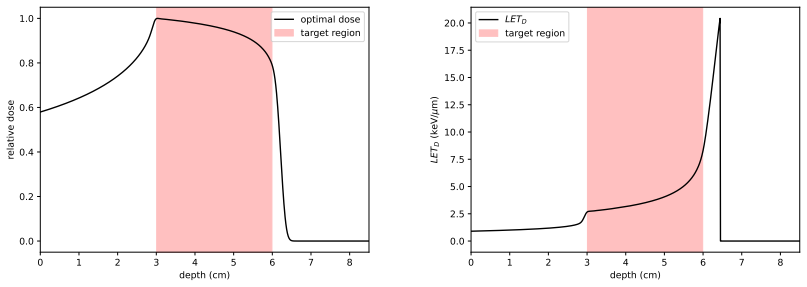
- The biological-dose optimization example produces a tapered distal dose with reduced healthy-tissue dose while keeping the biological effect uniform in the target, illustrating a concrete planning consequence.
Reading between the lines
- A natural next step beyond the paper is to concatenate one-dimensional slabs with different material parameters, which would extend the closed-form fluence to layered heterogeneous geometries while keeping the speed advantage.
- Because the model omits energy-loss straggling, it likely overpredicts the sharpness of the distal falloff; convolving Equation (16) with a Gaussian straggling kernel is a testable extension that would quantify the bias in the no-straggling assumption.
- The same characteristic solution generalizes to heavier ions by changing the exponent $p$, which could connect this framework to carbon-ion LET-weighted planning, though the linear-quadratic survival model would need the heavy-ion corrections the paper explicitly sets aside.
- The convex linearity of the biological dose in beam weights suggests the optimization could be embedded directly in existing inverse-planning solvers, a clinical integration the paper demonstrates in one dimension but does not develop for three-dimensional pencil-beam scanning.
Editorial analysis
A structured set of objections, weighed in public.
Referee Report
Summary. The paper develops a one-dimensional analytical model for proton transport in water-like geometries under continuous-slowing-down assumptions, yielding closed-form expressions for fluence, absorbed dose, track- and dose-averaged LET, and LET-dependent biological quantities such as survival fraction, RBE, and biological dose. The model is validated against MCsquare and TOPAS for a 62 MeV pristine Bragg peak, an uncertainty analysis of the Bragg-Kleeman parameters is carried out, and the framework is applied to treatment-planning optimisation with absorbed-dose and biological-dose objectives. The code used to generate the figures is made available via Zenodo.
Significance. If the internal sign inconsistency is corrected and the Monte Carlo comparison is made quantitative, the closed-form formulas would provide a genuinely fast and interpretable tool for exploring LET-weighted optimisation and variable RBE in one-dimensional geometries. The availability of code, the clear treatment of parameter uncertainty, and the explicit falsifiable predictions for dose and LET are strengths. However, as printed the central transport equation is inconsistent with the claimed solution, and the validation is single-case and qualitative, so the significance cannot be fully assessed without revision.
major comments (3)
- [§2.2, §2.5, Eqs (7), (14)–(15)] The sign in Eq (7) is inconsistent with the characteristic solution (14) and the closed-form fluence (15). Since Eq (13) defines S(E) > 0, physical energy loss requires dE/dz = -S(E), and the phase-space continuity equation is ∂_z u - ∂_E(S u) = 0. Equation (14), E^p = E_max^p - z/α, is exactly the characteristic of the minus-sign equation, and direct substitution of Eq (15) into Eq (7) leaves a nonzero residual; the proposed solution solves the minus-sign equation instead. Equation (1) carries the same plus-sign error. If the implementation behind Fig 6 uses the minus-sign equation, Eqs (1) and (7) should be corrected and the derivation revisited; if the implementation follows the printed plus-sign equation, the agreement in Fig 6 cannot be reproduced. This must be resolved before the transport model can be accepted.
- [§2.9, Fig 6] The Monte Carlo validation is a single-energy, qualitative comparison. Only the 62 MeV pristine Bragg peak is shown, no quantitative agreement metric (gamma index, range difference, dose difference) or Monte Carlo statistical uncertainty is reported, and LET—which the biological sections depend on—is not validated at all. The analytical model uses α = 2.2 × 10^-3 and p = 1.77 from [19], while Table 1 lists α = 0.00246 ± 0.00025 and p = 1.75 ± 0.02 for water; this discrepancy is not explained. The claim of high fidelity needs multi-energy comparisons with explicitly stated parameter choices and a quantitative agreement metric.
- [§2.5–2.6, Eq (16)] The energy interval I = [E_min, E_max] used in Eq (16) and in Figs 5–6 is never specified. At depth z > 0, characteristics with initial energies near E_min produce protons with energies below E_min, so the dose integral is truncated at E_min; depending on the chosen E_min this can remove a non-negligible low-energy contribution near the Bragg peak. The authors should report E_min and E_max and demonstrate that the dose and LET results are insensitive to E_min, for example by a convergence study in E_min.
minor comments (4)
- [§4.1, Fig 11 caption] The caption states σα = 0.01µα = 0.0175 and σp = 0.01µp = 0.0000246, which swaps the two parameters; the correct values should be σα = 0.0000246 and σp = 0.0175.
- [§5.1, Fig 17] The text for Examples 1 and 2 sets beam ranges equally spaced in [z_prox, z_dist + 1/4], while the Fig 17 caption states that ranges extend to z_dist + 0.15; the two choices should be reconciled.
- [§2.3, Eq (21)] Equation (21) is valid only for z ≤ α E_0^p; for larger depths the argument (E_0^p - z/α)^{1/p} is not real. The domain of validity should be stated.
- [§2.3, Eq (17)] Equation (12) defines S(z) := -dE/dx as a positive quantity, while Eq (17) writes L_Δ = dE_Δ/dz without a sign; since LET is a positive energy-deposition quantity, the sign convention should be clarified.
Circularity Check
No material circularity: the model uses externally calibrated empirical parameters and is validated against independent Monte Carlo codes; no prediction reduces to a fitted value or to the authors' own prior work.
full rationale
The paper's central claims are a closed-form fluence and dose from a continuous-slowing-down transport model, and biological metrics built on literature LQ parameters. The transport model takes the Bragg-Kleeman range-energy power law with alpha and p taken from published sources (Table 1 and [19]) as inputs; the closed-form solution (15) is obtained by the method of characteristics, and the dose (16) is the standard stopping-power-weighted fluence integral. Validation in Section 2.9 is against independent MCsquare and TOPAS simulations, not against the same data used to fit the model. The biological quantities use LQ coefficients, lambda, c_X-ray, and beta from Chaudhary et al. [8], which are external experimental fits; equations (24)-(27) and (29) are explicit definitions and standard modelling choices, not derivations whose conclusions are hidden in an assumption. No self-citation is load-bearing: the authors do not cite their own prior work as the basis for any premise, and no uniqueness theorem is invoked. The apparent sign inconsistency between Eq. (7) and the characteristic Eq. (14) is a mathematical-correctness concern, not a circular reduction, so it does not affect the circularity score.
Assumptions & free parameters
free parameters (4)
- Bragg-Kleeman coefficient α for water =
0.00246 cm MeV^-p (Table 1); 0.0022 used in Section 2.9
- Bragg-Kleeman exponent p for water =
1.75 (Table 1); 1.77 in Section 2.9
- LQ parameters c_X-ray, β, λ for AG01522 and U87 =
AG01522: 0.54, 0.051, 0.0451; U87: 0.11, 0.059, 0.0127
- Basis beam widths σ_i^2 =
1 for all i in Examples 1-3
assumptions (4)
- domain assumption Continuous energy loss without straggling
- domain assumption Angular scattering is minimal and nonelastic collisions are rare
- domain assumption Stopping power follows the Bragg-Kleeman power law over the whole energy interval
- domain assumption The linear-quadratic survival model holds, with α linear in LET and β constant
Cite this review
Pith. "Pith review of Efficient Proton Transport Modelling for Proton Beam Therapy and Biological Quantification." pith.science (2026). https://pith.science/paper/B6BO7H7B
@misc{pith2026241116735,
  author       = {Pith},
  title        = {Pith review of: Efficient Proton Transport Modelling for Proton Beam Therapy and Biological Quantification},
  year         = {2026},
  howpublished = {\url{https://pith.science/paper/B6BO7H7B}},
  note         = {Machine review of arXiv:2411.16735}
}
read the original abstract
In this work, we present a fundamental mathematical model for proton transport, tailored to capture the key physical processes underpinning Proton Beam Therapy (PBT). The model provides a robust and computationally efficient framework for exploring various aspects of PBT, including dose delivery, linear energy transfer, treatment planning and the evaluation of relative biological effectiveness. Our findings highlight the potential of this model as a complementary tool to more complex and computationally intensive simulation techniques currently used in clinical practice.
Figures


Figures from the paper (19 more)



Forward citations
Cited by 2 Pith papers
-
A nodally bound-preserving finite element method for hyperbolic convection-reaction problems
A SUPG finite element method with nodal bound constraints, solved as a variational inequality, is analyzed for hyperbolic convection-reaction problems and is shown to converge at the expected rates in the linear case.
-
Optimal control of a kinetic equation
Introduces a hypocoercive finite element framework for optimal control of the Kolmogorov equation, proving temporal stability and error bounds for the regularized problem.
Reference graph
Works this paper leans on
-
[19]
An analytical approximation of the Bragg curve for therapeutic proton beams
T. Bortfeld. “An analytical approximation of the Bragg curve for therapeutic proton beams”. In: Medical physics 24.12 (1997), pp. 2024–2033
work page 1997
-
[1]
Treatment planning for proton therapy: what is needed in the next 10 years?
H. Nystrom et al. “Treatment planning for proton therapy: what is needed in the next 10 years?” In: The British Journal of Radiology 93.1107 (2020), p. 20190304
work page 2020
-
[2]
A generic algorithm for Monte Carlo simulation of proton transport
F. Salvat. “A generic algorithm for Monte Carlo simulation of proton transport”. In: Nuclear Instruments and Methods in Physics Research Section B: Beam Interactions with Materials and Atoms 316 (2013), pp. 144–159
work page 2013
-
[3]
A fast Monte Carlo code for proton transport in radiation therapy based on MCNPX
K. Jabbari et al. “A fast Monte Carlo code for proton transport in radiation therapy based on MCNPX”. In: Journal of Medical Physics 39.3 (2014), pp. 156–163
work page 2014
-
[4]
Reoptimization of intensity modulated proton therapy plans based on linear energy transfer
J. Unkelbach et al. “Reoptimization of intensity modulated proton therapy plans based on linear energy transfer”. In: International Journal of Radiation Oncology* Biology* Physics 96.5 (2016), pp. 1097–1106
work page 2016
-
[5]
M. Frizzelle et al. “Using multi-centre data to train and validate a knowledge-based model for planning radiotherapy of the head and neck”. In: Physics and Imaging in Radiation Oncology 21 (2022), pp. 18–23
work page 2022
-
[6]
A.-M. Fanou et al. “Implementation, dosimetric assessment, and treatment validation of knowledge-based planning (KBP) models in VMAT head and neck radiation oncology”. In: Biomedicines 11.3 (2023), p. 762
work page 2023
-
[7]
An empirical method for deriving RBE values associated with electrons, photons and radionuclides
M. Bellamy et al. “An empirical method for deriving RBE values associated with electrons, photons and radionuclides”. In: Radiation protection dosimetry 167.4 (2015), pp. 664–670
work page 2015
Show all 47 references
-
[8]
Relative biological effectiveness variation along monoenergetic and mod- ulated Bragg peaks of a 62-MeV therapeutic proton beam: a preclinical assessment
P. Chaudhary et al. “Relative biological effectiveness variation along monoenergetic and mod- ulated Bragg peaks of a 62-MeV therapeutic proton beam: a preclinical assessment”. In: International Journal of Radiation Oncology* Biology* Physics 90.1 (2014), pp. 27–35. 27
2014
-
[9]
Relative biological effectiveness of proton beams in clinical therapy
L. E. Gerweck et al. “Relative biological effectiveness of proton beams in clinical therapy”. In: Radiotherapy and oncology 50.2 (1999), pp. 135–142
1999
-
[10]
Variable proton relative biological effectiveness: how do we move for- ward?
T. Underwood et al. “Variable proton relative biological effectiveness: how do we move for- ward?” In: International Journal of Radiation Oncology, Biology, Physics 95.1 (2016), pp. 56– 58
2016
-
[11]
Linear energy transfer-guided optimization in intensity modulated pro- ton therapy: feasibility study and clinical potential
D. Giantsoudi et al. “Linear energy transfer-guided optimization in intensity modulated pro- ton therapy: feasibility study and clinical potential”. In: International Journal of Radiation Oncology* Biology* Physics 87.1 (2013), pp. 216–222
2013
-
[12]
Report of the AAPM TG-256 on the relative biological effectiveness of proton beams in radiation therapy
H. Paganetti et al. “Report of the AAPM TG-256 on the relative biological effectiveness of proton beams in radiation therapy”. In: Medical physics 46.3 (2019), e53–e78
2019
-
[13]
Difference in the relative biological effectiveness and DNA damage repair processes in response to proton beam therapy according to the positions of the spread out Bragg peak
H. Hojo et al. “Difference in the relative biological effectiveness and DNA damage repair processes in response to proton beam therapy according to the positions of the spread out Bragg peak”. In: Radiation oncology 12 (2017), pp. 1–9
2017
-
[14]
Relative biological effectiveness (RBE) values for proton beam therapy. Vari- ations as a function of biological endpoint, dose, and linear energy transfer
H. Paganetti. “Relative biological effectiveness (RBE) values for proton beam therapy. Vari- ations as a function of biological endpoint, dose, and linear energy transfer”. In: Physics in Medicine & Biology 59.22 (2014), R419
2014
-
[15]
Optimal control for mathematical models of cancer therapies
H. Sch¨ attler et al. “Optimal control for mathematical models of cancer therapies”. In: An application of geometric methods (2015)
2015
-
[16]
Adaptive radiotherapy for anatomical changes
J.-J. Sonke et al. “Adaptive radiotherapy for anatomical changes”. In: Seminars in radiation oncology. Vol. 29. 3. Elsevier. 2019, pp. 245–257
2019
-
[17]
Accuracy of parameterized proton range models; a comparison
H. E. S. Pettersen et al. “Accuracy of parameterized proton range models; a comparison”. In: Radiation Physics and Chemistry 144 (2018), pp. 295–297
2018
-
[18]
Dosimetry and quality control of scanning proton beams
S. N. Boon. “Dosimetry and quality control of scanning proton beams”. In: (1998)
1998
-
[20]
Theoretical aspects of energy–range relations, stopping power and energy strag- gling of protons
W. Ulmer. “Theoretical aspects of energy–range relations, stopping power and energy strag- gling of protons”. In: Radiation physics and chemistry 76.7 (2007), pp. 1089–1107
2007
-
[21]
Review of particle physics
S. Navas et al. “Review of particle physics”. In: Physical Review D 110.3 (2024), p. 030001
2024
-
[22]
D. J. Thomas. ICRU report 85: fundamental quantities and units for ionizing radiation . 2012
2012
-
[23]
A systematic review on the usage of averaged LET in radiation biology for particle therapy
F. Kalholm et al. “A systematic review on the usage of averaged LET in radiation biology for particle therapy”. In: Radiotherapy and Oncology 161 (2021), pp. 211–221
2021
-
[24]
Fast multipurpose Monte Carlo simulation for proton therapy using multi-and many-core CPU architectures
K. Souris et al. “Fast multipurpose Monte Carlo simulation for proton therapy using multi-and many-core CPU architectures”. In: Medical physics 43.4 (2016), pp. 1700–1712
2016
-
[25]
The TOPAS tool for particle simulation, a Monte Carlo simulation tool for physics, biology and clinical research
B. Faddegon et al. “The TOPAS tool for particle simulation, a Monte Carlo simulation tool for physics, biology and clinical research”. In: Physica Medica 72 (2020), pp. 114–121
2020
-
[26]
Intensity modulated proton therapy
H. Kooy et al. “Intensity modulated proton therapy”. In: The British journal of radiology 88.1051 (2015), p. 20150195
2015
-
[27]
Biophysical characteristics of HIMAC clinical irradiation system for heavy- ion radiation therapy
T. Kanai et al. “Biophysical characteristics of HIMAC clinical irradiation system for heavy- ion radiation therapy”. In: International Journal of Radiation Oncology* Biology* Physics 44.1 (1999), pp. 201–210
1999
-
[28]
Reformulation of a clinical-dose system for carbon-ion radiotherapy treat- ment planning at the National Institute of Radiological Sciences, Japan
T. Inaniwa et al. “Reformulation of a clinical-dose system for carbon-ion radiotherapy treat- ment planning at the National Institute of Radiological Sciences, Japan”. In: Physics in Medicine & Biology 60.8 (2015), p. 3271
2015
-
[29]
RBE and related modeling in carbon-ion therapy
C. P. Karger et al. “RBE and related modeling in carbon-ion therapy”. In: Physics in Medicine & Biology 63.1 (2017), 01TR02. 28
2017
-
[30]
A simple method to assess clonogenic survival of irradiated cancer cells
I. Serrano-Mendioroz et al. “A simple method to assess clonogenic survival of irradiated cancer cells”. In: Methods in Cell Biology . Vol. 174. Elsevier, 2023, pp. 127–136
2023
-
[31]
A generalized formulation of dual radiation action
A. M. Kellerer et al. “A generalized formulation of dual radiation action”. In: Radiation research 75.3 (1978), pp. 471–488
1978
-
[32]
Cell-survival probability at large doses: an alternative to the linear-quadratic model
L. Hanin et al. “Cell-survival probability at large doses: an alternative to the linear-quadratic model”. In: Physics in Medicine & Biology 55.16 (2010), p. 4687
2010
-
[33]
Chadwick et al
K. Chadwick et al. Molecular theory of cell survival. Tech. rep. Instituut voor Toepassing van Atoomenergie in de Landbouw, Wageningen . . ., 1973
1973
-
[34]
A microdosimetric-kinetic theory of the dependence of the RBE for cell death on LET
R. B. Hawkins. “A microdosimetric-kinetic theory of the dependence of the RBE for cell death on LET”. In: Medical physics 25.7 (1998), pp. 1157–1170
1998
-
[35]
Direct comparison between protons and alpha-particles of the same LET: I. Irradiation methods and inactivation of asynchronous V79, HeLa and C3H 10T1 /2 cells
D. Goodhead et al. “Direct comparison between protons and alpha-particles of the same LET: I. Irradiation methods and inactivation of asynchronous V79, HeLa and C3H 10T1 /2 cells”. In: International journal of radiation biology 61.5 (1992), pp. 611–624
1992
-
[36]
Inactivation and mutation induction in V79 cells by low energy protons: re- evaluation of the results at the LNL facility
M. Belli et al. “Inactivation and mutation induction in V79 cells by low energy protons: re- evaluation of the results at the LNL facility”. In: International journal of radiation biology 63.3 (1993), pp. 331–337
1993
-
[37]
A phenomenological model for the relative biological effectiveness in ther- apeutic proton beams
J. Wilkens et al. “A phenomenological model for the relative biological effectiveness in ther- apeutic proton beams”. In: Physics in Medicine & Biology 49.13 (2004), p. 2811
2004
-
[38]
Radiobiological investigations of proton and light ion therapy
N. Tilly. “Radiobiological investigations of proton and light ion therapy”. PhD thesis. Stock- holm University, 2002
2002
-
[39]
Range uncertainty in proton therapy due to variable biological effectiveness
A. Carabe et al. “Range uncertainty in proton therapy due to variable biological effectiveness”. In: Physics in Medicine & Biology 57.5 (2012), p. 1159
2012
-
[40]
The incorporation of the concept of minimum RBE (RBE min) into the linear-quadratic model and the potential for improved radiobiological analysis of high-LET treatments
A. Carabe-Fernandez et al. “The incorporation of the concept of minimum RBE (RBE min) into the linear-quadratic model and the potential for improved radiobiological analysis of high-LET treatments”. In: International journal of radiation biology 83.1 (2007), pp. 27–39
2007
-
[41]
Variations in linear energy transfer within clinical proton therapy fields and the potential for biological treatment planning
C. Grassberger et al. “Variations in linear energy transfer within clinical proton therapy fields and the potential for biological treatment planning”. In: International Journal of Radiation Oncology* Biology* Physics 80.5 (2011), pp. 1559–1566
2011
-
[42]
A systematic review of let-guided treatment plan optimisation in proton therapy: identifying the current state and future needs
M. McIntyre et al. “A systematic review of let-guided treatment plan optimisation in proton therapy: identifying the current state and future needs”. In: Cancers 15.17 (2023), p. 4268
2023
-
[43]
T. J. Sullivan. Introduction to uncertainty quantification / [internet resource] . eng. 1st ed. 2015.. Texts in applied mathematics ; v. 63. Cham: Springer, 2015. isbn: 9783319233956
2015
-
[44]
A fast optimization algorithm for multicriteria intensity modulated proton therapy planning
W. Chen et al. “A fast optimization algorithm for multicriteria intensity modulated proton therapy planning”. In: Medical physics 37.9 (2010), pp. 4938–4945
2010
-
[45]
Beyond bixels: Generalizing the optimization parameters for intensity modulated radiation therapy
J. Markman et al. “Beyond bixels: Generalizing the optimization parameters for intensity modulated radiation therapy”. In: Medical Physics 29.10 (2002), pp. 2298–2304
2002
-
[46]
Variable RBE in proton therapy: comparison of different model predic- tions and their influence on clinical-like scenarios
G. Giovannini et al. “Variable RBE in proton therapy: comparison of different model predic- tions and their influence on clinical-like scenarios”. In: Radiation Oncology 11 (2016), pp. 1– 16
2016
-
[47]
Clinical commissioning of a pencil beam scanning treatment planning system for proton therapy
J. Saini et al. “Clinical commissioning of a pencil beam scanning treatment planning system for proton therapy”. In: International journal of particle therapy 3.1 (2016), pp. 51–60. 1Institute of Mathematical Innovation, University of Bath 29 2Department of Mathematical Scienc...
2016
Reviewed August 12, 2026 · model on record in the stance chip above.
Discussion (0). Continue with ORCID to comment.