REVIEW 4 major objections 6 minor 25 references
Joint estimation of activity, attenuation and motion in respiratory-self-gated time-of-flight PET
T0 review · 4 major / 6 minor · reviewed 2026-08-11 · deepseek-v4-flash
Pith's one-line read Respiratory self-gated TOF PET data contain enough information to jointly estimate activity, gate-matched attenuation, and motion, yielding a single motion- and attenuation-artifact-free image without external hardware.
desk verdict Useful methods paper: the phantom benchmark shows the joint MLACF+motion approach recovers static-reference lesion contrast, but the end-to-end 'no hardware' claim is only partially tested. read the letter →
The pith
A machine-rendered reading of the paper's core claim, the machinery that carries it, and where it could break.
The reading
What carries the argument
The central objects are the forward model $\bar{y}^k_{it}(\lambda) = a^k_i \big(P(S^k\lambda)\big)_{it} + r^k_{it}$ for each respiratory gate $k$, the factorization $a^k = g^k \tilde{a}$ of gate-matched attenuation sinograms into the static CT-based sinogram $\tilde{a}$ and gate-specific multiplicative correction factors $g^k$, and the MLACF update that estimates $g^k$ under an intensity prior favoring values close to 1. The factorization converts the ill-posed joint estimation of activity and attenuation into a well-scaled multiplicative correction problem, and the MLACF estimates are used both to define non-rigid image warps (via diffeomorphic demons registration of gate-by-gate reconstructions) and to provide gate-matched attenuation for the final joint reconstruction.
What would settle it
Acquire a moving phantom whose true attenuation is known at each respiratory gate (for example, by fast CT at each phase) alongside the PET list-mode data. If the jointly estimated gate-matched attenuation sinograms do not match the true phase-matched CT sinograms up to the known scaling ambiguity, or if the final activity image does not match a reconstruction that used the true gated attenuation, the central claim that the data-driven estimates are free of attenuation bias is contradicted.
Extended reading notes
Core claim
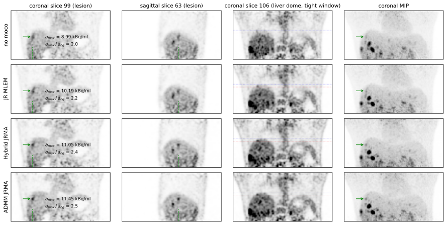
The central claim is that respiratory self-gated TOF PET data contain enough information to estimate, purely from the emission data, the respiratory phase-matched attenuation sinograms and the non-rigid motion between respiratory gates, and to combine all gates into a single high-quality activity image. The load-bearing idea is to write each gate's attenuation sinogram as a product of a breath-hold CT-based static attenuation sinogram and nonnegative gate-specific correction factors, estimated with MLACF updates and an intensity prior that keeps the correction factors close to unity; this resolves the scale ambiguity inherent in joint activity/attenuation estimation. Attenuation and motion estimates obtained either separately (Hybrid) or jointly (ADMM) are then used in a final joint-reconstruction OS-MLEM that uses all acquired counts. The paper reports that this restores phantom lesion contrast to the static-scan value and visibly removes motion and attenuation artifacts in patients.
Load-bearing premise
The whole pipeline presumes that the breath-hold CT-based static attenuation sinogram is a valid baseline and that every respiratory attenuation change can be represented by nonnegative multiplicative correction factors pulled toward 1 by a hand-tuned intensity prior, with a static scatter estimate treated as adequate.
Editorial extensions
If this is right
- A single activity image can be reconstructed from all respiratory gates, using all counts without discarding data, which minimizes noise while removing motion blur.
- The data-driven PCA-based respiratory signal correlates strongly with the true motion signal (Pearson correlation 0.91 in the phantom), so external gating hardware can be omitted.
- Correcting attenuation per gate removes the liver-dome banana artifact and improves lesion-to-background contrast beyond motion correction with static attenuation alone.
- The comparable performance of both methods across two scanner types (Siemens mCT and GE DMI) suggests the approach generalizes to different TOF PET/CT systems.
- The authors recommend Hybrid JRMA for clinical translation over ADMM JRMA because the latter is sensitive to the Lagrangian penalty parameter.
Reading between the lines
- If the multiplicative correction model and the intensity prior hold for other attenuation mismatches (for instance, patient positioning differences in PET/MRI), the same factorization could be applied as long as a reliable baseline attenuation image is available.
- The motion vector fields were underestimated (12.2 mm vs. the true 20 mm in the phantom), so residual bias in small moving-structure quantification is likely; clinical use would need to tolerate or compensate for that bias.
- The method's reliance on a static scatter estimate may limit accuracy at high scatter fractions; replacing it with gated scatter estimates is a testable extension.
- The hybrid pipeline's dependence on non-rigid registration of MLACF reconstructions means that registration errors propagate into the final image; learning-based motion priors could reduce that error.
Signed reviews
Editorial analysis
A structured set of objections, weighed in public.
Referee Report
Summary. The manuscript proposes two data-driven algorithms for jointly estimating activity, gate-dependent attenuation, and respiratory motion in respiratory self-gated TOF PET, with the goal of producing a single motion- and attenuation-corrected activity image without external respiratory hardware. The hybrid JRMA method estimates gate-by-gate MLACF attenuation sinograms and motion warping operators from gate-by-gate MLACF reconstructions, then performs a final JR OS-MLEM reconstruction; the ADMM JRMA method couples these estimates inside a modified ADMM framework. The methods are evaluated on a moving Wilhelm thorax phantom with a matching static reference acquisition and on three clinical FDG PET/CT datasets. The phantom experiment shows lesion-to-background contrast improving from 2.0 (no motion correction) to 5.2/5.3 with the proposed methods, matching the 5.2 contrast of the static reference, while patient datasets show reduced banana artifacts and improved lesion contrast on visual inspection.
Significance. If fully validated, this work would make respiratory motion and attenuation correction more practical by removing the need for external respiratory hardware and by estimating gate-matched attenuation without reconstructing attenuation images. The phantom experiment with an independent static reference is the strongest element, and the evaluation on two different scanner platforms supports generalizability. The appendix derivations are coherent, the MLACF-based updates are transparent, and the authors honestly recommend the simpler hybrid method over the more complex ADMM variant. The principal quantitative result, contrast recovery matching the static phantom, is compelling. However, the headline claims are not yet directly supported: the only ground-truth phantom experiment used the actor-based gating signal rather than the data-driven signal, and the attenuation model is a regularized multiplicative correction to a breath-hold CT sinogram, so the 'free from attenuation artifacts' claim is conditional on the CT prior and on the manually tuned regularization strength. The paper is a solid proof-of-concept but needs additional validation before the stated guarantees can be accepted.
major comments (4)
- [Section 4, Fig. 2] The quantitative Wilhelm phantom experiment uses the actor-based ground-truth gating signal for gate definition rather than the PCA data-driven signal, as stated in Section 4: 'we decided to use actor-based ground-truth signal for the gate definition in the Wilhelm acquisition.' Because the headline claim is operation without external hardware, the only ground-truth experiment does not test that claim. Please repeat the phantom evaluation with data-driven gating, or quantitatively assess the sensitivity of lesion contrast and artifact reduction to the choice of gating signal.
- [Section A.2, Eqs. (21)-(24); Table 2] The gate-dependent attenuation is modeled exclusively as nonnegative multiplicative correction factors g_k applied to the breath-hold CT attenuation sinogram, regularized toward unity with gamma = 0.2 mean(emission sinogram). The claim that the reconstructed images are 'free from attenuation artifacts' is therefore conditional on the CT-derived baseline being accurate and on gamma being appropriately tuned. No experiment perturbs the CT prior (e.g., misregistration, truncation, or bias) or varies gamma. Because a biased CT prior cannot be fully corrected by g_k under the strong regularization, please add robustness tests over both the CT prior and gamma, or explicitly reformulate the claim to reflect this dependence.
- [Section 4, Fig. 4] The estimated peak displacement between gate 1 and gate 6 is 12.2 mm against the true 20 mm, a 39% underestimation, and the corresponding JR MLEM estimate is 9.3 mm. The paper offers plausible explanations (intra-gate motion, 5 mm slices, regularization) but provides no quantitative evidence that this residual motion error does not affect the final activity image or the reported contrast recovery. Please quantify residual motion after correction in the static-reference phantom, or provide a motion-error sensitivity analysis, to support the claim that the methods produce a motion-corrected image.
- [Section 5.2; Eq. (23)] The MLACF attenuation update in Eq. (23) uses the expected scattered and random coincidences r_k in the numerator, but the scatter estimate is static and not gate-matched, as acknowledged in Section 5.2. Since the attenuation correction factors g_k are estimated jointly with the emission data, a gate-dependent scatter mismatch could bias the estimated attenuation sinograms. Please add a sensitivity analysis or a brief quantitative argument showing that smooth, static scatter does not materially bias g_k in the tested count statistics.
minor comments (6)
- [Abstract and Fig. 1] The acronym is used inconsistently: 'JMRA' appears in Figure 1 and in some text passages, while the title and most of the manuscript use 'JRMA.' Please standardize the acronym throughout.
- [Fig. 5 caption] The caption contains a typo: 'reconstructon' should be 'reconstruction.'
- [Section 3.2.2] The injected activity is written as '4.25 ˙MBq/kg' with an unusual dot before 'MBq'; this should be '4.25 MBq/kg.'
- [Section 2.4] The text says the algorithm is divided into 'four simpler subproblems' but then lists five subproblems (subproblems 1 through 5). Please renumber or reword the introduction to this subsection.
- [Appendix A.3, Eqs. (25)-(27)] The replacement of the exact ADMM subproblem (25) with the denoising problem (26)-(27) using the average of inverse-warped gate images is an approximation that is not covered by standard ADMM convergence theory. Because this is a heuristic modification, it should be flagged as such in the main text rather than only in the appendix.
- [Section 3.3] The paper does not state whether the reconstruction code or phantom data will be made available. A data/code availability statement would improve reproducibility, especially given the complexity of the proposed pipelines.
Circularity Check
No circularity: the reported contrasts are measured against an independent static acquisition, and the MLACF/TOF attenuation machinery is external, not fitted to the claimed endpoints.
full rationale
The derivation chain is self-contained rather than circular. The forward model (Eq. 1) and the joint optimization (Eq. 3) are standard Poisson-model formulations; the attenuation sinograms are estimated from the gated emission data via MLACF updates (Eq. 23), not by fitting the lesion contrast or any reported endpoint. The phantom validation compares against a separate 10-minute static acquisition with phase-matched CT attenuation, which is an independent external benchmark: the lesion-to-background contrast of 5.2 is measured, not enforced by construction. The paper does rely on prior work by overlapping authors for the MLACF algorithm [5] and for the TOF attenuation-identification theory [3], [4], but those are published, externally established results and are not invoked as a substitute for the current experiment. The passages that weaken the claims are limitations, not circular steps: Section 4 states that the Wilhelm gate definition used the actor-based 'ground-truth' signal rather than the data-driven signal, and Section 5.2 concedes a static scatter estimate and hand-tuned parameters (γ and ρ). These affect generalizability and robustness, but no equation reduces to its own input and no fitted parameter is renamed as a prediction. Therefore the circularity score is 0.
Assumptions & free parameters
free parameters (4)
- Intensity prior weight gamma =
gamma = 0.2 x mean(emission sinogram)
- ADMM penalty parameter rho =
rho = 1e-6 after rescaling forward operators to unit norm
- Total variation weight beta =
beta = 7e-5
- Number of respiratory gates =
6
assumptions (6)
- domain assumption A breath-hold CT/MR attenuation image is available and all respiratory attenuation changes can be represented as nonnegative multiplicative correction factors on its sinogram.
- domain assumption TOF-PET emission data determine the attenuation sinogram up to a global constant, as established in the cited literature.
- domain assumption Each respiratory gate is an independent Poisson realization with known expected scatter and random coincidences.
- domain assumption Respiratory motion is modeled by invertible diffeomorphic warps that can be recovered by demons registration of gated reconstructions.
- ad hoc to paper The Poisson likelihood in the attenuation update can be replaced by a non-TOF weighted Gaussian likelihood to obtain an analytic MLACF update.
- ad hoc to paper ADMM subproblem 3 can be approximated by a single-denoising problem using the average of inverse-warped gate images.
Cite this review
Pith. "Pith review of Joint estimation of activity, attenuation and motion in respiratory-self-gated time-of-flight PET." pith.science (2026). https://pith.science/paper/7K3CYB2E
@misc{pith2026241215018,
  author       = {Pith},
  title        = {Pith review of: Joint estimation of activity, attenuation and motion in respiratory-self-gated time-of-flight PET},
  year         = {2026},
  howpublished = {\url{https://pith.science/paper/7K3CYB2E}},
  note         = {Machine review of arXiv:2412.15018}
}
read the original abstract
Whole-body PET imaging is often hindered by respiratory motion during acquisition, causing significant degradation in the quality of reconstructed activity images. An additional challenge in PET/CT imaging arises from the respiratory phase mismatch between CT-based attenuation correction and PET acquisition, leading to attenuation artifacts. To address these issues, we propose two new, purely data-driven methods for the joint estimation of activity, attenuation, and motion in respiratory self-gated TOF PET. These methods enable the reconstruction of a single activity image free from motion and attenuation artifacts. The proposed methods were evaluated using data from the anthropomorphic Wilhelm phantom acquired on a Siemens mCT PET/CT system, as well as 3 clinical FDG PET/CT datasets acquired on a GE DMI PET/CT system. Image quality was assessed visually to identify motion and attenuation artifacts. Lesion uptake values were quantitatively compared across reconstructions without motion modeling, with motion modeling but static attenuation correction, and with our proposed methods. For the Wilhelm phantom, the proposed methods delivered image quality closely matching the reference reconstruction from a static acquisition. The lesion-to-background contrast for a liver dome lesion improved from 2.0 (no motion correction) to 5.2 (proposed methods), matching the contrast from the static acquisition (5.2). In contrast, motion modeling with static attenuation correction yielded a lower contrast of 3.5. In patient datasets, the proposed methods successfully reduced motion artifacts in lung and liver lesions and mitigated attenuation artifacts, demonstrating superior lesion to background separation. Our proposed methods enable the reconstruction of a single, high-quality activity image that is motion-corrected and free from attenuation artifacts, without the need for external hardware.
Figures


Figures from the paper (5 more)


Reference graph
Works this paper leans on
-
[1]
PET respiratory motion correction: quo vadis?
F. Lamare, A Bousse, K Thielemans, et al., “PET respiratory motion correction: quo vadis?”Physics in Medicine & Biology, vol. 67, no. 3, 03TR02, 2022. doi: 10.1088/1361-6560/ac43fc. 17
-
[2]
Device-less gating for PET/CT using PCA,
K. Thielemans, S. Rathore, F. Engbrant, and P. Razifar, “Device-less gating for PET/CT using PCA,” in 2011 IEEE Nuclear Science Symposium Conference Record, IEEE, 2011. doi: 10.1109/ NSSMIC.2011.6153742
-
[3]
Time-of-flight pet data determine the attenuation sinogram up to a constant,
M. Defrise, A. Rezaei, and J. Nuyts, “Time-of-flight pet data determine the attenuation sinogram up to a constant,” Physics in Medicine & Biology, vol. 57, no. 4, p. 885, 2012. doi: 10.1088/0031- 9155/57/4/885
doi:10.1088/0031- 2012
-
[4]
Simultaneous reconstruction of activity and attenuation in time-of-flight PET,
A. Rezaei, M. Defrise, G. Bal, et al., “Simultaneous reconstruction of activity and attenuation in time-of-flight PET,” IEEE transactions on medical imaging, vol. 31, no. 12, pp. 2224–2233, 2012. doi: 10.1109/TMI.2012.2212719
arXiv 2012
-
[5]
ML-reconstruction for TOF-PET with simultaneous estima- tion of the attenuation factors,
A. Rezaei, M. Defrise, and J. Nuyts, “ML-reconstruction for TOF-PET with simultaneous estima- tion of the attenuation factors,” IEEE Transactions on Medical Imaging, vol. 33, no. 7, pp. 1563– 1572, 2014. doi: 10.1109/TMI.2014.2318175
-
[6]
S. Boyd, N. Parikh, E. Chu, B. Peleato, J. Eckstein, et al., “Distributed optimization and statistical learning via the alternating direction method of multipliers,” Foundations and Trends in Machine learning, vol. 3, no. 1, pp. 1–122, 2011. doi: 10.1561/2200000016
-
[7]
Anthropomorphic thorax phantom for cardio-respiratory motion simulation in tomographic imaging,
K. Bolwin, B. Czekalla, L. J. Frohwein, F. B¨ uther, and K. P. Sch¨ afers, “Anthropomorphic thorax phantom for cardio-respiratory motion simulation in tomographic imaging,” Physics in Medicine & Biology, 2018. doi: 10.1088/1361-6560/aaa201
-
[8]
Noise properties of motion-compensated tomographic image recon- struction methods,
S. Y. Chun and J. A. Fessler, “Noise properties of motion-compensated tomographic image recon- struction methods,” IEEE Transactions on Medical Imaging, vol. 32, no. 2, pp. 141–152, 2013. doi: 10.1109/TMI.2012.2206604
Show all 25 references
-
[9]
Non-rigid summing of gated pet via optical flow,
G. Klein, B. Reutter, and R. Huesman, “Non-rigid summing of gated pet via optical flow,” IEEE Transactions on Nuclear Science, vol. 44, no. 4, pp. 1509–1512, 1997. doi: 10.1109/23.632704
1997 doi
-
[10]
Lung motion correction on respiratory gated 3-d pet/ct images,
M. Dawood, N. Lang, X. Jiang, and K. Schafers, “Lung motion correction on respiratory gated 3-d pet/ct images,” IEEE Transactions on Medical Imaging, vol. 25, no. 4, pp. 476–485, 2006. doi: 10.1109/TMI.2006.870892
2006
-
[11]
Regularized b-spline deformable registration for respiratory motion cor- rection in PET images,
W. Bai and M. Brady, “Regularized b-spline deformable registration for respiratory motion cor- rection in PET images,” en, Physics in Medicine and Biology, vol. 54, no. 9, pp. 2719–2736, May
-
[12]
Respiratory motion compensation for PET/CT with motion information derived from matched attenuation-corrected gated PET data,
Y. Lu, K. Fontaine, T. Mulnix, et al., “Respiratory motion compensation for PET/CT with motion information derived from matched attenuation-corrected gated PET data,” en, J Nucl Med, vol. 59, no. 9, pp. 1480–1486, Sep. 2018. doi: 10.2967/jnumed.117.203000
2018 doi
-
[13]
Maximum-likelihood joint image reconstruction/motion estimation in attenuation-corrected respiratory gated pet/ct using a single attenuation map,
A. Bousse, O. Bertolli, D. Atkinson, et al., “Maximum-likelihood joint image reconstruction/motion estimation in attenuation-corrected respiratory gated pet/ct using a single attenuation map,” IEEE Transactions on Medical Imaging, vol. 35, no. 1, pp. 217–228, 2016. doi: 10 . 1...
2016
-
[14]
Evaluation of a direct motion estimation/correction method in respiratory-gated PET/MRI with motion-adjusted attenuation,
A. Bousse, R. Manber, B. F. Holman, et al., “Evaluation of a direct motion estimation/correction method in respiratory-gated PET/MRI with motion-adjusted attenuation,” en, Medical Physics, vol. 44, no. 6, pp. 2379–2390, Jun. 2017. doi: 10.1002/mp.12253
2017 doi
-
[15]
Maximum-likelihood joint image reconstruction and motion estimation with misaligned attenuation in TOF-PET/CT,
A. Bousse, O. Bertolli, D. Atkinson, et al., “Maximum-likelihood joint image reconstruction and motion estimation with misaligned attenuation in TOF-PET/CT,” en, Physics in Medicine and Biology, vol. 61, no. 3, pp. L11–9, Feb. 2016. doi: 10.1088/0031-9155/61/3/L11
2016 doi
-
[16]
Joint estimation of activity, attenuation and motion in respiratory-gated time-of-flight pet,
F. Jolivet, A. Rezaei, G. Schramm, et al., “Joint estimation of activity, attenuation and motion in respiratory-gated time-of-flight pet,” in 2022 IEEE Nuclear Science Symposium and Medical Imaging Conference (NSS/MIC), 2022, pp. 1–3. doi: 10.1109/NSS/MIC44845.2022.10399268
2022
-
[17]
A modified expectation maximization algorithm for penalized likelihood estima- tion in emission tomography,
A. R. De Pierro, “A modified expectation maximization algorithm for penalized likelihood estima- tion in emission tomography,” IEEE transactions on medical imaging, vol. 14, no. 1, pp. 132–137,
-
[18]
Performance evaluation of the biograph mCT flow PET/CT system according to the NEMA NU2-2012 standard,
I. Rausch, J. Cal-Gonz´ alez, D. Dapra, et al., “Performance evaluation of the biograph mCT flow PET/CT system according to the NEMA NU2-2012 standard,” EJNMMI Physics, vol. 2, no. 1, p. 26, Dec. 2015. doi: 10.1186/s40658-015-0132-1
2012 doi
-
[19]
Studies of a next-generation silicon-photomultiplier-based time-of-flight PET/CT system,
D. F. C. Hsu, E. Ilan, W. T. Peterson, J. Uribe, M. Lubberink, and C. S. Levin, “Studies of a next-generation silicon-photomultiplier-based time-of-flight PET/CT system,” Journal of Nuclear Medicine, vol. 58, no. 9, pp. 1511–1518, Sep. 2017. doi: 10.2967/jnumed.117.189514
2017 doi
-
[20]
ADMM penalty parameter selection by residual balancing,
B. Wohlberg, “ADMM penalty parameter selection by residual balancing,” arXiv preprint arXiv:1704.06209,
-
[21]
Fast gradient-based algorithms for constrained total variation image de- noising and deblurring problems,
A. Beck and M. Teboulle, “Fast gradient-based algorithms for constrained total variation image de- noising and deblurring problems,” IEEE transactions on image processing, vol. 18, no. 11, pp. 2419– 2434, 2009. doi: 10.1109/TIP.2009.2028250
2009
-
[22]
Diffeomorphic demons: Efficient non- parametric image registration,
T. Vercauteren, X. Pennec, A. Perchant, and N. Ayache, “Diffeomorphic demons: Efficient non- parametric image registration,” NeuroImage, vol. 45, no. 1, S61–S72, 2009. doi: 10 . 1016 / j . neuroimage.2008.10.040. 19
2009
-
[1995]
doi: 10.1109/42.370409. 18
-
[2009]
doi: 10.1088/0031-9155/54/9/008
- [2017]
Reviewed August 11, 2026 · model on record in the stance chip above.
Discussion (0). Continue with ORCID to comment.