REVIEW 5 major objections 6 minor 53 references
Iterative Collaboration Network Guided By Reconstruction Prior for Medical Image Super-Resolution
T0 review · 5 major / 6 minor · reviewed 2026-08-16 · deepseek-v4-flash
Pith's one-line read The paper claims that medical image super-resolution improves when an artifact-cleaning reconstruction branch and an upscaling branch exchange information iteratively, with every intermediate stage supervised.
desk verdict A real architecture in need of stronger baselines and error bars before the superiority claim holds up. read the letter →
The pith
A machine-rendered reading of the paper's core claim, the machinery that carries it, and where it could break.
The reading
What carries the argument
The load-bearing mechanism is the iterative collaboration loop itself: reconstruction output feeds super-resolution, and super-resolved output feeds the next reconstruction stage, so the two tasks refine each other. Inside each branch, the residual spatial-channel feature learning (RSCFL) module combines a vision Mamba block, which scans image patches in four directions with a linear-complexity selective state-space model to capture long-range spatial dependencies, with a channel ranking block, which reweights feature channels to suppress background noise. The SR-Rec fusion module computes semantic attention scores between SR and reconstruction features and adaptively combines them, so reconstruction information is transferred where it is useful. Multi-stage L1 losses supervise every intermediate output, decomposing a 4x upsampling into two supervised 2x steps.
What would settle it
Construct low-resolution medical images through a different realistic degradation (for example, Gaussian blur plus noise, or undersampled k-space reconstruction), and retrain the same network; if its PSNR/SSIM advantage over T2Net disappears or reverses, the iterative reconstruction-prior claim is specific to the synthetic degradation rather than general.
Extended reading notes
Core claim
In the paper's own terms, the discovery is that reconstruction prior, iteratively fused into the super-resolution branch, progressively improves image quality more than either single-task super-resolution or parallel multi-task learning. The reconstruction branch generates an artifact-free image Rec1 from LR; the SR branch upsamples it to SR2x; the SR-Rec fusion module transfers reconstruction features into the SR branch via semantic attention; then SR2x is re-fed into the reconstruction branch to produce Rec2, which guides the final SR4x. Multi-stage L1 supervision on each reconstruction and SR output stabilizes this loop. The reported quantitative tables show the best PSNR/SSIM among the compared methods on IXI and fastMRI at 2x and 4x, and on Hecktor against T2Net, and the ablation study attributes the gains to the reconstruction branch, the fusion module, and multi-stage supervision.
Load-bearing premise
The reported gains depend on the low-resolution images being produced by a specific synthetic frequency-domain degradation model; if that model does not resemble real clinical acquisition, the improvements may not transfer.
Editorial extensions
If this is right
- If the central claim holds, serial coupling of reconstruction and super-resolution should be preferred over parallel shared-feature coupling; the paper's ablation reports 33.76 vs 33.41 PSNR at 2x on IXI.
- Linear-complexity state-space feature extraction can replace Transformers for medical super-resolution at lower parameter and FLOP counts; the paper reports 0.996M params and 9.61G FLOPs for vision Mamba vs 1.260M and 20.66G for SwinIR at similar or better PSNR.
- Decomposing high upsampling into supervised 2x stages reduces error accumulation; multi-stage supervision improves 4x PSNR from 31.02 to 31.24 dB over single-stage supervision in the ablation.
- Super-resolved images produced under reconstruction guidance improve downstream tumor segmentation; the paper reports Dice 0.5265 vs 0.5019 with T2Net-generated images.
Reading between the lines
- A natural extension, not tested in the paper, is whether the loop still wins when low-resolution images come from real accelerated acquisitions or other degradation kernels; all benchmark gains are measured on images synthesized with one frequency-domain degradation model.
- The same iterative clean-then-upscale loop could transfer to other restoration pairs, such as denoising plus super-resolution or inpainting plus super-resolution, where one branch removes a corruption and the other enlarges.
- The downstream segmentation experiment suggests that task-based evaluation could be more clinically meaningful than PSNR/SSIM alone, though the reported Dice gap is small and measured in one segmentation setting.
Signed reviews
Editorial analysis
A structured set of objections, weighed in public.
Referee Report
Summary. The paper proposes ICONet, an iterative collaboration network for medical image super-resolution that combines a reconstruction branch, a super-resolution branch, and an SR-Rec fusion module. The reconstruction branch generates an artifact-free image that guides the super-resolution branch, and the two branches interact iteratively across multiple upsampling stages. The feature extraction backbone is a residual spatial-channel feature learning (RSCFL) module built on vision Mamba blocks and a channel ranking block. Experiments are reported on IXI (brain MRI), fastMRI (knee MRI), and Hecktor (PET) datasets under 2x and 4x upsampling, plus a downstream brain tumor segmentation task. The paper reports the best PSNR/SSIM among the compared methods and provides ablations showing contributions from the reconstruction branch, fusion module, loss weight, number of blocks, and multi-stage supervision.
Significance. If the reported results hold, ICONet would be a meaningful contribution to medical image super-resolution by demonstrating that iterative multi-task collaboration with a Mamba-based backbone can improve both performance and efficiency relative to Transformer-based alternatives. The design idea of progressively propagating reconstruction priors into super-resolution is clearly articulated and the ablation study gives initial evidence that each component contributes. The inclusion of a downstream segmentation evaluation is a positive step toward clinical relevance. However, the strength of the headline claim is currently limited by the narrow baseline set, the absence of statistical rigor, and the reliance on a single synthetic degradation model, which together prevent the paper from fully supporting its claim of outperforming state-of-the-art approaches.
major comments (5)
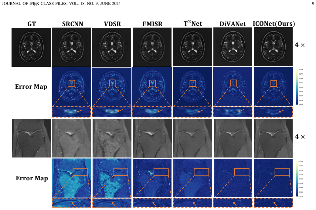
- [IV-C, Tables I-III] The state-of-the-art comparison includes only SRCNN, VDSR, FMISR, T2Net, and DiVANet. Recent medical SR methods (e.g., TransMRSR [13], MFTN [33], Deform-Mamba [41], and the authors' own self-prior guided Mamba network [42]) are omitted from the quantitative tables, even though they are cited in the related work. Furthermore, no error bars, seeds, or significance tests are reported. On fastMRI, the margins over T2Net/DiVANet are 0.12-0.14 dB at 2x and 0.07-0.14 dB at 4x, which are within the range of typical run-to-run variation. The claim that ICONet outperforms most state-of-the-art approaches is therefore not established. Please add the missing baselines, at least three seeds with mean and standard deviation, and appropriate statistical tests.
- [IV-B] Training details are insufficient for reproducibility. The authors report the optimizer, learning rate, feature dimension, and loss weights, but omit the number of training epochs, batch size, patch size, data augmentation, learning rate schedule, and the exact procedure for generating low-resolution images with the frequency-domain degradation model of Ref. [50]. Without these details (and ideally code release), the experimental numbers cannot be independently reproduced. Please provide a complete training configuration and make the code publicly available.
- [IV-B and evaluation setup] All experiments use a single synthetic degradation model from Ref. [50]. The statement that this model 'helps generate LR images that more closely resemble the characteristics of real-world scenarios' is not supported by any experiment. Because the method is intended for clinical imaging, the authors should evaluate at least one additional degradation model (e.g., k-space undersampling with noise for MRI) or a real paired LR/HR dataset to demonstrate that the reported gains are not an artifact of the specific synthetic degradation.
- [IV-D6, Table IX] The comparison with SwinIR appears to use a non-standard configuration: 1.260M parameters and 20.66 GFLOPs, whereas standard SwinIR typically has about 11.5M parameters. This may put SwinIR at a disadvantage. The authors should specify the exact SwinIR configuration, use a recognized standard baseline, and also compare with other Transformer-based SR models (e.g., RCAN or SwinIR-light) to substantiate the efficiency claim.
- [IV-E, Table X] The downstream segmentation experiment reports a single Dice score for each method without variance or significance testing. The setup also includes high-resolution T1 images as auxiliary inputs, but the segmentation training protocol is not described in detail. This makes it difficult to attribute the improvement to the SR method rather than to random variation or the additional T1 input. Please report multiple runs, standard deviations, and a statistical test, and clarify the segmentation training procedure.
minor comments (6)
- [Introduction, Section I] There is a placeholder citation in the text 'traditional super-resolution approaches [ ?], [12]'; the question mark should be replaced with the appropriate reference.
- [Section IV-B] The degradation model from Ref. [50] is referenced but not described; please summarize the model and how it is applied to each dataset, especially for PET images where the acquisition physics differs from MRI.
- [Section III-C, Eq. (4)-(7)] The notation for the semantic attention block is dense; in particular, the symmetric computation for F_Rec_SAB is mentioned in the text but not written out. For clarity, define both directions explicitly or state that they follow the same form.
- [Section IV-D2] The phrase 'black box-like sharable features' is vague; please provide a more precise explanation of why parallel multi-task sharing underperforms the serial framework.
- [Section IV-C and Conclusion] The abstract and Section I say ICONet 'outperforms most state-of-the-art approaches,' while the conclusion says it 'greatly outperforms state-of-the-art medical image super-resolution approaches.' This inconsistency should be resolved after the comparison set is expanded.
- [General] The reconstruction branch is trained on the same ground-truth images as the super-resolution branch, so the resulting 'prior' is a learned self-prior rather than an external or physics-based constraint. The paper should explicitly acknowledge this and discuss its implications for the method's generalizability.
Circularity Check
No significant circularity: empirical SR claims are tested on held-out public benchmarks; self-citations are contextual and not load-bearing.
full rationale
No significant circularity was found. The paper's central claim is an empirical performance claim: ICONet achieves the best PSNR/SSIM on held-out test portions of IXI, fastMRI, and Hecktor (Tables I-III) compared with SRCNN, VDSR, FMISR, T2Net, and DiVANet. The reconstruction prior is generated by a learned branch trained on the same ground-truth images as the SR branch, but this is a joint training objective rather than a fitted parameter that is later renamed as a prediction; the contribution of that branch is separately tested by ablations (Table IV) on held-out data. The multi-stage loss (Eq. 8), loss-weight search (Table VI), and RSCFL-count selection (Table VII) are hyperparameter/architecture choices, not quantities that by construction force the reported SR scores. Self-citations ([35], [36], [41], [42]) appear only as related-work context and as prior Mamba/multi-contrast SR methods; they are not used to justify a uniqueness theorem or to license an unverified ansatz, so they are not load-bearing. Baseline-selection and statistical-significance weaknesses (small baseline set, no error bars) are comparison-quality issues, not circularity. The headline results are evaluated against external public benchmarks and do not reduce to the paper's own inputs by definition.
Assumptions & free parameters
free parameters (4)
- Loss weight alpha =
0.9
- Number of RSCFL blocks N =
7
- Learning rate =
1e-4
- Feature dimension =
96
assumptions (4)
- domain assumption The frequency-domain degradation model of Ref. [50] produces low-resolution images representative of real-world medical imaging conditions.
- domain assumption The reconstruction branch output is an artifact-free image that can serve as a useful prior for super-resolution.
- domain assumption Mamba's four-directional selective scanning captures spatial dependencies in medical images better than content-similarity attention.
- standard math L1 loss yields better performance and convergence than L2 for image restoration.
Cite this review
Pith. "Pith review of Iterative Collaboration Network Guided By Reconstruction Prior for Medical Image Super-Resolution." pith.science (2026). https://pith.science/paper/T2NVT26C
@misc{pith2026250416958,
  author       = {Pith},
  title        = {Pith review of: Iterative Collaboration Network Guided By Reconstruction Prior for Medical Image Super-Resolution},
  year         = {2026},
  howpublished = {\url{https://pith.science/paper/T2NVT26C}},
  note         = {Machine review of arXiv:2504.16958}
}
read the original abstract
High-resolution medical images can provide more detailed information for better diagnosis. Conventional medical image super-resolution relies on a single task which first performs the extraction of the features and then upscaling based on the features. The features extracted may not be complete for super-resolution. Recent multi-task learning,including reconstruction and super-resolution, is a good solution to obtain additional relevant information. The interaction between the two tasks is often insufficient, which still leads to incomplete and less relevant deep features. To address above limitations, we propose an iterative collaboration network (ICONet) to improve communications between tasks by progressively incorporating reconstruction prior to the super-resolution learning procedure in an iterative collaboration way. It consists of a reconstruction branch, a super-resolution branch, and a SR-Rec fusion module. The reconstruction branch generates the artifact-free image as prior, which is followed by a super-resolution branch for prior knowledge-guided super-resolution. Unlike the widely-used convolutional neural networks for extracting local features and Transformers with quadratic computational complexity for modeling long-range dependencies, we develop a new residual spatial-channel feature learning (RSCFL) module of two branches to efficiently establish feature relationships in spatial and channel dimensions. Moreover, the designed SR-Rec fusion module fuses the reconstruction prior and super-resolution features with each other in an adaptive manner. Our ICONet is built with multi-stage models to iteratively upscale the low-resolution images using steps of 2x and simultaneously interact between two branches in multi-stage supervisions.
Figures


Figures from the paper (6 more)




Reference graph
Works this paper leans on
-
[13]
Transmrsr: transformer-based self-distilled generative prior for brain MRI super-resolution,
S. Huang, X. Liu, T. Tan, M. Hu, X. Wei, T. Chen, and B. Sheng, “Transmrsr: transformer-based self-distilled generative prior for brain MRI super-resolution,” Vis. Comput. , vol. 39, no. 8, pp. 3647–3659, 2023
work page 2023
-
[33]
MFTN: multi-level feature transfer network based on mri-transformer for MR image super-resolution,
S. Huang, G. Chen, Y . Yang, X. Wang, and C. Liang, “MFTN: multi-level feature transfer network based on mri-transformer for MR image super-resolution,” in Thirty-Eighth AAAI Conference on Artificial Intelligence, AAAI, 2024, pp. 2366–2373
work page 2024
-
[41]
Deform-mamba network for MRI super-resolution,
Z. Ji, B. Zou, X. Kui, P. Vera, and S. Ruan, “Deform-mamba network for MRI super-resolution,” in Medical Image Computing and Computer Assisted Intervention - MICCAI , vol. 15007, 2024, pp. 242–252
work page 2024
-
[42]
Z. Ji, B. Zou, X. Kui, H. Li, P. Vera, and S. Ruan, “Generation of super- resolution for medical image via a self-prior guided mamba network with edge-aware constraint,” Pattern Recognit. Lett. , vol. 187, pp. 93– 99, 2025
work page 2025
-
[50]
Multi-contrast super-resolution MRI through a progressive network,
Q. Lyu, H. Shan, C. Steber, C. Helis, C. Whitlow, M. Chan, and G. Wang, “Multi-contrast super-resolution MRI through a progressive network,” IEEE Trans. Medical Imaging, vol. 39, no. 9, pp. 2738–2749, 2020
work page 2020
-
[1]
Cross-scope spatial-spectral informa- tion aggregation for hyperspectral image super-resolution,
S. Chen, L. Zhang, and L. Zhang, “Cross-scope spatial-spectral informa- tion aggregation for hyperspectral image super-resolution,” IEEE Trans. Image Process., vol. 33, pp. 5878–5891, 2024
work page 2024
-
[2]
Hyperspectral image super-resolution based on spatial-spectral feature extraction network,
Y . Li, S. Chen, W. Luo, L. Zhou, and W. Xie, “Hyperspectral image super-resolution based on spatial-spectral feature extraction network,” Chinese Journal of Electronics , vol. 32, no. 3, pp. 415–428, 2023
work page 2023
-
[3]
Nonlocal low-rank tensor completion for visual data,
L. Zhang, L. Song, B. Du, and Y . Zhang, “Nonlocal low-rank tensor completion for visual data,” IEEE Trans. Cybern. , vol. 51, no. 2, pp. 673–685, 2021
work page 2021
Show all 53 references
-
[4]
Image super-resolution using deep convolutional networks,
C. Dong, C. C. Loy, K. He, and X. Tang, “Image super-resolution using deep convolutional networks,” IEEE Trans. Pattern Anal. Mach. Intell. , vol. 38, no. 2, pp. 295–307, 2016
2016
-
[5]
Accurate image super-resolution using very deep convolutional networks,
J. Kim, J. K. Lee, and K. M. Lee, “Accurate image super-resolution using very deep convolutional networks,” in IEEE Conference on Computer Vision and Pattern Recognition, CVPR , 2016, pp. 1646–1654
2016
-
[6]
Super-resolution reconstruction of knee magnetic resonance imaging based on deep learning,
D. Qiu, S. Zhang, Y . Liu, J. Zhu, and L. Zheng, “Super-resolution reconstruction of knee magnetic resonance imaging based on deep learning,” Comput. Methods Programs Biomed. , vol. 187, p. 105059, 2020
2020
-
[7]
A trusted medical image super-resolution method based on feedback adaptive weighted dense network,
L. Chen, X. Yang, G. Jeon, M. Anisetti, and K. Liu, “A trusted medical image super-resolution method based on feedback adaptive weighted dense network,” Artif. Intell. Medicine , vol. 106, p. 101857, 2020
2020
-
[8]
Task transformer network for joint MRI reconstruction and super-resolution,
C. Feng, Y . Yan, H. Fu, L. Chen, and Y . Xu, “Task transformer network for joint MRI reconstruction and super-resolution,” in Medical Image Computing and Computer Assisted Intervention, MICCAI , vol. 12906, 2021, pp. 307–317
2021
-
[9]
Enhanced deep residual networks for single image super-resolution,
B. Lim, S. Son, H. Kim, S. Nah, and K. M. Lee, “Enhanced deep residual networks for single image super-resolution,” in IEEE Conference on Computer Vision and Pattern Recognition, CVPR, 2017, pp. 1132–1140
2017
-
[10]
PET image super- resolution using generative adversarial networks,
T. Song, S. R. Chowdhury, F. Yang, and J. Dutta, “PET image super- resolution using generative adversarial networks,” Neural Networks, vol. 125, pp. 83–91, 2020. JOURNAL OF LATEX CLASS FILES, VOL. 18, NO. 9, JUNE 2024 12
2020
-
[11]
Arbitrary- scale image super-resolution via degradation perception,
W. Wan, Z. Wang, Z. Wang, L. Gu, J. Sun, and Q. Wang, “Arbitrary- scale image super-resolution via degradation perception,” IEEE Trans. Computational Imaging, vol. 10, pp. 666–676, 2024
2024
-
[12]
Single image superresolu- tion based on gradient profile sharpness,
Q. Yan, Y . Xu, X. Yang, and T. Q. Nguyen, “Single image superresolu- tion based on gradient profile sharpness,” IEEE Trans. Image Process. , vol. 24, no. 10, pp. 3187–3202, 2015
2015
-
[14]
Deep residual learning for image recognition,
K. He, X. Zhang, S. Ren, and J. Sun, “Deep residual learning for image recognition,” in IEEE Conference on Computer Vision and Pattern Recognition, CVPR. IEEE Computer Society, 2016, pp. 770–778
2016
-
[15]
An image is worth 16x16 words: Trans- formers for image recognition at scale,
A. Dosovitskiy, L. Beyer, A. Kolesnikov, D. Weissenborn, X. Zhai, T. Unterthiner, M. Dehghani, M. Minderer, G. Heigold, S. Gelly, J. Uszkoreit, and N. Houlsby, “An image is worth 16x16 words: Trans- formers for image recognition at scale,” in International Conference on Learni...
2021
-
[16]
Combining recurrent, convolutional, and continuous-time models with linear state space layers,
A. Gu, I. Johnson, K. Goel, K. Saab, T. Dao, A. Rudra, and C. R ´e, “Combining recurrent, convolutional, and continuous-time models with linear state space layers,” in Advances in Neural Information Processing Systems, NeurIPS, 2021, pp. 572–585
2021
-
[17]
Efficiently modeling long sequences with structured state spaces,
A. Gu, K. Goel, and C. R ´e, “Efficiently modeling long sequences with structured state spaces,” in The Tenth International Conference on Learning Representations, ICLR , 2022
2022
-
[18]
Mamba: Linear-time sequence modeling with selective state spaces,
A. Gu and T. Dao, “Mamba: Linear-time sequence modeling with selective state spaces,” arXiv preprint arXiv:2312.00752 , 2023
2023 arXiv
-
[19]
Segmamba: Long-range sequential modeling mamba for 3d medical image segmentation,
Z. Xing, T. Ye, Y . Yang, G. Liu, and L. Zhu, “Segmamba: Long-range sequential modeling mamba for 3d medical image segmentation,” arXiv preprint arXiv:2401.13560, 2024
2024 arXiv
-
[20]
Vm-unet: Vision mamba unet for medical image segmentation,
J. Ruan and S. Xiang, “Vm-unet: Vision mamba unet for medical image segmentation,” CoRR, vol. abs/2402.02491, 2024
2024 arXiv
-
[21]
Vision mamba: Efficient visual representation learning with bidirectional state space model,
L. Zhu, B. Liao, Q. Zhang, X. Wang, W. Liu, and X. Wang, “Vision mamba: Efficient visual representation learning with bidirectional state space model,” arXiv preprint arXiv:2401.09417 , 2024
2024 arXiv
-
[22]
Selective structured state-spaces for long-form video understanding,
J. Wang, W. Zhu, P. Wang, X. Yu, L. Liu, M. Omar, and R. Hamid, “Selective structured state-spaces for long-form video understanding,” in IEEE Conference on Computer Vision and Pattern Recognition, CVPR , 2023, pp. 6387–6397
2023
-
[23]
Vmamba: Visual state space model,
Y . Liu, Y . Tian, Y . Zhao, H. Yu, L. Xie, Y . Wang, Q. Ye, and Y . Liu, “Vmamba: Visual state space model,” CoRR, vol. abs/2401.10166, 2024
2024 arXiv
-
[24]
Cubic convolution interpolation for digital image processing,
R. Keys, “Cubic convolution interpolation for digital image processing,” IEEE Trans. Acoust. Speech Signal Process. , vol. 29, no. 6, pp. 1153– 1160, 1981
1981
-
[25]
High-resolution image recovery from image- plane arrays, using convex projections,
H. Stark and P. Oskoui, “High-resolution image recovery from image- plane arrays, using convex projections,” J. Opt. Soc. Am. A. , vol. 6, no. 11, pp. 1715–1726, 1989
1989
-
[26]
Lrtv: Mr image super-resolution with low-rank and total variation regularizations,
F. Shi, J. Cheng, L. Wang, P.-T. Yap, and D. Shen, “Lrtv: Mr image super-resolution with low-rank and total variation regularizations,” IEEE Trans. Medical Imaging, vol. 34, no. 12, pp. 2459–2466, 2015
2015
-
[27]
Coupled dictionary training for image super-resolution,
J. Yang, Z. Wang, Z. Lin, S. Cohen, and T. S. Huang, “Coupled dictionary training for image super-resolution,” IEEE Trans. Image Process., vol. 21, no. 8, pp. 3467–3478, 2012
2012
-
[28]
Image super-resolution via sparse representation,
J. Yang, J. Wright, T. S. Huang, and Y . Ma, “Image super-resolution via sparse representation,” IEEE Trans. Image Process., vol. 19, no. 11, pp. 2861–2873, 2010
2010
-
[29]
Mapanet: A multi-scale attention-guided progressive aggregation network for multi-contrast MRI super-resolution,
L. Liu, T. Liu, W. Zhou, Y . Wang, and M. Liu, “Mapanet: A multi-scale attention-guided progressive aggregation network for multi-contrast MRI super-resolution,” IEEE Trans. Computational Imaging, vol. 10, pp. 928– 940, 2024
2024
-
[30]
Cytopathology image super-resolution of portable microscope based on convolutional window-integration transformer,
J. Zhang, S. Cheng, X. Liu, N. Li, G. Rao, and S. Zeng, “Cytopathology image super-resolution of portable microscope based on convolutional window-integration transformer,” IEEE Trans. Computational Imaging , vol. 11, pp. 77–88, 2025
2025
-
[31]
A fast medical image super resolution method based on deep learning network,
S. Zhang, G. Liang, S. Pan, and L. Zheng, “A fast medical image super resolution method based on deep learning network,” IEEE Access, vol. 7, pp. 12 319–12 327, 2019
2019
-
[32]
Photo-realistic single image super-resolution using a generative adversarial network,
C. Ledig, L. Theis, F. Huszar, J. Caballero, A. Cunningham, A. Acosta, A. P. Aitken, A. Tejani, J. Totz, Z. Wang, and W. Shi, “Photo-realistic single image super-resolution using a generative adversarial network,” in IEEE Conference on Computer Vision and Pattern Recognition, ...
2017
-
[34]
Multi-contrast brain magnetic resonance image super-resolution using the local weight similarity,
H. Zheng, X. Qu, Z. Bai, Y . Liu, D. Guo, J. Dong, X. Peng, and Z. Chen, “Multi-contrast brain magnetic resonance image super-resolution using the local weight similarity,” BMC Medical Imaging , vol. 17, no. 1, pp. 6:1–6:13, 2017
2017
-
[35]
Wavelet-aware transformer network for multi-contrast knee MRI super-resolution,
Z. Ji, X. Kui, S. Liao, C. Zhu, Y . Li, Y . Dai, and B. Zou, “Wavelet-aware transformer network for multi-contrast knee MRI super-resolution,” in IEEE International Conference on Bioinformatics and Biomedicine, BIBM, 2023, pp. 3037–3044
2023
-
[36]
Multi-scale deformable transformer for multi-contrast knee MRI super-resolution,
B. Zou, Z. Ji, C. Zhu, Y . Dai, W. Zhang, and X. Kui, “Multi-scale deformable transformer for multi-contrast knee MRI super-resolution,” Biomed. Signal Process. Control. , vol. 79, no. Part, p. 104154, 2023
2023
-
[37]
Deep face super- resolution with iterative collaboration between attentive recovery and landmark estimation,
C. Ma, Z. Jiang, Y . Rao, J. Lu, and J. Zhou, “Deep face super- resolution with iterative collaboration between attentive recovery and landmark estimation,” in IEEE Conference on Computer Vision and Pattern Recognition, CVPR, 2020, pp. 5568–5577
2020
-
[38]
Progressive semantic-aware style transformation for blind face restoration,
C. Chen, X. Li, L. Yang, X. Lin, L. Zhang, and K. K. Wong, “Progressive semantic-aware style transformation for blind face restoration,” in IEEE Conference on Computer Vision and Pattern Recognition, CVPR , 2021, pp. 11 896–11 905
2021
-
[39]
Analysis of zero-order holder discretization of two-dimensional sliding-mode control systems,
Z. Galias and X. Yu, “Analysis of zero-order holder discretization of two-dimensional sliding-mode control systems,” IEEE Trans. Circuits Syst. II Express Briefs , vol. 55-II, no. 12, pp. 1269–1273, 2008
2008
-
[40]
U-mamba: Enhancing long-range depen- dency for biomedical image segmentation,
J. Ma, F. Li, and B. Wang, “U-mamba: Enhancing long-range depen- dency for biomedical image segmentation,” CoRR, vol. abs/2401.04722, 2024
2024 arXiv
-
[43]
Squeeze-and-excitation networks,
J. Hu, L. Shen, and G. Sun, “Squeeze-and-excitation networks,” in IEEE Conference on Computer Vision and Pattern Recognition, CVPR , 2018, pp. 7132–7141
2018
-
[44]
Loss functions for image restoration with neural networks,
H. Zhao, O. Gallo, I. Frosio, and J. Kautz, “Loss functions for image restoration with neural networks,” IEEE Trans. Computational Imaging, vol. 3, no. 1, pp. 47–57, 2017
2017
-
[45]
fastmri: An open dataset and benchmarks for accelerated mri,
J. Zbontar, F. Knoll, A. Sriram, T. Murrell, Z. Huang, M. J. Muck- ley, A. Defazio, R. Stern, P. Johnson, M. Bruno et al. , “fastmri: An open dataset and benchmarks for accelerated mri,” arXiv preprint arXiv:1811.08839, 2018
2018 arXiv
-
[46]
fastmri: A publicly available raw k-space and dicom dataset of knee images for ac- celerated mr image reconstruction using machine learning,
F. Knoll, J. Zbontar, A. Sriram, M. J. Muckley, M. Bruno, A. Defazio, M. Parente, K. J. Geras, J. Katsnelson, H. Chandarana et al., “fastmri: A publicly available raw k-space and dicom dataset of knee images for ac- celerated mr image reconstruction using machine learning,” Ra...
2020
-
[47]
Head and neck tumor segmentation in PET/CT: the HECKTOR challenge,
V . Oreiller, V . Andrearczyk, M. Jreige, S. Boughdad, H. Elhalawani, J. Castelli, M. Valli `eres, S. Zhu, J. Xie, Y . Peng, A. Iantsen, M. Hatt, Y . Yuan, J. Ma, X. Yang, C. Rao, S. Pai, K. Ghimire, X. Feng, M. A. Naser, C. D. Fuller, F. Yousefirizi, A. Rahmim, H. Chen, L. Wa...
2022
-
[48]
Image quality assessment: from error visibility to structural similarity,
Z. Wang, A. C. Bovik, H. R. Sheikh, and E. P. Simoncelli, “Image quality assessment: from error visibility to structural similarity,” IEEE Trans. Image Process., vol. 13, no. 4, pp. 600–612, 2004
2004
-
[49]
Single image super-resolution based on directional vari- ance attention network,
P. Behjati, P. Rodr ´ıguez, C. Fern ´andez, I. Hupont, A. Mehri, and J. Gonz `alez, “Single image super-resolution based on directional vari- ance attention network,” Pattern Recognit., vol. 133, p. 108997, 2023
2023
-
[51]
Swinir: Image restoration using swin transformer,
J. Liang, J. Cao, G. Sun, K. Zhang, L. V . Gool, and R. Timofte, “Swinir: Image restoration using swin transformer,” in IEEE International Con- ference on Computer Vision Workshops, ICCVW , 2021, pp. 1833–1844
2021
-
[52]
Swin transformer: Hierarchical vision transformer using shifted windows,
Z. Liu, Y . Lin, Y . Cao, H. Hu, Y . Wei, Z. Zhang, S. Lin, and B. Guo, “Swin transformer: Hierarchical vision transformer using shifted windows,” in IEEE International Conference on Computer Vision, ICCV, 2021, pp. 9992–10 002
2021
-
[53]
Attention u-net: Learning where to look for the pancreas,
O. Oktay, J. Schlemper, L. L. Folgoc, M. C. H. Lee, M. P. Heinrich, K. Misawa, K. Mori, S. G. McDonagh, N. Y . Hammerla, B. Kainz, B. Glocker, and D. Rueckert, “Attention u-net: Learning where to look for the pancreas,” CoRR, vol. abs/1804.03999, 2018
2018 arXiv
Reviewed August 16, 2026 · model on record in the stance chip above.
Discussion (0). Continue with ORCID to comment.