REVIEW 3 major objections 5 minor 1 cited by
HeartSimSage: Attention-Enhanced Graph Neural Networks for Accelerating Cardiac Mechanics Modeling
T0 review · 3 major / 5 minor · reviewed 2026-08-16 · deepseek-v4-flash
Pith's one-line read HeartSimSage trains a graph neural network to emulate cardiac FEA, claiming 0.13% averaged displacement error and a 13,000x GPU speedup while accepting variable patient mesh topology.
desk verdict A credible, well-engineered biventricular GNN emulator whose headline accuracy number does not reproduce from its own Table 5, and whose generalization claim may be inflated by an unclearly stratified train/test split. read the letter →
The pith
A machine-rendered reading of the paper's core claim, the machinery that carries it, and where it could break.
The reading
What carries the argument
The architecture's load-bearing pieces are three. First, a set of Laplace-Dirichlet solutions on each geometry, with potentials such as $\Phi_{\mathrm{AB}}$ (apex-to-base), $\Phi_{\mathrm{EP}}$ (transmural), and $\Phi_{\mathrm{LV-RV}}$ (septal), encode every node's relative position and also feed a rule-based generator for fiber and sheet orientations. Second, a neighbor-selection scheme partitions candidate neighbors into five distance shells, from the nearest 0.2% out to the 10th-percentile distance, and samples roughly 12 neighbors per node, keeping both local and mid-range information in the message-passing. Third, an eight-head transformer-style attention layer reweights those edge embeddings before average pooling and a residual addition to the node embedding. Global inputs, namely the Holzapfel-Ogden material parameters, chamber pressures, and forty shape descriptors, are encoded separately and concatenated with the node embedding, and three MLP heads decode the x, y, and z displacement components. Subset training on 300 randomly chosen nodes per sample per epoch keeps the training cost low while still covering the whole domain across epochs.
What would settle it
Regroup the 5,000 cases so that all augmented geometries from the same source heart stay in the same fold, retrain HeartSimSage, and report test displacement errors; if the mean error rises well above 0.13%, the current test error contains geometry leakage. A complementary check is to train on the 4,850 augmented cases only and test on the 150 original, unaugmented segmentations, which isolates whether the model has learned anatomy transfer rather than interpolation between augmented siblings.
Extended reading notes
Core claim
The paper's central claim is that an attention-enhanced GNN using Laplace-Dirichlet spatial encoding, distance-partitioned neighbor sampling, and per-axis decoders can faithfully emulate passive biventricular FEA across variable geometries, pressures, and orthotropic material parameters. On the test set, the averaged component-wise displacement errors are at most 0.15% ± 0.14%, pointwise differences stay below 0.4 mm in nearly all cases, and the LV and RV cavity volume errors are 0.20% ± 0.17% and 0.52% ± 0.36%. Prediction takes 0.0084 seconds per case on an L40S GPU and 0.6 seconds on a CPU, versus 112 ± 35 seconds for one FEA solve on the same CPU, which yields the reported 13,000x and 190x speedups. The paper further argues that the Laplace-Dirichlet features beat one-hot boundary encodings, that attention weights concentrate on the nearest neighbors while mid-range nodes still contribute, and that the model transfers to a published left-ventricle dataset with accuracy close to the earlier GNN emulator.
Load-bearing premise
The generalization claim rests on the train/test split of the 5,000 simulated cases, but 4,850 of those cases are augmented variants of just 150 source hearts and the paper does not say the split is patient-level; if meshes derived from the same source segmentation appear in both training and test sets, the reported 0.13% error overstates accuracy on a truly new patient.
Editorial extensions
If this is right
- Patient-specific parameter estimation becomes a search problem rather than a bottleneck: hundreds of candidate material-parameter sets could be evaluated in seconds, which is what digital-twin calibration currently lacks.
- Dynamic node topology means a model trained once on a population of meshes can be applied to a new patient's mesh without remeshing, reordering, or retraining, a prerequisite for real clinical adoption.
- The Laplace-Dirichlet spatial encoding is geometry-agnostic, so the same recipe can be carried to atrial, whole-heart, or other organ mechanics where boundary-relative position matters.
- Attention-weight analysis yields a practical design rule for other mesh-based emulators: sample neighbors mostly from the nearest shells while keeping a few mid-range nodes, rather than using uniform random sampling.
- If the accuracy holds under patient-level evaluation, interactive cardiac simulation becomes feasible for surgical planning and near-real-time monitoring.
Reading between the lines
- The 13,000x number compares a single GPU inference to a single CPU FEA solve; the end-to-end economy of the method also depends on the one-time cost of simulating and storing the 5,000-case training corpus, which the paper does not amortize into the speedup.
- A patient-level split is the sharpest test the authors could run next, and the paper's own description of the augmentation pipeline makes such leakage a live possibility rather than a settled issue.
- The attention weights already show a steep distance decay, so a simple variant that replaces learned attention with a fixed distance-based weighting would reveal how much of the gain comes from attention per se and how much from the multiscale neighbor sampling.
- Because all inputs are static and the cardiac cycle is dynamic, extending the architecture with a temporal decoder for time-varying pressures is the natural next step, and nothing in the described design blocks that extension.
Signed reviews
Editorial analysis
A structured set of objections, weighed in public.
Referee Report
Summary. The paper presents HeartSimSage, an attention-enhanced graph neural network emulator of passive biventricular cardiac finite element mechanics. The model takes as input a patient-specific biventricular mesh, Laplace-Dirichlet spatial encodings, fiber orientations, material parameters, chamber pressures, and shape descriptors, and outputs nodal displacement fields. The authors train on 5,000 simulated cases derived from 150 segmented image volumes plus geometric augmentation, and report a GPU inference speedup of roughly 13,000x and CPU speedup of 190x over FEA, together with a nominal averaged displacement error of 0.13% ± 0.12%. The manuscript includes sensitivity studies of learning rate, batch size, Laplace encoding, subset node count, neighbor connection strategy, and attention mechanism, plus a comparison with a published left-ventricular GNN emulator.
Significance. If the accuracy and speed claims hold, HeartSimSage would be a useful surrogate for passive biventricular FEA in applications that require many simulations, such as parameter estimation or digital twin workflows. The architecture is sensibly engineered: Laplace-Dirichlet features are a natural spatial encoding for irregular cardiac meshes; the subset-based training and GraphSAGE-inspired neighbor partitioning address scalability; and the attention analysis in Section 3.3 provides interpretable evidence that the model weights local neighbors more strongly. The authors also ship a code link and compare against a reimplementation of a published LV emulator, which is commendable. However, the central accuracy claim is currently not verifiable from the paper's own Table 5, and the train/test split is not shown to be patient-independent. These issues are fixable but they block acceptance as written.
major comments (3)
- [§3.2, Table 5; Abstract; §5] The headline accuracy figure '0.13% ± 0.12%' is not reproducible from Table 5. Averaging the three component-wise means gives (0.15 + 0.11 + 0.10)/3 = 0.12%, not 0.13%; averaging the reported standard deviations gives (0.14 + 0.10 + 0.10)/3 ≈ 0.11%, not 0.12%. A pooled computation over all nodes and components would give yet another value, and the paper does not state which aggregation was used. Because the abstract and conclusion both advertise this number, the central accuracy claim as written is not verifiable from the paper's own reported data. Please specify the exact error metric used for the headline number (e.g., mean over nodes of per-node Euclidean displacement error normalized per geometry, or mean over components) and recalculate all reported accuracies consistently.
- [§2.4 and §2.6] The train/validation/test split is not stratified at the patient or source-segmentation level. Section 2.4 generates 4,850 pseudo-geometries from 150 source segmentations, and Section 2.6 splits the resulting 5,000 cases into 4,000 training, 500 validation, and 500 test cases without stating whether augmented meshes derived from the same source geometry are confined to one split. If augmented cases from the same source appear in both training and test sets, the reported 0.13% error would be inflated by geometry leakage and would not support the claimed generalization to new patients. Please either demonstrate that no augmented test geometry shares a source segmentation with any training geometry, or perform a source-level split and report test metrics under that split.
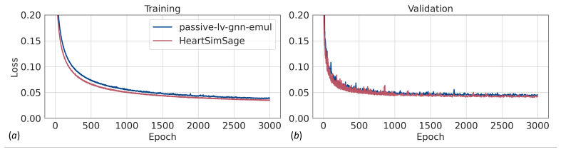
- [§4.7, Table 14] The external 'validation' on the LV mechanics dataset reports only training and validation losses; no held-out test displacement or volume errors are given for either HeartSimSage or the passive-lv-gnn-emul baseline. The abstract's statement that the model was 'validated' on a published LV dataset is therefore stronger than the evidence presented. Please add test-set metrics and specify the split used for the LV dataset, ideally at the patient level.
minor comments (5)
- [§3.2, Fig. 6] The sentence 'A node-wise difference ... is below 0.4 mm for all cases except case 5' refers to the nine randomly selected cases shown in Fig. 6, but as written it could be misread as a claim about the entire test set. Please clarify that this statement concerns the displayed cases only.
- [§2.5.2, Eq. (9)] The notation in the neighbor subset definitions, e.g., 'k∈V nSC1', is ambiguous because the subscript is not defined as a separate set or constant. Please clarify whether these are all nodes in the mesh or a candidate pool of a specific size.
- [§2.6] The code repository link is useful, but the manuscript should state a versioned commit or release and a license, since the reproducibility claim depends on the exact implementation.
- [§3.2] The GPU is described as an 'Nvidia Titan L40S'; the correct product name is usually 'NVIDIA L40S' (not 'Titan'). Please correct this label.
- [§3.3, Table 6] The one-way ANOVA result is reported only as p < 0.05; please state the number of samples used in the test and whether multiple-comparison correction was applied across the five distance bins.
Circularity Check
No significant circularity: the emulator is benchmarked against the independent FEA solver it imitates, with an external LV dataset check; self-citations are contextual, not load-bearing.
full rationale
The central claim is that HeartSimSage reproduces FEA displacement fields for passive biventricular mechanics. The ground-truth labels are FEA solutions from an in-house svFSI-derived solver, and the network maps geometry, fiber orientations, Laplace-Dirichlet features, material parameters, and pressures to displacements (Eqs. 5-18). No parameter is fitted to the test error and then renamed a prediction; the reported errors (Eq. 20) compare network outputs to independent FEA outputs, which is the correct external target for an emulator. The external LV dataset comparison (Sec. 4.7, Table 14) further benchmarks the model against the published GNN of Dalton et al. [34], so the accuracy claim is not sustained solely by self-citation. The citations to the authors' prior work [29,85,86] supply the mechanics formulation, parameter ranges, and solver benchmarking context; they are not invoked as a uniqueness theorem or to forbid alternative architectures, and the sensitivity analyses (Sec. 4) compare architectural choices on validation loss rather than importing a forced result. Two caveats are noted but are not circularity. First, the train/test split in Sec. 2.4-2.6 is not described as patient-stratified, so augmented pseudo-geometries from the same 150 source segmentations could appear in both training and test, which would inflate generalization claims; this is a potential data-leakage risk, not a reduction of the prediction to its inputs by construction. Second, the abstract's '0.13% +- 0.12%' does not obviously follow from Table 5's component-wise means and standard deviations, since simple averaging of the three component means gives 0.12% and of the standard deviations gives about 0.11%, and the paper does not state the aggregation rule; this is an internal reporting inconsistency that affects verifiability, not a circular derivation. Because the derivation is a standard supervised emulation pipeline with an independent target and an external check, no circular step is present.
Assumptions & free parameters
free parameters (4)
- Learned network weights (all MLP and attention parameters) =
Not enumerated; optimized on 4,000 training cases
- Subset size nS =
300 nodes per sample per epoch
- Neighbor count nNB =
12
- Fiber angle rule parameters =
Endocardial 40 degrees to epicardial -50 degrees; sheet angle -65 to 25 degrees
assumptions (4)
- domain assumption The svFSI-based stabilized finite element solver produces accurate ground truth displacements for passive biventricular mechanics.
- domain assumption Rule-based fiber orientation with fixed angle ranges is adequate for patient-specific fiber architecture.
- domain assumption Augmented pseudo-geometries represent anatomical variability of real patients.
- domain assumption Randomly sampling 300 nodes per case per epoch gives sufficiently representative training gradients for the full displacement field.
Cite this review
Pith. "Pith review of HeartSimSage: Attention-Enhanced Graph Neural Networks for Accelerating Cardiac Mechanics Modeling." pith.science (2026). https://pith.science/paper/676CO5ZO
@misc{pith2026250418968,
  author       = {Pith},
  title        = {Pith review of: HeartSimSage: Attention-Enhanced Graph Neural Networks for Accelerating Cardiac Mechanics Modeling},
  year         = {2026},
  howpublished = {\url{https://pith.science/paper/676CO5ZO}},
  note         = {Machine review of arXiv:2504.18968}
}
read the original abstract
Finite element analysis (FEA) forms the cornerstone of modeling cardiac biomechanics but is computationally expensive, limiting its clinical application for digital twin creation, which often requires tens to hundreds of simulations to estimate tissue parameters. We developed an attention-enhanced graph neural network (GNN)-based FEA emulator, HeartSimSage, to rapidly predict passive biventricular myocardial displacements from patient-specific geometries, chamber pressures, and material properties. HeartSimSage addresses the limitations of current emulators by effectively handling diverse three-dimensional (3D) biventricular geometries, mesh topologies, fiber directions, structurally based constitutive models, and physiological boundary conditions. It supports flexible mesh structures with variable node counts, orderings, and element connectivity. To optimize information propagation, we designed a neighboring connection strategy inspired by Graph Sample and Aggregate (GraphSAGE) that prioritizes local interactions while maintaining mid-to-long-range dependencies. We further incorporated Laplace-Dirichlet solutions for enhanced spatial encoding and employed subset-based training for improved efficiency. By integrating an attention mechanism, HeartSimSage adaptively weighs neighbor contributions and filters irrelevant information, enhancing prediction accuracy. HeartSimSage achieves approximately 13,000x speedup on GPU and 190x on CPU compared to traditional FEA, while maintaining a nominal averaged error of 0.13% +- 0.12% in predicting biventricular displacements. We validated our model using a published left ventricle dataset and conducted sensitivity analyses on hyperparameters, neighboring strategies, and the attention mechanism.
Figures


Figures from the paper (12 more)


Forward citations
Cited by 1 Pith paper
-
HeartUnloadNet: A Weakly-Supervised Cycle-Consistent Graph Network for Predicting Unloaded Cardiac Geometry from Diastolic States
HeartUnloadNet maps end-diastolic left-ventricular meshes to unloaded geometries in milliseconds on synthetic finite element data, but the reported DSC and sample-efficiency numbers are internally inconsistent.
Reference graph
Works this paper leans on
-
[1]
N. A. Trayanova, J. Constantino, V. Gurev, Electromechanical models of the ventricles, American Journal of Physiology-Heart and Circula- tory Physiology 301 (2011) H279–H286
2011
-
[2]
S. G. Campbell, A. D. McCulloch, Multi-scale computational models of familial hypertrophic cardiomyopathy: genotype to phenotype, Journal of The Royal Society Interface 8 (2011) 1550–1561
2011
-
[3]
A. L. Marsden, M. Esmaily-Moghadam, Multiscale modeling of cardio- vascular flows for clinical decision support, Applied Mechanics Reviews 67 (2015) 030804
2015
-
[4]
Palit, S
A. Palit, S. K. Bhudia, T. N. Arvanitis, G. A. Turley, M. A. Williams, Computational modelling of left-ventricular diastolic mechanics: Effect of fibre orientation and right-ventricle topology, Journal of biomechan- ics 48 (2015) 604–612
2015
-
[5]
Mittal, J
R. Mittal, J. H. Seo, V. Vedula, Y. J. Choi, H. Liu, H. H. Huang, S. Jain, L. Younes, T. Abraham, R. T. George, Computational model- ing of cardiac hemodynamics: current status and future outlook, Jour- nal of Computational Physics 305 (2016) 1065–1082
2016
-
[6]
Sotiropoulos, T
F. Sotiropoulos, T. B. Le, A. Gilmanov, Fluid mechanics of heart valves and their replacements, Annual Review of Fluid Mechanics 48 (2016) 259–283
2016
-
[7]
Vedula, J
V. Vedula, J. Lee, H. Xu, C.-C. J. Kuo, T. K. Hsiai, A. L. Marsden, A method to quantify mechanobiologic forces during zebrafish cardiac de- velopment using 4-d light sheet imaging and computational modeling, PLoS computational biology 13 (2017) e1005828. 41
2017
-
[8]
Finsberg, C
H. Finsberg, C. Xi, J. L. Tan, L. Zhong, M. Genet, J. Sundnes, L. C. Lee, S. T. Wall, Efficient estimation of personalized biventricular me- chanical function employing gradient-based optimization, International journal for numerical methods in biomedical engineering 34 (2018) e2982
2018
Show all 119 references
-
[9]
J. Lee, V. Vedula, K. I. Baek, J. Chen, J. J. Hsu, Y. Ding, C.-C. Chang, H. Kang, A. Small, P. Fei, et al., Spatial and temporal variations in hemodynamic forces initiate cardiac trabeculation, JCI insight 3 (2018)
2018
-
[10]
M. R. Pfaller, J. M. H¨ ormann, M. Weigl, A. Nagler, R. Chabiniok, C. Bertoglio, W. A. Wall, The importance of the pericardium for cardiac biomechanics: from physiology to computational modeling, Biomechanics and modeling in mechanobiology 18 (2019) 503–529
2019
-
[11]
Yoshida, J
K. Yoshida, J. W. Holmes, Computational models of cardiac hypertro- phy, Progress in biophysics and molecular biology 159 (2021) 75–85
2021
-
[12]
E. L. Schwarz, L. Pegolotti, M. R. Pfaller, A. L. Marsden, Beyond cfd: Emerging methodologies for predictive simulation in cardiovascu- lar health and disease, Biophysics Reviews 4 (2023)
2023
-
[13]
Green, W
L. Green, W. X. Chan, A. Tulzer, G. Tulzer, C. H. Yap, Myocardial biomechanical effects of fetal aortic valvuloplasty, Biomechanics and Modeling in Mechanobiology 23 (2024) 1433–1448
2024
-
[14]
Corral-Acero, F
J. Corral-Acero, F. Margara, M. Marciniak, C. Rodero, F. Loncaric, Y. Feng, A. Gilbert, J. F. Fernandes, H. A. Bukhari, A. Wajdan, et al., The ‘digital twin’to enable the vision of precision cardiology, European heart journal 41 (2020) 4556–4564
2020
-
[15]
Qureshi, G
A. Qureshi, G. Y. Lip, D. A. Nordsletten, S. E. Williams, O. Aslanidi, A. De Vecchi, Imaging and biophysical modelling of thrombogenic mechanisms in atrial fibrillation and stroke, Frontiers in Cardiovascular Medicine 9 (2023) 1074562
2023
-
[16]
Qureshi, M
A. Qureshi, M. Balmus, G. Lip, S. Williams, D. Nordsletten, O. Aslanidi, A. De Vecchi, Mechanistic modelling of virchows triad to assess thrombogenicity and stroke risk in atrial fibrillation patients, European Heart Journal-Digital Health 3 (2022) ztac076–2788. 42
2022
-
[17]
M. S. Bazzi, R. Balouchzadeh, S. N. Pavey, J. D. Quirk, H. Yanagi- sawa, V. Vedula, J. E. Wagenseil, V. H. Barocas, Experimental and mouse-specific computational models of the fbln4 smko mouse to iden- tify potential biomarkers for ascending thoracic aortic aneurysm, Car- diov...
2022
-
[18]
Crozier, C
A. Crozier, C. Augustin, A. Neic, A. Prassl, M. Holler, T. Fastl, A. Hen- nemuth, K. Bredies, T. Kuehne, M. Bishop, et al., Image-based person- alization of cardiac anatomy for coupled electromechanical modeling, Annals of biomedical engineering 44 (2016) 58–70
2016
-
[19]
L. Marx, J. A. Niestrawska, M. A. Gsell, F. Caforio, G. Plank, C. M. Augustin, Robust and efficient fixed-point algorithm for the inverse elastostatic problem to identify myocardial passive material parameters and the unloaded reference configuration, Journal of computational ...
2022
-
[20]
Balaban, H
G. Balaban, H. Finsberg, H. H. Odland, M. E. Rognes, S. Ross, J. Sundnes, S. Wall, High-resolution data assimilation of cardiac me- chanics applied to a dyssynchronous ventricle, International journal for numerical methods in biomedical engineering 33 (2017) e2863
2017
-
[21]
Gonzalo, C
A. Gonzalo, C. M. Augustin, S. F. Bifulco, ˚A. Telle, Y. Chahine, A. Kassar, M. Guerrero-Hurtado, E. Dur´ an, P. Mart´ ınez-Legazpi, O. Flores, et al., Multiphysics simulations reveal haemodynamic im- pacts of patient-derived fibrosis-related changes in left atrial tissue me- ...
2024
-
[22]
Mangion, H
K. Mangion, H. Gao, D. Husmeier, X. Luo, C. Berry, Advances in computational modelling for personalised medicine after myocardial infarction, Heart 104 (2018) 550–557
2018
-
[23]
P. M. Boyle, T. Zghaib, S. Zahid, R. L. Ali, D. Deng, W. H. Franceschi, J. B. Hakim, M. J. Murphy, A. Prakosa, S. L. Zimmerman, et al., Computationally guided personalized targeted ablation of persistent atrial fibrillation, Nature biomedical engineering 3 (2019) 870–879
2019
-
[24]
S. A. Niederer, M. S. Sacks, M. Girolami, K. Willcox, Scaling digi- tal twins from the artisanal to the industrial, Nature Computational Science 1 (2021) 313–320. 43
2021
-
[25]
Stimm, D
J. Stimm, D. A. Nordsletten, J. Jilberto, R. Miller, E. Berbero˘ glu, S. Kozerke, C. T. Stoeck, Personalization of biomechanical simula- tions of the left ventricle by in-vivo cardiac dti data: Impact of fiber interpolation methods, Frontiers in Physiology 13 (2022) 2485
2022
-
[26]
H. J. Arevalo, F. Vadakkumpadan, E. Guallar, A. Jebb, P. Malamas, K. C. Wu, N. A. Trayanova, Arrhythmia risk stratification of patients after myocardial infarction using personalized heart models, Nature communications 7 (2016) 11437
2016
-
[27]
Gillette, M
K. Gillette, M. A. Gsell, M. Strocchi, T. Grandits, A. Neic, M. Man- ninger, D. Scherr, C. H. Roney, A. J. Prassl, C. M. Augustin, et al., A personalized real-time virtual model of whole heart electrophysiology, Frontiers in Physiology (2022) 1860
2022
-
[28]
A. L. Brown, M. Salvador, L. Shi, M. R. Pfaller, Z. Hu, K. E. Harold, T. Hsiai, V. Vedula, A. L. Marsden, A modular framework for implicit 3d–0d coupling in cardiac mechanics, Computer Methods in Applied Mechanics and Engineering 421 (2024) 116764
2024
-
[29]
L. Shi, I. Y. Chen, H. Takayama, V. Vedula, An optimization frame- work to personalize passive cardiac mechanics, Computer Methods in Applied Mechanics and Engineering 432 (2024) 117401
2024
-
[30]
Jiang, Z
Y. Jiang, Z. Yan, X. Wang, R. Chen, X.-C. Cai, A highly parallel algorithm for simulating the elastodynamics of a patient-specific human heart with four chambers using a heterogeneous hyperelastic model, Journal of Computational Physics 508 (2024) 113027
2024
-
[31]
L. Feng, H. Gao, X. Luo, Whole-heart modelling with valves in a fluid–structure interaction framework, Computer Methods in Applied Mechanics and Engineering 420 (2024) 116724
2024
-
[32]
C. M. Augustin, A. Neic, M. Liebmann, A. J. Prassl, S. A. Niederer, G. Haase, G. Plank, Anatomically accurate high resolution modeling of human whole heart electromechanics: a strongly scalable algebraic multigrid solver method for nonlinear deformation, Journal of compu- tati...
2016
-
[33]
Bucelli, A
M. Bucelli, A. Zingaro, P. C. Africa, I. Fumagalli, L. Dede’, A. Quar- teroni, A mathematical model that integrates cardiac electrophysiol- ogy, mechanics, and fluid dynamics: Application to the human left heart, International journal for numerical methods in biomedical engi- ...
2023
-
[34]
Dalton, H
D. Dalton, H. Gao, D. Husmeier, Emulation of cardiac mechanics using graph neural networks, Computer Methods in Applied Mechanics and Engineering 401 (2022) 115645
2022
-
[35]
Salvador, F
M. Salvador, F. Regazzoni, A. Quarteroni, et al., Fast and robust parameter estimation with uncertainty quantification for the cardiac function, Computer Methods and Programs in Biomedicine 231 (2023) 107402
2023
-
[36]
Salvador, F
M. Salvador, F. Kong, M. Peirlinck, D. W. Parker, H. Chubb, A. M. Dubin, A. L. Marsden, Digital twinning of cardiac electrophysiology for congenital heart disease, Journal of the Royal Society Interface 21 (2024) 20230729
2024
-
[37]
Yun, H.-C
Y. Yun, H.-C. Kim, S. Y. Shin, J. Lee, A. D. Deshpande, C. Kim, Sta- tistical method for prediction of gait kinematics with gaussian process regression, Journal of biomechanics 47 (2014) 186–192
2014
-
[38]
A. B. Tepole, J. Zhang, H. Gomez, Data-driven methods in biome- chanics, Journal of biomechanical engineering 144 (2022) 120301
2022
-
[39]
Quarteroni, P
A. Quarteroni, P. Gervasio, F. Regazzoni, Combining physics-based and data-driven models: advancing the frontiers of research with sci- entific machine learning, arXiv preprint arXiv:2501.18708 (2025)
2025 arXiv
-
[40]
Peirlinck, J
M. Peirlinck, J. A. Hurtado, M. K. Rausch, A. B. Tepole, E. Kuhl, A universal material model subroutine for soft matter systems, Engineer- ing with Computers (2024) 1–23
2024
-
[41]
Sahli Costabal, Y
F. Sahli Costabal, Y. Yang, P. Perdikaris, D. E. Hurtado, E. Kuhl, Physics-informed neural networks for cardiac activation mapping, Frontiers in Physics 8 (2020) 42. 45
2020
-
[42]
Melis, R
A. Melis, R. H. Clayton, A. Marzo, Bayesian sensitivity analysis of a 1d vascular model with gaussian process emulators, International Journal for Numerical Methods in Biomedical Engineering 33 (2017) e2882
2017
-
[43]
Ranftl, T
S. Ranftl, T. S. M¨ uller, U. Windberger, G. Brenn, W. von der Linden, A bayesian approach to blood rheological uncertainties in aortic hemo- dynamics, International Journal for Numerical Methods in Biomedical Engineering 39 (2023) e3576
2023
-
[44]
Khatibisepehr, B
S. Khatibisepehr, B. Huang, S. Khare, Design of inferential sensors in the process industry: A review of bayesian methods, Journal of Process Control 23 (2013) 1575–1596
2013
-
[45]
Liu, Bayesian Deep Learning With Random Feature-Based Gaussian Processes, Ph.D
Y. Liu, Bayesian Deep Learning With Random Feature-Based Gaussian Processes, Ph.D. thesis, State University of New York at Stony Brook, 2023
2023
-
[46]
M. Sena, M. S. Erkilinc, T. Dippon, B. Shariati, R. Emmerich, J. K. Fischer, R. Freund, Bayesian optimization for nonlinear system identi- fication and pre-distortion in cognitive transmitters, Journal of Light- wave Technology 39 (2021) 5008–5020
2021
-
[47]
Dalton, D
D. Dalton, D. Husmeier, H. Gao, Physics-informed graph neural net- work emulation of soft-tissue mechanics, Computer Methods in Applied Mechanics and Engineering 417 (2023) 116351
2023
-
[48]
Regazzoni, M
F. Regazzoni, M. Salvador, L. Ded` e, A. Quarteroni, A machine learning method for real-time numerical simulations of cardiac electromechanics, Computer methods in applied mechanics and engineering 393 (2022) 114825
2022
-
[49]
Cicci, S
L. Cicci, S. Fresca, A. Manzoni, A. Quarteroni, Efficient approxima- tion of cardiac mechanics through reduced-order modeling with deep learning-based operator approximation, International Journal for Nu- merical Methods in Biomedical Engineering 40 (2024) e3783
2024
-
[50]
Liang, M
L. Liang, M. Liu, J. Elefteriades, W. Sun, Synergistic integration of deep neural networks and finite element method with applications of nonlinear large deformation biomechanics, Computer Methods in Ap- plied Mechanics and Engineering 416 (2023) 116347. 46
2023
-
[51]
Maher, N
G. Maher, N. Wilson, A. Marsden, Accelerating cardiovascular model building with convolutional neural networks, Medical & biological en- gineering & computing 57 (2019) 2319–2335
2019
-
[52]
Su, J.-M
B. Su, J.-M. Zhang, H. Zou, D. Ghista, T. T. Le, C. Chin, Generating wall shear stress for coronary artery in real-time using neural networks: Feasibility and initial results based on idealized models, Computers in biology and medicine 126 (2020) 104038
2020
-
[53]
Ronneberger, P
O. Ronneberger, P. Fischer, T. Brox, U-net: Convolutional net- works for biomedical image segmentation, in: Medical Image Comput- ing and Computer-Assisted Intervention–MICCAI 2015: 18th Interna- tional Conference, Munich, Germany, October 5-9, 2015, Proceedings, Part III 18, ...
2015
-
[54]
Y. H. Liu, Feature extraction and image recognition with convolutional neural networks, in: Journal of Physics: Conference Series, volume 1087, IOP Publishing, 2018, p. 062032
2018
-
[55]
Y. Chen, H. Jiang, C. Li, X. Jia, P. Ghamisi, Deep feature extraction and classification of hyperspectral images based on convolutional neu- ral networks, IEEE transactions on geoscience and remote sensing 54 (2016) 6232–6251
2016
-
[56]
Sveinsson Cepero, S
N. Sveinsson Cepero, S. C. Shadden, Seqseg: Learning local segments for automatic vascular model construction, Annals of Biomedical En- gineering (2024) 1–22
2024
-
[57]
P. Du, M. H. Parikh, X. Fan, X.-Y. Liu, J.-X. Wang, Conditional neural field latent diffusion model for generating spatiotemporal turbulence, Nature Communications 15 (2024) 10416
2024
-
[58]
Kadry, S
K. Kadry, S. Gupta, J. Sogbadji, M. Schaap, K. Petersen, T. Mizukami, C. Collet, F. R. Nezami, E. R. Edelman, A diffusion model for simu- lation ready coronary anatomy with morpho-skeletal control, in: Eu- ropean Conference on Computer Vision, Springer, 2024, pp. 396–412
2024
-
[59]
Y. Guo, Y. Li, L. Chen, D. Sui, Y. Chen, S. Li, A diffusion model approach for solving the inverse problem between cardiac electrical- physiology and electrocardiograph, in: 2024 IEEE International Con- 47 ference on Bioinformatics and Biomedicine (BIBM), IEEE, 2024, pp. 3244–3247
2024
-
[60]
Kissas, Y
G. Kissas, Y. Yang, E. Hwuang, W. R. Witschey, J. A. Detre, P. Perdikaris, Machine learning in cardiovascular flows modeling: Pre- dicting arterial blood pressure from non-invasive 4d flow mri data using physics-informed neural networks, Computer Methods in Applied Me- chanics...
2020
-
[61]
Buoso, T
S. Buoso, T. Joyce, S. Kozerke, Personalising left-ventricular biophys- ical models of the heart using parametric physics-informed neural net- works, Medical Image Analysis 71 (2021) 102066
2021
-
[62]
Caforio, F
F. Caforio, F. Regazzoni, S. Pagani, E. Karabelas, C. Augustin, G. Haase, G. Plank, A. Quarteroni, Physics-informed neural network estimation of material properties in soft tissue nonlinear biomechanical models, Computational Mechanics (2024) 1–27
2024
-
[63]
Arzani, J.-X
A. Arzani, J.-X. Wang, R. M. D’Souza, Uncovering near-wall blood flow from sparse data with physics-informed neural networks, Physics of Fluids 33 (2021)
2021
-
[64]
P. Perdikaris, Probabilistic data fusion and physics-informed machine learning: A new paradigm for modeling under uncertainty, and its application to accelerating the discovery of new materials., Technical Report, Univ. of Pennsylvania, Philadelphia, PA (United States), 2024
2024
-
[65]
Raissi, P
M. Raissi, P. Perdikaris, N. Ahmadi, G. E. Karniadakis, Physics-informed neural networks and extensions, arXiv preprint arXiv:2408.16806 (2024)
2024 arXiv
-
[66]
Garay, J
J. Garay, J. Dunstan, S. Uribe, F. S. Costabal, Physics-informed neural networks for parameter estimation in blood flow models, Computers in Biology and Medicine 178 (2024) 108706
2024
-
[67]
H. Hu, L. Qi, X. Chao, Physics-informed neural networks (pinn) for computational solid mechanics: Numerical frameworks and applica- tions, Thin-Walled Structures (2024) 112495. 48
2024
-
[68]
Haghighat, M
E. Haghighat, M. Raissi, A. Moure, H. Gomez, R. Juanes, A deep learning framework for solution and discovery in solid mechanics, arXiv preprint arXiv:2003.02751 (2020)
2020 arXiv
-
[69]
Haghighat, M
E. Haghighat, M. Raissi, A. Moure, H. Gomez, R. Juanes, A physics- informed deep learning framework for inversion and surrogate modeling in solid mechanics, Computer Methods in Applied Mechanics and En- gineering 379 (2021) 113741
2021
-
[70]
Aghaee, M
A. Aghaee, M. O. Khan, Pinning down the accuracy of physics- informed neural networks under laminar and turbulent-like aortic blood flow conditions, Computers in Biology and Medicine 185 (2025) 109528
2025
-
[71]
D. H. Pak, M. Liu, T. Kim, L. Liang, A. Caballero, J. Onofrey, S. S. Ahn, Y. Xu, R. McKay, W. Sun, et al., Patient-specific heart geometry modeling for solid biomechanics using deep learning, IEEE transactions on medical imaging (2023)
2023
-
[72]
Saleh, M
M. Saleh, M. Sommersperger, N. Navab, F. Tombari, Physics-encoded graph neural networks for deformation prediction under contact, arXiv preprint arXiv:2402.03466 (2024)
2024 arXiv
-
[73]
Salehi, D
Y. Salehi, D. Giannacopoulos, Physgnn: A physics–driven graph neural network based model for predicting soft tissue deformation in image– guided neurosurgery, Advances in Neural Information Processing Sys- tems 35 (2022) 37282–37296
2022
-
[74]
Vaswani, Attention is all you need, Advances in Neural Information Processing Systems (2017)
A. Vaswani, Attention is all you need, Advances in Neural Information Processing Systems (2017)
2017
-
[75]
J. Liu, G. P. Ong, X. Chen, Graphsage-based traffic speed forecasting for segment network with sparse data, IEEE Transactions on Intelligent Transportation Systems 23 (2020) 1755–1766
2020
-
[76]
J. Oh, K. Cho, J. Bruna, Advancing graphsage with a data-driven node sampling, arXiv preprint arXiv:1904.12935 (2019)
2019 arXiv
-
[77]
Hamilton, Z
W. Hamilton, Z. Ying, J. Leskovec, Inductive representation learning on large graphs, Advances in neural information processing systems 30 (2017). 49
2017
-
[78]
S. Gao, H. Zhou, Y. Gao, X. Zhuang, Bayeseg: Bayesian modeling for medical image segmentation with interpretable generalizability, Medi- cal Image Analysis 89 (2023) 102889
2023
-
[79]
Zhuang, Multivariate mixture model for myocardial segmentation combining multi-source images, IEEE transactions on pattern analysis and machine intelligence 41 (2018) 2933–2946
X. Zhuang, Multivariate mixture model for myocardial segmentation combining multi-source images, IEEE transactions on pattern analysis and machine intelligence 41 (2018) 2933–2946
2018
-
[80]
Karim, L.-E
R. Karim, L.-E. Blake, J. Inoue, Q. Tao, S. Jia, R. J. Housden, P. Bha- girath, J.-L. Duval, M. Varela, J. M. Behar, et al., Algorithms for left atrial wall segmentation and thickness–evaluation on an open-source ct and mri image database, Medical image analysis 50 (2018) 36–53
2018
-
[81]
Tobon-Gomez, A
C. Tobon-Gomez, A. J. Geers, J. Peters, J. Weese, K. Pinto, R. Karim, M. Ammar, A. Daoudi, J. Margeta, Z. Sandoval, et al., Benchmark for algorithms segmenting the left atrium from 3d ct and mri datasets, IEEE transactions on medical imaging 34 (2015) 1460–1473
2015
-
[82]
J. M. Wolterink, T. Leiner, B. D. De Vos, J.-L. Coatrieux, B. M. Kelm, S. Kondo, R. A. Salgado, R. Shahzad, H. Shu, M. Snoeren, et al., An evaluation of automatic coronary artery calcium scoring methods with cardiac ct using the orcascore framework, Medical physics 43 (2016) 2361–2373
2016
-
[83]
Updegrove, N
A. Updegrove, N. M. Wilson, J. Merkow, H. Lan, A. L. Marsden, S. C. Shadden, Simvascular: an open source pipeline for cardiovascular sim- ulation, Annals of biomedical engineering 45 (2017) 525–541
2017
-
[84]
Hang, Tetgen, a delaunay-based quality tetrahedral mesh generator, ACM Trans
S. Hang, Tetgen, a delaunay-based quality tetrahedral mesh generator, ACM Trans. Math. Softw 41 (2015) 11
2015
-
[85]
Ar´ ostica, D
R. Ar´ ostica, D. Nolte, A. Brown, A. Gebauer, E. Karabelas, J. Jilberto, M. Salvador, M. Bucelli, R. Piersanti, K. Osouli, et al., A software benchmark for cardiac elastodynamics, Computer Methods in Applied Mechanics and Engineering 435 (2025) 117485
2025
-
[86]
L. Shi, I. Y. Chen, V. Vedula, Personalized multiscale modeling of left atrial mechanics and blood flow, 2025. in preparation. 50
2025
-
[87]
J. Liu, A. L. Marsden, A unified continuum and variational multiscale formulation for fluids, solids, and fluid–structure interaction, Computer methods in applied mechanics and engineering 337 (2018) 549–597
2018
-
[88]
G. A. Holzapfel, R. W. Ogden, Constitutive modelling of passive my- ocardium: a structurally based framework for material characteriza- tion, Philosophical Transactions of the Royal Society A: Mathematical, Physical and Engineering Sciences 367 (2009) 3445–3475
2009
-
[89]
D. R. Nolan, A. L. Gower, M. Destrade, R. W. Ogden, J. McGarry, A robust anisotropic hyperelastic formulation for the modelling of soft tissue, Journal of the mechanical behavior of biomedical materials 39 (2014) 48–60
2014
-
[90]
J. D. Bayer, R. C. Blake, G. Plank, N. A. Trayanova, A novel rule-based algorithm for assigning myocardial fiber orientation to computational heart models, Annals of biomedical engineering 40 (2012) 2243–2254
2012
-
[91]
P. Y. Simard, D. Steinkraus, J. C. Platt, et al., Best practices for convolutional neural networks applied to visual document analysis., in: Icdar, volume 3, Edinburgh, 2003
2003
-
[92]
J. J. Beauchamp, J. S. Olson, Corrections for bias in regression esti- mates after logarithmic transformation, Ecology 54 (1973) 1403–1407
1973
-
[93]
URL: https://www
MSD Manual, Normal pressures in the heart and great vessels, 2024. URL: https://www. msdmanuals.com/en-nz/professional/multimedia/table/ normal-pressures-in-the-heart-and-great-vessels , [Accessed September 4, 2024]
2024
-
[94]
C. Zhu, V. Vedula, D. Parker, N. Wilson, S. Shadden, A. Marsden, svfsi: a multiphysics package for integrated cardiac modeling, Journal of Open Source Software 7 (2022) 4118
2022
-
[95]
Deshai, A
N. Deshai, A. Ambikapathy, R. Kumar, Deep Learning And Its Appli- cations, Cipher Publisher, 2024
2024
-
[96]
Huang, J
L. Huang, J. Qin, Y. Zhou, F. Zhu, L. Liu, L. Shao, Normalization tech- niques in training dnns: Methodology, analysis and application, IEEE 51 transactions on pattern analysis and machine intelligence 45 (2023) 10173–10196
2023
-
[97]
Huang, Normalization Techniques in Deep Learning, Springer, 2022
L. Huang, Normalization Techniques in Deep Learning, Springer, 2022
2022
-
[98]
G. E. Hinton, Learning multiple layers of representation, Trends in cognitive sciences 11 (2007) 428–434
2007
-
[99]
LeCun, Y
Y. LeCun, Y. Bengio, G. Hinton, Deep learning, nature 521 (2015) 436–444
2015
-
[100]
P. J. M. Ali, R. H. Faraj, E. Koya, P. J. M. Ali, R. H. Faraj, Data normalization and standardization: a technical report, Mach Learn Tech Rep 1 (2014) 1–6
2014
-
[101]
Goodfellow, Y
I. Goodfellow, Y. Bengio, A. Courville, Y. Bengio, Deep learning, vol- ume 1, MIT press Cambridge, 2016
2016
-
[102]
G. E. Hinton, A. Krizhevsky, S. D. Wang, Transforming auto-encoders, in: Artificial Neural Networks and Machine Learning–ICANN 2011: 21st International Conference on Artificial Neural Networks, Espoo, Finland, June 14-17, 2011, Proceedings, Part I 21, Springer, 2011, pp. 44–51
2011
-
[103]
K. He, X. Zhang, S. Ren, J. Sun, Deep residual learning for image recognition, in: Proceedings of the IEEE conference on computer vision and pattern recognition, 2016, pp. 770–778
2016
-
[104]
S. Kou, L. Caballero, R. Dulgheru, D. Voilliot, C. De Sousa, G. Kachar- ava, G. D. Athanassopoulos, D. Barone, M. Baroni, N. Cardim, et al., Echocardiographic reference ranges for normal cardiac chamber size: results from the norre study, European Heart Journal–Cardiovascular ...
2014
-
[105]
normal values
N. Kawel-Boehm, S. J. Hetzel, B. Ambale-Venkatesh, G. Captur, C. W. Chin, C. J. Fran¸ cois, M. Jerosch-Herold, J. M. Luu, Z. Raisi-Estabragh, J. Starekova, et al., Reference ranges (“normal values”) for cardiovas- cular magnetic resonance (cmr) in adults and children: 2025 upd...
2025
-
[106]
S. A. CHEEMA, A. Jalal, N. FEROZE, J. S. Khan, Dimensions of mitral valve of normal human hearts in pakistani subjects, Pakistan Heart Journal 29 (1996)
1996
-
[107]
D. W. Kitzman, D. G. Scholz, P. T. Hagen, D. M. Ilstrup, W. D. Edwards, Age-related changes in normal human hearts during the first 10 decades of life. part ii (maturity): a quantitative anatomic study of 765 specimens from subjects 20 to 99 years old, in: Mayo Clinic Proceedi...
1988
-
[108]
S. Wold, K. Esbensen, P. Geladi, Principal component analysis, Chemometrics and intelligent laboratory systems 2 (1987) 37–52
1987
-
[109]
Kherif, A
F. Kherif, A. Latypova, Principal component analysis, in: Machine learning, Elsevier, 2020, pp. 209–225
2020
-
[110]
Greenacre, P
M. Greenacre, P. J. Groenen, T. Hastie, A. I. d’Enza, A. Markos, E. Tuzhilina, Principal component analysis, Nature Reviews Methods Primers 2 (2022) 100
2022
-
[111]
Mrowca, C
D. Mrowca, C. Zhuang, E. Wang, N. Haber, L. F. Fei-Fei, J. Tenen- baum, D. L. Yamins, Flexible neural representation for physics predic- tion, Advances in neural information processing systems 31 (2018)
2018
-
[112]
Y. Li, J. Wu, R. Tedrake, J. B. Tenenbaum, A. Torralba, Learning particle dynamics for manipulating rigid bodies, deformable objects, and fluids, arXiv preprint arXiv:1810.01566 (2018)
2018 arXiv
-
[113]
Moln´ ar, L
S. Moln´ ar, L. Tam´ as, Representation learning for point clouds with variational autoencoders, in: European conference on computer vision, Springer, 2022, pp. 727–737
2022
-
[114]
Girin, S
L. Girin, S. Leglaive, X. Bie, J. Diard, T. Hueber, X. Alameda-Pineda, Dynamical variational autoencoders: A comprehensive review, arXiv preprint arXiv:2008.12595 (2020)
2020 arXiv
-
[115]
Zhang, H
Y. Zhang, H. Wang, G. Lin, V. C. H. Nicholas, Z. Shen, C. Miao, Starnet: Style-aware 3d point cloud generation, arXiv preprint arXiv:2303.15805 (2023). 53
2023 arXiv
-
[116]
Motiwale, W
S. Motiwale, W. Zhang, M. S. Sacks, High-speed high-fidelity cardiac simulations using a neural network finite element approach, in: Inter- national Conference on Functional Imaging and Modeling of the Heart, Springer, 2023, pp. 537–544
2023
-
[117]
Motiwale, W
S. Motiwale, W. Zhang, R. Feldmeier, M. S. Sacks, A neural network finite element approach for high speed cardiac mechanics simulations, Computer Methods in Applied Mechanics and Engineering 427 (2024) 117060
2024
-
[118]
Goodbrake, S
C. Goodbrake, S. Motiwale, M. S. Sacks, A neural network finite el- ement method for contact mechanics, Computer Methods in Applied Mechanics and Engineering 419 (2024) 116671
2024
-
[119]
J. C. Simo, R. L. Taylor, Quasi-incompressible finite elasticity in prin- cipal stretches. continuum basis and numerical algorithms, Computer methods in applied mechanics and engineering 85 (1991) 273–310. Appendix A. Passive mechanics formulation Let ΩXΩXΩX and ΩxΩxΩx be boun...
1991
Reviewed August 16, 2026 · model on record in the stance chip above.
Discussion (0). Continue with ORCID to comment.