REVIEW 3 major objections 5 minor 50 references
Inline calibration of spatial light modulators in nonlinear microscopy
T0 review · 3 major / 5 minor · reviewed 2026-08-07 · deepseek-v4-flash
Pith's one-line read This paper presents an inline calibration method that recovers a phase-only spatial light modulator's phase and amplitude response inside a multi-photon excitation fluorescence microscope, using only the microscope itself, with a phase…
desk verdict Useful inline SLM calibration for nonlinear microscopy with real code and data; the untested spatial-uniformity assumption is the main caveat, not a fatal flaw. read the letter →
The pith
A machine-rendered reading of the paper's core claim, the machinery that carries it, and where it could break.
The reading
What carries the argument
The central object is a two-group interference signal model: after splitting the SLM into pixel groups A and B, the focal intensity is modeled as $I(t)=|a E(g_A(t))+b E(g_B(t))|^2$, and the detected fluorescence as $\hat S(t)=\eta(t) I(t)^N + \hat S_{\rm bg}$. Here $E(g)$ is the unknown complex field response of the SLM as a function of gray value $g$, $\eta(t)$ is a photobleaching efficiency factor, and $N$ is the nonlinear order. A weighted least-squares fit of this model to measurements taken over many $(g_A,g_B)$ pairs recovers $E(g)$ in both phase and amplitude, without any external interferometer.
What would settle it
Repeat the calibration with the two pixel groups placed at different, non-overlapping locations on the SLM, such as left-versus-right and top-versus-bottom; if the recovered response $E(g)$ changes with location by more than the quoted 0.03 rad in phase or 2% in amplitude, the uniformity assumption that the method relies on is violated and the recovered curve does not represent the SLM as a whole.
Extended reading notes
Core claim
The central claim is that the phase and amplitude response of a phase-only spatial light modulator can be fully recovered from the nonlinear fluorescence signal of the microscope itself, with no additional hardware. The detected signal is modeled as $\hat S(t)=\eta(t)\,|aE(g_A(t))+bE(g_B(t))|^{2N}+\hat S_{\rm bg}$, where $E(g)$ is the unknown SLM response, $a$ and $b$ are complex transmission coefficients from the two pixel groups to the focus, $\eta(t)$ is a photobleaching efficiency factor, and $N$ is the effective nonlinear order. After fitting the noise, photobleaching, and prefactors with a weighted least-squares loss and the AMSGrad optimizer, the single complex curve $E(g)$ emerges, carrying both phase and amplitude. Repeated runs on different sample locations give an average phase precision of 0.03 rad and amplitude precision of 2%; comparison with a Twyman-Green interferometer shows the same response shape, with differences up to 0.4 rad that the authors attribute to the higher laser power and pulsed-mode operation used during the inline measurement.
Load-bearing premise
The method assumes the SLM's phase and amplitude response is identical across the whole chip; if different regions of the SLM respond differently, the single measured response curve will be a blend and the calibration will be wrong.
Editorial extensions
If this is right
- Multi-photon microscopes can recalibrate their SLM at operational laser power and wavelength, without moving to a separate interferometer.
- A full calibration uses only the microscope's own fluorescence signal and takes about 17 minutes of measurement plus under a minute of computation, making routine recalibration practical.
- The recovered complex field response includes amplitude information and any unmodulated bias (for example a front-surface reflection), giving a diagnostic of the SLM as a byproduct.
- Because the model explicitly fits the nonlinear order $N$, the approach applies to multi-PEF signals generally, not only to the two-photon case demonstrated here.
Reading between the lines
- A natural extension the authors do not pursue is to run the same fit on endogenous or structural fluorescence already present in the sample, eliminating the need for a dedicated calibration bead.
- Because the method records the response under the exact laser settings of the experiment, it could be used to track thermal drift of the SLM over time and to separate that drift from permanent aging.
- The two-group interference idea should transfer to other nonlinear contrast mechanisms (e.g. second- or third-harmonic generation) whenever the detected signal is a known power of the focal intensity, though the paper only demonstrates two-photon fluorescence.
Editorial analysis
A structured set of objections, weighed in public.
Referee Report
Summary. The manuscript presents an inline method for calibrating the phase and amplitude response E(g) of a phase-only SLM in a multi-photon excitation fluorescence microscope. The SLM is split into two pixel groups A and B; the two-photon fluorescence signal from a bead is recorded for combinations of gray values g_A and g_B. A weighted least-squares fit of a model S(t)=η(t)|aE(g_A)+bE(g_B)|^{2N}+S_bg recovers E(g), the complex transmission coefficients a,b, the background, and the nonlinear order N. Noise is characterized from the pixel-to-pixel variance of each image, and photobleaching is modeled by an empirical efficiency factor η(t)=exp(-P∫S dt) whose rate P is fit from flat-wavefront measurements. The method is demonstrated on a 2PEF microscope with a Meadowlark SLM; repeated runs give phase standard deviations of 0.03 rad and amplitude uncertainties of 2%. A comparison with a Twyman-Green interferometer shows a similar phase-response shape with differences up to 0.4 rad, which the authors attribute to differing laser operating conditions.
Significance. If the method is accepted, it provides a hardware-free calibration route for SLMs in multi-PEF microscopes, directly in operating conditions, and it explicitly addresses low SNR and photobleaching. The manuscript is unusually complete in data availability: code is on GitHub and raw data on 4TU.ResearchData; the photobleaching correction is derived from a fluorophore-population model in the supplement; the weighted-residual analysis is careful; repeated runs demonstrate high precision. These strengths make the work a useful contribution to the SLM-calibration literature, provided the uniformity and validation concerns are resolved.
major comments (3)
- [Section 2] The assumption that E(g) is uniform over the SLM is load-bearing but untested. The method fits a single complex response per gray value from the interference of two large pixel groups, so any spatial variation of the phase or amplitude response across the SLM—a known effect for LCoS devices (refs. [10,23,24])—will bias the recovered E(g) and also the flat-wavefront photobleaching calibration in Section 2.3. Please add a uniformity test, for example by repeating the calibration with different spatial partitions or with small regions of the SLM, and quantify how much spatial variation would be needed to affect the stated 0.03 rad and 2% precision.
- [Section 3 / Fig. 5] The Twyman-Green comparison is not a same-condition accuracy validation: the interferometer measurements were taken in CW mode at 0.14 W while the inline measurements used pulsed mode at 2.6 W, and the 0.4 rad difference is attributed to this difference. This supports consistency of shape but not that the inline method is accurate to 0.03 rad. Please validate against a reference measurement under identical laser settings and SLM temperature, or, if that is impossible, explicitly label the comparison as an environmental-dependence check and provide another accuracy benchmark such as a known phase pattern or a second inline method.
- [Section 2.3 / Eq. (6)] The photobleaching-rate fit assumes that all flat-wavefront measurements (g_A=g_B) have the same excitation intensity because a phase-only SLM 'does not significantly modulate the amplitude.' However, the method simultaneously fits an amplitude response |E(g)|, and if |E(g)| varies with gray value, the flat-wavefront points at different gray values have different intensities, biasing P and S_0. Please test this assumption using the fitted |E(g)|, for example by including only flat points with similar fitted |E(g)| and checking the stability of P, or replace the assumption by an explicit intensity correction.
minor comments (5)
- [Eq. (1)] The summation symbol renders as '/summationdisplay.' with a stray dot; please fix the LaTeX so the loss function is typeset correctly.
- [Reference [1]] Reference [1] lists affiliations as if they were author names ('Department of Precision Instruments, Tsinghua University...' and 'School of Physics, University of the Witwatersrand'); reformat the reference to separate authors and affiliations.
- [Section 2.4 / Fig. 5] The model has a global phase and scale gauge invariance (E(g) -> λE(g), a -> a/λ, b -> b/λ), so the absolute phase of E(g) is arbitrary; state how the curves in Fig. 5(a) were aligned (for example, by removing a constant phase offset) so the comparison is reproducible.
- [Sections 1 and 3] The spelling 'Twymann-Green' should be 'Twyman-Green' for consistency with the literature and with Ref. [16].
- [Section 2.4] Please specify the initialization distribution for the random complex E(g) (for example, uniform phase with unit amplitude) and state whether multiple random starts were used to assess convergence of the AMSGrad fit.
Circularity Check
No significant circularity: the response E(g) is the fitted target, benchmarked against an independent Twyman-Green interferometer; self-citations are not load-bearing.
full rationale
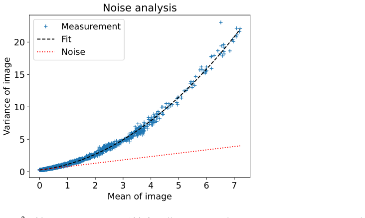
The derivation chain is self-contained in the relevant sense. The target quantity E(g) is estimated by weighted least-squares minimization of the loss (Eq. 1) against the measured multi-PEF signal, using the forward model S(t)=eta(t)|aE(g_A)+bE(g_B)|^{2N}+S_bg (Eq. 8). E(g) is the fitted quantity, not an input derived from itself, so no prediction is being claimed from a quantity that was defined in terms of it. The photobleaching factor eta(t) is estimated from the g_A=g_B subset (flat-wavefront measurements, Eq. 6) and then applied as a nuisance correction; although Eq. 5 uses the measured signal integral, this does not define E(g) in terms of itself. The noise model (Eq. 3) and nonlinear order N are also fitted nuisance parameters rather than predictions. The external Twyman-Green interferometer comparison is independent of the inline fit, and the agreement in response shape is genuine supporting evidence. Self-citations (OpenWFS, the prior setup schematic) are used for software and experimental details, not as load-bearing justification. The uniformity assumption E(g) uniform over the SLM is an untested limitation, and the absolute phase and overall amplitude of E(g) are gauge-ambiguous, but these are identifiability and accuracy concerns, not circularity. No circular step is exhibited.
Assumptions & free parameters
free parameters (7)
- E(g) complex response (256 gray values) =
256 complex values, normalized to mean amplitude 1
- a, b (complex transmission coefficients) =
two complex numbers
- S_bg (background signal) =
scalar (not specified)
- N (nonlinear order) =
near 2, slightly lower
- S0 (unbleached peak signal) =
scalar
- P (photobleaching rate) =
scalar
- Noise model coefficients (sigma_r^2, c_s, c_t) =
sigma_r^2=0.27, c_s=0.52, c_t=0.35
assumptions (6)
- domain assumption SLM field response E(g) is uniform across the SLM
- domain assumption SLM is conjugated to the back pupil of the objective
- domain assumption Two-photon signal is modeled as S = eta(t) |a E_A + b E_B|^(2N) + S_bg
- domain assumption Photobleaching efficiency factor eta(t) = exp(-P ∫ S dt)
- domain assumption Phase-only SLM has negligible amplitude modulation for flat-wavefront points
- domain assumption Noise variance is the sum of read noise and shot noise
Cite this review
Pith. "Pith review of Inline calibration of spatial light modulators in nonlinear microscopy." pith.science (2026). https://pith.science/paper/DBKPMCG4
@misc{pith2026250522482,
  author       = {Pith},
  title        = {Pith review of: Inline calibration of spatial light modulators in nonlinear microscopy},
  year         = {2026},
  howpublished = {\url{https://pith.science/paper/DBKPMCG4}},
  note         = {Machine review of arXiv:2505.22482}
}
read the original abstract
We present a method for calibrating the response of a phase-only spatial light modulator in nonlinear microscopy. Our method uses the microscope image itself as calibration measurement and requires no additional hardware components. Our method is adapted to the nonlinear signals encountered in multi-photon excitation fluorescence microscopes, and works well even under low light conditions and with strong photobleaching.
Figures


Figures from the paper (3 more)


Reference graph
Works this paper leans on
-
[1]
A review of liquid crystal spatial light modulators: Devices and applications
Yiqian Yang, Andrew Forbes, Liangcai Cao, Department of Precision Instruments, Tsinghua University, Beijing 100084, China , and School of Physics, University of the Witwatersrand, Wits, South Africa . A review of liquid crystal spatial light modulators: Devices and applications. Opto-Electronic Science , 2(8):230026--230026, 2023. http://www.oejournal.org...
arXiv 2023
-
[2]
C. Maurer, A. Jesacher, S. Bernet, and M. Ritsch-Marte . What spatial light modulators can do for optical microscopy. Laser & Photonics Reviews , 5(1):81--101, 2011. https://onlinelibrary.wiley.com/doi/abs/10.1002/lpor.200900047
-
[3]
Wavefront Shaping for Biomedical Imaging
Joel Kubby, Sylvain Gigan, and Meng Cui. Wavefront Shaping for Biomedical Imaging . Cambridge University Press, June 2019
work page 2019
-
[4]
Cox, Valeria Rodr \'i guez-Fajardo , and Andrew Forbes
Jonathan Pinnell, Isaac Nape, Bereneice Sephton, Mitchell A. Cox, Valeria Rodr \'i guez-Fajardo , and Andrew Forbes. Modal analysis of structured light with spatial light modulators: A practical tutorial. JOSA A , 37(11):C146--C160, November 2020. https://opg.optica.org/josaa/abstract.cfm?uri=josaa-37-11-C146
work page 2020
-
[5]
Applications of Spatial Light Modulators in Raman Spectroscopy
Faris Sinjab, Zhiyu Liao, and Ioan Notingher. Applications of Spatial Light Modulators in Raman Spectroscopy . Applied Spectroscopy , 73(7):727--746, July 2019. https://doi.org/10.1177/0003702819834575
-
[6]
Advanced optical trapping by complex beam shaping
Mike Woerdemann, Christina Alpmann, Michael Esseling, and Cornelia Denz. Advanced optical trapping by complex beam shaping. Laser & Photonics Reviews , 7(6):839--854, 2013. https://onlinelibrary.wiley.com/doi/abs/10.1002/lpor.201200058
-
[7]
Abderrahmen Trichili, Ki-Hong Park, Mourad Zghal, Boon S. Ooi, and Mohamed-Slim Alouini. Communicating Using Spatial Mode Multiplexing : Potentials , Challenges , and Perspectives . IEEE Communications Surveys & Tutorials , 21(4):3175--3203, 2019. https://ieeexplore.ieee.org/abstract/document/8710274
-
[8]
Focusing and compression of ultrashort pulses through scattering media
Ori Katz, Eran Small, Yaron Bromberg, and Yaron Silberberg. Focusing and compression of ultrashort pulses through scattering media. Nature Photonics , 5(6):372--377, June 2011. https://www.nature.com/articles/nphoton.2011.72
work page 2011
Show all 50 references
-
[9]
Germain, and Meng Cui
Jianyong Tang, Ronald N. Germain, and Meng Cui. Superpenetration optical microscopy by iterative multiphoton adaptive compensation technique. Proceedings of the National Academy of Sciences , 109(22):8434--8439, May 2012. https://www.pnas.org/doi/abs/10.1073/pnas.1119590109
2012 doi
-
[10]
Progress in Phase Calibration for Liquid Crystal Spatial Light Modulators
Rujia Li and Liangcai Cao. Progress in Phase Calibration for Liquid Crystal Spatial Light Modulators . Applied Sciences , 9(10):2012, January 2019. https://www.mdpi.com/2076-3417/9/10/2012
2012
-
[11]
High- Precision Calibration of Phase-Only Spatial Light Modulators
Yicheng Zhao, Wenxiang Yan, Yuan Gao, Zheng Yuan, Zhi-Cheng Ren, Xi-Lin Wang, Jianping Ding, and Hui-Tian Wang. High- Precision Calibration of Phase-Only Spatial Light Modulators . IEEE Photonics Journal , 14(1):1--8, February 2022. https://ieeexplore.ieee.org/document/9623471
2022
-
[12]
Xiaodong Xun and Robert W. Cohn. Phase calibration of spatially nonuniform spatial light modulators. Applied Optics , 43(35):6400--6406, December 2004. https://opg.optica.org/ao/abstract.cfm?uri=ao-43-35-6400
2004
-
[13]
Evaluation of phase-only liquid crystal spatial light modulator for phase modulation performance using a Twyman -- Green interferometer
Hongxin Zhang, Jian Zhang, and Liying Wu. Evaluation of phase-only liquid crystal spatial light modulator for phase modulation performance using a Twyman -- Green interferometer. Measurement Science and Technology , 18(6):1724, May 2007. https://dx.doi.org/10.1088/0957-0233/18/6/S09
2007 doi
-
[14]
Polarization phase shifting interferometric technique for phase calibration of a reflective phase spatial light modulator
Somparna Mukhopadhyay, Sanjukta Sarkar, Kallol Bhattacharya, and Lakshminarayan Hazra. Polarization phase shifting interferometric technique for phase calibration of a reflective phase spatial light modulator. Optical Engineering , 52(3):035602, March 2013. https://www.spiedig...
2013 doi
-
[15]
A compensation method for the full phase retardance nonuniformity in phase-only liquid crystal on silicon spatial light modulators
Long Teng, Mike Pivnenko, Brian Robertson, Rong Zhang, and Daping Chu. A compensation method for the full phase retardance nonuniformity in phase-only liquid crystal on silicon spatial light modulators. Optics Express , 22(21):26392--26402, October 2014. https://opg.optica.org...
2014
-
[16]
Yuanyuan Dai, Jacopo Antonello, and Martin J. Booth. Calibration of a phase-only spatial light modulator for both phase and retardance modulation. Optics Express , 27(13):17912--17926, June 2019. https://opg.optica.org/oe/abstract.cfm?uri=oe-27-13-17912
2019
-
[17]
Simple and fast calibration method for phase-only spatial light modulators
Minchol Lee, Donghoon Koo, and Jeongmin Kim. Simple and fast calibration method for phase-only spatial light modulators. Optics Letters , 48(1):5--8, January 2023. https://opg.optica.org/ol/abstract.cfm?uri=ol-48-1-5
2023
-
[18]
Improved method to fully compensate the spatial phase nonuniformity of LCoS devices with a Fizeau interferometer
Qiang Lu, Lei Sheng, Fei Zeng, Shijie Gao, and Yanfeng Qiao. Improved method to fully compensate the spatial phase nonuniformity of LCoS devices with a Fizeau interferometer. Applied Optics , 55(28):7796--7802, October 2016. https://opg.optica.org/ao/abstract.cfm?uri=ao-55-28-7796
2016
-
[19]
Zheng Zhang, Guowen Lu, and Francis T. S. Yu. Simple method for measuring phase modulation in liquid crystal televisions. Optical Engineering , 33(9):3018--3022, September 1994. https://www.spiedigitallibrary.org/journals/optical-engineering/volume-33/issue-9/0000/Simple-metho...
1994 doi
-
[20]
Arsenault, and Michel Doucet
Alain Bergeron, Jonny Gauvin, Fran c ois Gagnon, Denis Gingras, Henri H. Arsenault, and Michel Doucet. Phase calibration and applications of a liquid-crystal spatial light modulator. Applied Optics , 34(23):5133--5139, August 1995. https://opg.optica.org/ao/abstract.cfm?uri=ao...
1995
-
[21]
Alfonso Serrano-Heredia , Guowen Lu, Purwadi Purwosumarto, and Francis T. S. Yu. Measurement of the phase modulation in liquid crystal television based on the fractional- Talbot effect. Optical Engineering , 35(9):2680--2684, September 1996. https://www.spiedigitallibrary.org/...
1996 doi
-
[22]
Crossland, and Daping Chu
Zichen Zhang, Haining Yang, Brian Robertson, Maura Redmond, Mike Pivnenko, Neil Collings, William A. Crossland, and Daping Chu. Diffraction based phase compensation method for phase-only liquid crystal on silicon devices in operation. Applied Optics , 51(17):3837--3846, June 2...
2012
-
[23]
o m, Martin Persson, J \
David Engstr \"o m, Martin Persson, J \"o rgen Bengtsson, and Mattias Goks \"o r. Calibration of spatial light modulators suffering from spatially varying phase response. Optics Express , 21(13):16086--16103, July 2013. https://opg.optica.org/oe/abstract.cfm?uri=oe-21-13-16086
2013
-
[24]
Spatially resolved phase-response calibration of liquid-crystal-based spatial light modulators
Stephan Reichelt. Spatially resolved phase-response calibration of liquid-crystal-based spatial light modulators. Applied Optics , 52(12):2610--2618, April 2013. https://opg.optica.org/ao/abstract.cfm?uri=ao-52-12-2610
2013
-
[25]
Fern \'a ndez, Pedro M
Jos \'e Luis Mart \'i nez Fuentes, Enrique J. Fern \'a ndez, Pedro M. Prieto, and Pablo Artal. Interferometric method for phase calibration in liquid crystal spatial light modulators using a self-generated diffraction-grating. Optics Express , 24(13):14159--14171, June 2016. h...
2016
-
[26]
An interferometric method for local phase modulation calibration of LC-SLM using self-generated phase grating
Zixin Zhao, Zhaoxian Xiao, Yiying Zhuang, Hangying Zhang, and Hong Zhao. An interferometric method for local phase modulation calibration of LC-SLM using self-generated phase grating. Review of Scientific Instruments , 89(8):083116, August 2018. https://doi.org/10.1063/1.5031938
2018 doi
-
[27]
Self-referenced multiple-beam interferometric method for robust phase calibration of spatial light modulator
Yunhui Gao, Rujia Li, and Liangcai Cao. Self-referenced multiple-beam interferometric method for robust phase calibration of spatial light modulator. Optics Express , 27(23):34463--34471, November 2019. https://opg.optica.org/oe/abstract.cfm?uri=oe-27-23-34463
2019
-
[28]
Remulla and Nathaniel Hermosa
Katherine Isabel T. Remulla and Nathaniel Hermosa. Spatial light modulator phase calibration based on spatial mode projection. Applied Optics , 58(21):5624--5630, July 2019. https://opg.optica.org/ao/abstract.cfm?uri=ao-58-21-5624
2019
-
[29]
Two- Shot Calibration Method for Phase-Only Spatial Light Modulators with Generalized Spatial Differentiator
Junyi Huang, Tengfeng Zhu, and Zhichao Ruan. Two- Shot Calibration Method for Phase-Only Spatial Light Modulators with Generalized Spatial Differentiator . Physical Review Applied , 14(5):054040, November 2020. https://link.aps.org/doi/10.1103/PhysRevApplied.14.054040
2020 doi
-
[30]
Absolutely interferometric calibration of phase liquid crystal spatial light modulators using honeycomb gratings composited with Billet-split Fresnel zone plates
Chi Wang, Jian Shan, and Junyong Zhang. Absolutely interferometric calibration of phase liquid crystal spatial light modulators using honeycomb gratings composited with Billet-split Fresnel zone plates. Applied Optics , 63(4):1105--1109, February 2024. https://opg.optica.org/a...
2024
-
[31]
Calibration of liquid crystal spatial light modulators by using the double-phase method
Luis Ord \'o \ n ez, Erick Ipus, and Omel Mendoza-Yero . Calibration of liquid crystal spatial light modulators by using the double-phase method. Applied Optics , 63(36):9232, December 2024. https://opg.optica.org/abstract.cfm?URI=ao-63-36-9232
2024
-
[32]
Data Fitting and Uncertainty: A Practical Introduction to Weighted Least Squares and Beyond
Tilo Strutz. Data Fitting and Uncertainty: A Practical Introduction to Weighted Least Squares and Beyond . Vieweg + Teubner, Wiesbaden, 1. aufl edition, 2011
2011
-
[33]
Janesick
James R. Janesick. Photon Transfer: DN -- > [Lambda] . Number PM170 in SPIE Press Monograph. SPIE, Bellingham, Wash. < 1000 20th St. Bellingham WA 98225-6705 USA > , 2007
2007
-
[34]
Martin McClain
W. Martin McClain. Two-photon molecular spectroscopy. Accounts of Chemical Research , 7(5):129--135, May 1974. https://doi.org/10.1021/ar50077a001
1974 doi
-
[35]
Reddi, Satyen Kale, and Sanjiv Kumar
Sashank J. Reddi, Satyen Kale, and Sanjiv Kumar. On the Convergence of Adam and Beyond . In International Conference on Learning Representations , February 2018
2018
-
[36]
Lakowicz
Joseph R. Lakowicz. Nonlinear and Two-Photon-Induced Fluorescence . Number Volume 5 in Topics in Fluorescence Spectroscopy . Kluwer Academic Publishers, Boston, MA, 2002
2002
-
[37]
Horton, Ke Wang, Shean-Jen Chen, and Chris Xu
Li-Chung Cheng, Nicholas G. Horton, Ke Wang, Shean-Jen Chen, and Chris Xu. Measurements of multiphoton action cross sections for multiphoton microscopy. Biomedical Optics Express , 5(10):3427--3433, October 2014
2014
-
[38]
Paudel, Dimitre G
David Sinefeld, Hari P. Paudel, Dimitre G. Ouzounov, Thomas G. Bifano, and Chris Xu. Adaptive optics in multiphoton microscopy: Comparison of two, three and four photon fluorescence. Optics Express , 23(24):31472--31483, November 2015. https://www.ncbi.nlm.nih.gov/pmc/articles...
2015
-
[39]
Noninvasive nonlinear focusing and imaging through strongly scattering turbid layers
Ori Katz, Eran Small, Yefeng Guan, and Yaron Silberberg. Noninvasive nonlinear focusing and imaging through strongly scattering turbid layers. Optica , 1(3):170--174, September 2014
2014
-
[40]
Complex Quadratic Optimization and Semidefinite Programming
Shuzhong Zhang and Yongwei Huang. Complex Quadratic Optimization and Semidefinite Programming . SIAM Journal on Optimization , 16(3):871--890, January 2006. https://epubs.siam.org/doi/abs/10.1137/04061341X
2006 doi
-
[41]
Sidiropoulos, Xiao Fu, and Ananthram Swami
John Tranter, Nicholas D. Sidiropoulos, Xiao Fu, and Ananthram Swami. Fast Unit-Modulus Least Squares With Applications in Beamforming . IEEE Transactions on Signal Processing , 65(11):2875--2887, June 2017. http://ieeexplore.ieee.org/document/7849224/
2017
-
[42]
OpenWFS on Github - A framework for conducting and simulating wavefront shaping experiments in Python , July 2023
Ivo M Vellekoop. OpenWFS on Github - A framework for conducting and simulating wavefront shaping experiments in Python , July 2023. https://github.com/IvoVellekoop/openwfs. For this manuscript version 1.0.0 was used
2023
-
[43]
OpenWFS ---a library for conducting and simulating wavefront shaping experiments
Jeroen H Doornbos, Dani \"e l W S Cox, Tom Knop, Harish Sasikumar, and Ivo M Vellekoop. OpenWFS ---a library for conducting and simulating wavefront shaping experiments. Journal of Physics: Photonics , 7(1):015016, January 2025
2025
-
[44]
Inline SLM Calibration - Self-referencing Phase-Response Calibration Method Using Non-Linear Feedback
Daniel W S Cox and Ivo M Vellekoop. Inline SLM Calibration - Self-referencing Phase-Response Calibration Method Using Non-Linear Feedback . https://github.com/dedean16/inline_slm_calibration, February 2025
2025
-
[45]
Dani \"e l Cox, Harish Sasikumar, and I. M. (Ivo) Vellekoop. Raw measurement data of SLM calibration: Inline calibration of spatial light modulators in nonlinear microscopy. https://data.4tu.nl/datasets/e06926cc-5f6e-4fc0-8170-16d71d5b1c1e, March 2025
2025
-
[46]
Dani \"e l W. S. Cox and Ivo M. Vellekoop. Orthonormalization of phase-only basis functions. Optics Express , 33(2):2427--2436, January 2025. https://opg.optica.org/oe/abstract.cfm?uri=oe-33-2-2427
2025
-
[47]
Koester, Dagmar Baur, Rainer Uhl, and Stefan W
Helmut J. Koester, Dagmar Baur, Rainer Uhl, and Stefan W. Hell. Ca2+ Fluorescence Imaging with Pico- and Femtosecond Two-Photon Excitation : Signal and Photodamage . Biophysical Journal , 77(4):2226--2236, October 1999. https://www.cell.com/biophysj/abstract/S0006-3495(99)77063-3
1999
-
[48]
Patterson and David W
George H. Patterson and David W. Piston. Photobleaching in Two-Photon Excitation Microscopy . Biophysical Journal , 78(4):2159--2162, April 2000
2000
-
[49]
Highly Nonlinear Photodamage in Two-Photon Fluorescence Microscopy
Alexander Hopt and Erwin Neher. Highly Nonlinear Photodamage in Two-Photon Fluorescence Microscopy . Biophysical Journal , 80(4):2029--2036, April 2001. https://www.cell.com/biophysj/abstract/S0006-3495(01)76173-5
2001
-
[50]
Cranfill, Brittney R
Paula J. Cranfill, Brittney R. Sell, Michelle A. Baird, John R. Allen, Zeno Lavagnino, H. Martijn de Gruiter , Gert-Jan Kremers, Michael W. Davidson, Alessandro Ustione, and David W. Piston. Quantitative assessment of fluorescent proteins. Nature Methods , 13(7):557--562, July 2016
2016
Reviewed August 7, 2026 · model on record in the stance chip above.
Discussion (0). Sign in to comment.