REVIEW 5 major objections 5 minor 164 references
ConfLUNet: Multiple sclerosis lesion instance segmentation in presence of confluent lesions
T0 review · 5 major / 5 minor · reviewed 2026-08-07 · deepseek-v4-flash
Pith's one-line read ConfLUNet, the first end-to-end instance segmentation framework for MS lesions, significantly outperforms connected components and automated confluent lesion splitting on the held-out test set in panoptic quality, lesion detection F1, and…
desk verdict A solid, transparent instance-segmentation paper for MS lesions whose main risk is the acknowledged single-rater ground truth, but which deserves peer review. read the letter →
The pith
A machine-rendered reading of the paper's core claim, the machinery that carries it, and where it could break.
The reading
What carries the argument
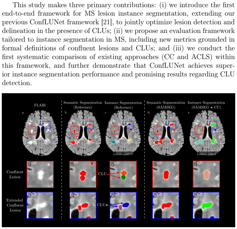
The argument rests on two pieces of machinery. One is formal definition: a confluent lesion is a connected component that overlaps at least two reference lesion instances, and each of those instances is a confluent lesion unit (CLU); dilating the semantic mask before the same test defines extended CLU+, capturing near-touch lesions. These definitions convert a clinically messy phenomenon into countable objects that can be scored. The other is ConfLUNet's architecture, which adds two output heads to a standard self-configuring 3D encoder-decoder segmentation backbone: a heatmap of lesion centers (Gaussian-smoothed centers of mass) and a 3D offset vector field pointing from each voxel to its lesion center. At inference, voxels are displaced by their predicted offsets and assigned to the nearest detected center, so lesion boundaries reflect learned geometric evidence rather than pure spatial connectivity.
What would settle it
Re-annotate the 13 test-set patients with several independent raters who each mark CLU instances inside confluent regions, then recompute ConfLUNet's CLU detection F1 against each rater and against a multi-rater consensus reference; if ConfLUNet's advantage over connected components shrinks or disappears under these alternative references, the reported gains are an artifact of a single annotator's judgment rather than a property of the method.
Extended reading notes
Core claim
On its own terms, the paper's central claim is that an end-to-end model that simultaneously learns what is lesion, where lesion centers are, and which voxel belongs to which center can separate confluent MS lesion units better than deriving instances from a semantic mask by connected components or by statistical splitting. The evidence is the held-out comparison: ConfLUNet reaches Panoptic Quality 42.0% against 37.5% for CC and 36.8% for ACLS (p = 0.017 and 0.005), lesion-wise F1 67.3% against 61.6% and 59.9% (p = 0.028 and 0.013), and CLU F1 81.5%, with statistically significant recall gains over CC (+12.5%) and precision gains over ACLS (+31.2%). The paper also establishes the failure modes of the existing methods: CC systematically undercounts CLUs while ACLS systematically oversplits lesions, and these behaviors hold across three different semantic segmentation tools, supporting the claim that the default evaluation practice is misaligned with clinical needs.
Load-bearing premise
The whole evaluation stands on the single-rater manual annotations being a correct partition of confluent lesions into units, yet the paper itself acknowledges that in large confluent lesions distinguishing individual units is extremely difficult, if not impossible, for human raters.
Editorial extensions
If this is right
- Connected components, the default post-processing across the MS lesion literature, systematically underestimates confluent lesion unit counts and should be reconsidered for instance-level evaluation.
- ACLS overestimates lesion counts and oversplits, so its high recall comes at a precision cost that the new metrics expose.
- ConfLUNet offers a balanced precision-recall operating point, which the authors argue is closer to clinical needs for lesion counting and lesion-level biomarkers.
- The formal CLU and CLU+ definitions and metrics give future work a shared reference for measuring instance segmentation in MS, replacing the current reliance on CC-derived counts.
- Because ConfLUNet needs only a single FLAIR volume, the approach is a practical candidate for clinical pipelines that do not routinely acquire research-grade multi-contrast scans.
Reading between the lines
- If the single-rater ground truth is noisy in exactly the confluent regions the method targets, the reported advantage might change in either direction; a multi-rater study would tell whether ConfLUNet is exploiting signal or label bias.
- The CLU+ dilation-based definition effectively forgives boundary disagreements of one voxel, which suggests the practical near-term role of such a model may be to flag candidate confluent regions and their units for human confirmation.
- The same center-and-offset formulation could be tested on longitudinal data, since the authors note that temporal appearance can disambiguate confluent lesions; that would be a natural and testable extension for CLU separation.
- Reporting patient-wise averages hides the clinical worst case; the lesion-wise analysis in the paper shows CC's CLU recall falling to 41.5%, implying future evaluations should report both patient-wise and lesion-wise CLU metrics.
Editorial analysis
A structured set of objections, weighed in public.
Referee Report
Summary. The paper addresses the problem of instance segmentation of multiple sclerosis (MS) lesions, with a focus on confluent lesions that appear as merged clusters on MRI. The authors formalize definitions of confluent lesions and confluent lesion units (CLUs), propose CLU-aware evaluation metrics, systematically compare two existing post-processing approaches (connected components, CC, and automated confluent lesion splitting, ACLS), and introduce ConfLUNet, an end-to-end 3D instance segmentation network with semantic, center, and offset heads. On a held-out test set of 13 patients, ConfLUNet reportedly achieves significantly higher Panoptic Quality (42.0% vs 37.5%/36.8%) and lesion detection F1 (67.3% vs 61.6%/59.9%) than CC and ACLS, and the highest F1CLU (81.5%). The paper also provides open-source code and a detailed evaluation framework.
Significance. If the results hold, the paper makes a valuable contribution by formalizing the CLU evaluation problem, providing reproducible metrics, and demonstrating that an end-to-end model can achieve a better precision-recall balance than connectivity-based or center-splitting post-processing. The experimental design is careful in several respects: a held-out test set, 5-fold cross-validation, an architecture-matched baseline (3D U-Net with the same backbone), paired Wilcoxon tests, Holm-Bonferroni-corrected analyses, and explicit disclosure of limitations. The release of code and containers supports reproducibility. The main significance is in the evaluation framework and in establishing a concrete baseline for future instance segmentation methods in MS. However, the strength of the central claim is tempered by the heavy reliance on single-rater manual annotations for confluent regions, which the paper itself acknowledges are extremely difficult to label, and by the small test set with very few confluent cases.
major comments (5)
- [Section 4.1.2, Appendix A, Limitations] The evaluation rests entirely on manual instance annotations made by a single neurobiologist and reviewed by a single neurologist. The paper itself states in Appendix A (Fig. A.10) that for large confluent lesions it is 'extremely difficult, if not impossible, to distinguish individual lesion units,' and the Limitations section concedes that 'single-rater annotations may introduce bias.' Because the reference instances define the CLU sets L_CLU and L_CLU+ (Eqs. 3–4) and the center/offset training targets (Section 3.1), all reported metrics (PQ, F1, F1CLU) and the ranking of ConfLUNet versus CC/ACLS inherit this potential bias. Without inter-rater reproducibility data or at least a sensitivity analysis on a subset of images re-annotated by a second rater, the central claim that ConfLUNet better separates true confluent lesion units is not fully established.
- [Section 5.3, Table 5] The paper highlights F1CLU as a key result, but the difference between ConfLUNet (81.5%) and CC (79.8%) is not reported as statistically significant, and the F1CLU+ improvement over CC is marginal (p = 0.046) and may not survive the Holm-Bonferroni correction shown in Appendix D. The CLU-specific comparisons are further limited to the 7 test patients with at least two CLUs (Section 5.3), giving very low statistical power. The claims about improved CLU detection should be softened accordingly, and the exact p-values (including non-significant ones) for the F1CLU comparisons should be reported.
- [Section 4.1.1, Section 5.3] The test set is small (n=13, with only 7 patients exhibiting at least two CLUs), and the two patients with the most extreme confluency were excluded by design because their confluent lesions were deemed indiscernible. Excluding the cases where the ground truth is most ambiguous removes exactly the cases most relevant to the method's intended use case, which narrows the external validity of the reported gains. The authors acknowledge this in the Limitations, but they do not quantify how the conclusions would change if those two patients were included (even with their uncertain labels). Reporting performance on those two patients as a sensitivity analysis would help assess robustness.
- [Abstract, Section 3, Reference [21]] The abstract and Section 3 describe ConfLUNet as 'the first end-to-end instance segmentation framework for MS lesions,' but the authors' own prior work, reference [21] (ISBI 2024), already proposes ConfLUNet for confluent lesion identification. The present manuscript should explicitly state the architectural and methodological differences from [21] and adjust the novelty claim accordingly. As written, the 'first' claim is at odds with the cited prior work and could be misleading to readers.
- [Section 4.2, FPCLU definition] The construction of FPCLU (predicted lesions that match a reference lesion but are not the best match for that reference) implies that any method that never oversplits, such as CC, will automatically achieve PrecisionCLU = 100% regardless of how many reference CLUs it merges. Table 5 confirms that CC reaches 100% PrecisionCLU. As a result, F1CLU comparisons between CC and splitting methods are essentially recall-only comparisons, and the proposed metric does not directly penalize the merging failure mode that motivates the paper. The authors should justify this asymmetry or introduce an additional metric that penalizes merging explicitly.
minor comments (5)
- [Abstract] There is a formatting error in the abstract: 'textF 1CLU' should be 'F1CLU'.
- [Throughout] The capitalization of the method name is inconsistent: the text uses both 'ConfLUNet' and 'ConfLUnet' (e.g., Figure D.12), and 'ConfLUnet' appears in some figures and tables. The authors should standardize to one spelling.
- [Section 4.3, Reference list] The text cites 'SAMSEG [114]' for the SAMSEG tool, but the main reference list contains only [1]–[47]; [114] appears in the Appendix E reference list (Cerri et al.). This citation mismatch should be fixed so that the main text uses the correct reference number.
- [Section 2.1, References] The ACLS description cites '[20, 120]' and 'our previous work [29]', but [120] is only in the Appendix E bibliography, not in the main reference list. The authors should ensure all citations in the main text point to the main reference list or move the relevant entries.
- [Section 4.2] The matching procedure uses a low IoU threshold (λ = 0.1) and mutual-best matching. The authors should briefly justify why this threshold is appropriate for 3D lesions and discuss its impact on reported PQ and F1 values, given that small spatial errors can cause a predicted lesion to fall below the threshold.
Circularity Check
No significant circularity: ConfLUNet's performance claims rest on held-out test comparisons, and the CLU definitions/metrics are evaluation instruments rather than fitted inputs.
full rationale
The paper's central claims are empirical: ConfLUNet is trained on 50 patients and evaluated on a held-out set of 13 patients against CC and ACLS, with PQ, F1, and CLU-aware metrics computed from test-set predictions. No equation in the paper reduces a reported prediction to the input by construction. The reference centers used to supervise the center/offset heads (Section 3.1) are derived from the manual instance annotations, but this is standard supervised training; the held-out split means the reported numbers are not fitted. The CLU and CLU+ definitions (Eqs. 2-4) are used to define evaluation sets and metrics, not to generate ConfLUNet's outputs, and validation-based model selection used panoptic quality (Section 3.2), not CLU-specific metrics, so no target metric was directly optimized. Self-citations to the authors' prior ConfLUNet work [21] and ISMRM comparison [29] are motivational and are re-validated on new data here; no load-bearing argument depends on an unverified self-cited theorem. The acknowledged limitation of single-rater annotations (Section 6, Appendix A) is a validity and generalizability concern about the ground truth, not a circularity in the derivation chain: even if the labels are imperfect, the methods are compared against the same labels in an externally falsifiable way. No claim reduces to its own assumptions by definition or by self-citation, so the appropriate circularity score is 0.
Assumptions & free parameters
free parameters (7)
- Matching IoU threshold lambda =
0.1
- Semantic probability threshold =
0.5
- Size filter for predicted lesions =
<3mm along any axis or <14mm3
- Dilation iterations for CLU+ definition =
1
- Center heatmap Gaussian sigma =
2 voxels
- Loss weights alpha and beta =
alpha=100, beta=1
- Connectivity structures for CC and ACLS =
6-connectivity for CC; 26-connectivity for ACLS center detection
assumptions (5)
- domain assumption Manual instance annotations are a reliable ground truth for CLUs despite the difficulty of separating confluent lesions.
- domain assumption CLUs are visible in a single cross-sectional FLAIR image (with MPRAGE/EPI consultation for raters); temporal information is not required.
- domain assumption Each lesion has a well-defined center of mass that serves as the instance anchor; learning offset vectors to this center is sufficient to separate confluent lesions.
- domain assumption Connected component analysis on reference semantic masks (Eq. 2) and the dilation-based CLU+ definition capture the clinically meaningful notion of confluence.
- standard math Standard connected components, Gaussian filtering, and IoU computations behave as expected.
Cite this review
Pith. "Pith review of ConfLUNet: Multiple sclerosis lesion instance segmentation in presence of confluent lesions." pith.science (2026). https://pith.science/paper/MKGONOT3
@misc{pith2026250522537,
  author       = {Pith},
  title        = {Pith review of: ConfLUNet: Multiple sclerosis lesion instance segmentation in presence of confluent lesions},
  year         = {2026},
  howpublished = {\url{https://pith.science/paper/MKGONOT3}},
  note         = {Machine review of arXiv:2505.22537}
}
read the original abstract
Accurate lesion-level segmentation on MRI is critical for multiple sclerosis (MS) diagnosis, prognosis, and disease monitoring. However, current evaluation practices largely rely on semantic segmentation post-processed with connected components (CC), which cannot separate confluent lesions (aggregates of confluent lesion units, CLUs) due to reliance on spatial connectivity. To address this misalignment with clinical needs, we introduce formal definitions of CLUs and associated CLU-aware detection metrics, and include them in an exhaustive instance segmentation evaluation framework. Within this framework, we systematically evaluate CC and post-processing-based Automated Confluent Splitting (ACLS), the only existing methods for lesion instance segmentation in MS. Our analysis reveals that CC consistently underestimates CLU counts, while ACLS tends to oversplit lesions, leading to overestimated lesion counts and reduced precision. To overcome these limitations, we propose ConfLUNet, the first end-to-end instance segmentation framework for MS lesions. ConfLUNet jointly optimizes lesion detection and delineation from a single FLAIR image. Trained on 50 patients, ConfLUNet significantly outperforms CC and ACLS on the held-out test set (n=13) in instance segmentation (Panoptic Quality: 42.0% vs. 37.5%/36.8%; p = 0.017/0.005) and lesion detection (F1: 67.3% vs. 61.6%/59.9%; p = 0.028/0.013). For CLU detection, ConfLUNet achieves the highest F1[CLU] (81.5%), improving recall over CC (+12.5%, p = 0.015) and precision over ACLS (+31.2%, p = 0.003). By combining rigorous definitions, new CLU-aware metrics, a reproducible evaluation framework, and the first dedicated end-to-end model, this work lays the foundation for lesion instance segmentation in MS.
Figures


Figures from the paper (6 more)


Reference graph
Works this paper leans on
-
[21]
M. Wynen, M. Istasse, P. M. Gordaliza, A. Stölting, P. Maggi, B. Macq, M. B. Cuadra, Conflunet: Improving Confluent Lesion Identification In Multiple Sclerosis With Instance Segmentation, in: 2024 IEEE Interna- tional Symposium on Biomedical Imaging (ISBI), 2024, pp. 1–5, iSSN: 1945-8452. doi:10.1109/ISBI56570.2024.10635707. URL https://ieeexplore.ieee.or...
arXiv 2024
-
[1]
D. S. Reich, C. F. Lucchinetti, P. A. Calabresi, Multiple Sclerosis, The New England Journal of Medicine 378 (2) (2018) 169–180. doi:10. 1056/NEJMra1401483
2018
-
[2]
A. J. Thompson, et al., Diagnosis of multiple sclerosis: 2017 revisions of the McDonald criteria, The Lancet. Neurology 17 (2) (2018) 162–173. doi:10.1016/S1474-4422(17)30470-2
-
[3]
on behalf of the MAGNIMS study group, MAGNIMS consensus guidelines on the use of MRI in multiple sclerosis—establishing disease prognosis and monitoring patients, Nature Reviews Neurology 11 (10) (2015) 597–606. doi:10.1038/nrneurol.2015.157. URL https://www.nature.com/articles/nrneurol.2015.157
-
[4]
P. A. Brex, O. Ciccarelli, J. I. O’Riordan, M. Sailer, A. J. Thompson, D. H. Miller, A longitudinal study of abnormalities on MRI and dis- ability from multiple sclerosis, The New England Journal of Medicine 346 (3) (2002) 158–164.doi:10.1056/NEJMoa011341
-
[5]
S. J. Khoury, C. R. Guttmann, E. J. Orav, M. J. Hohol, S. S. Ahn, L. Hsu, R. Kikinis, G. A. Mackin, F. A. Jolesz, H. L. Weiner, Longitud- inal MRI in multiple sclerosis: correlation between disability and lesion burden, Neurology 44 (11) (1994) 2120–2124. doi:10.1212/wnl.44. 11.2120
-
[6]
R. A. Rudick, J.-C. Lee, J. Simon, E. Fisher, Significance of T2 lesions in multiple sclerosis: A 13-year longitudinal study, Annals of Neurology 60 (2) (2006) 236–242.doi:10.1002/ana.20883. 27
-
[7]
Montalban, Revised McDonald criteria 2023, in: 2024 40th Con- gress of the European Committe for Treatment and Research in Multiple Sclerosis (ECTRIMS 2024), 2024, pp
X. Montalban, Revised McDonald criteria 2023, in: 2024 40th Con- gress of the European Committe for Treatment and Research in Multiple Sclerosis (ECTRIMS 2024), 2024, pp. 0–0
2023
Show all 164 references
-
[8]
Maggi, M
P. Maggi, M. Absinta, M. Grammatico, L. Vuolo, G. Emmi, G. Carlucci, G. Spagni, A. Barilaro, A. M. Repice, L. Emmi, D. Prisco, V. Mar- tinelli, R. Scotti, N. Sadeghi, G. Perrotta, P. Sati, B. Dachy, D. S. Reich, M. Filippi, L. Massacesi, Central vein sign differentiates Multip...
2018 doi
-
[9]
Absinta, P
M. Absinta, P. Sati, F. Masuzzo, G. Nair, V. Sethi, H. Kolb, J. Ohayon, T. Wu, I. C. M. Cortese, D. S. Reich, Association of Chronic Active Multiple Sclerosis Lesions With Disability In Vivo, JAMA neurology 76 (12) (2019) 1474–1483.doi:10.1001/jamaneurol.2019.2399
2019
-
[10]
Elliott, J
C. Elliott, J. S. Wolinsky, S. L. Hauser, L. Kappos, F. Barkhof, C. Bernasconi, W. Wei, S. Belachew, D. L. Arnold, Slowly ex- panding/evolving lesions as a magnetic resonance imaging marker of chronic active multiple sclerosis lesions, Multiple Sclerosis (Hound- mills, Basings...
2019
-
[11]
La Rosa, M
F. La Rosa, M. Wynen, O. Al-Louzi, E. S. Beck, T. Huelnhagen, P. Maggi, J.-P. Thiran, T. Kober, R. T. Shinohara, P. Sati, others, Cor- tical lesions, central vein sign, and paramagnetic rim lesions in multiple sclerosis: Emerging machine learning techniques and future avenues,...
2022
-
[12]
G. Barquero, et al., RimNet: A deep 3D multimodal MRI architecture for paramagnetic rim lesion assessment in multiple sclerosis, NeuroIm- age: Clinical 28 (2020) 102412.doi:10.1016/j.nicl.2020.102412. URL http://www.sciencedirect.com/science/article/pii/ S2213158220302497
2020
-
[13]
Maggi, M
P. Maggi, M. J. Fartaria, J. Jorge, F. L. Rosa, M. Absinta, P. Sati, R. Meuli, R. D. Pasquier, D. S. Reich, M. B. Cuadra, C. Granziera, J. Richiardi, T. Kober, CVSnet: A machine learning approach for automated central vein sign assessment in multiple sclerosis, NMR in Biomedic...
2020 doi
-
[15]
Zhang, T
H. Zhang, T. D. Nguyen, J. Zhang, M. Marcille, P. Spincemaille, Y. Wang, S. A. Gauthier, E. M. Sweeney, QSMRim-Net: Imbalance- aware learning for identification of chronic active multiple sclerosis le- sions on quantitative susceptibility maps, NeuroImage : Clinical 34 (2022) ...
2022
-
[16]
Wynen, F
M. Wynen, F. La Rosa, A. Sellimi, G. Barquero, G. Perrotta, V. Lolli, V. Van Pesch, C. Granziera, T. Kober, P. Sati, others, Longitudinal automated assessment of paramagnetic rim lesions in multiple sclerosis using RimNet, in: ISMRM, 2021, pp. 0–0
2021
-
[17]
Zivadinov, M
R. Zivadinov, M. Zorzon, R. De Masi, D. Nasuelli, G. Cazzato, Effect of intravenous methylprednisolone on the number, size and confluence of plaques in relapsing–remitting multiple sclerosis, Journal of the Neurolo- gical Sciences 267 (1) (2008) 28–35.doi:10.1016/j.jns.2007.09...
2008 doi
-
[18]
Lassmann, Multiple sclerosis: Lessons from molecular neuropathology, Experimental Neurology 262 (2014) 2–7
H. Lassmann, Multiple sclerosis: Lessons from molecular neuropathology, Experimental Neurology 262 (2014) 2–7. doi:10.1016/j.expneurol.2013.12.003. URL https://www.sciencedirect.com/science/article/pii/ S0014488613003610
2014 doi
-
[19]
J. O. Harris, J. A. Frank, N. Patronas, D. E. McFarlin, H. F. McFar- land, Serial gadolinium-enhanced magnetic resonance imaging scans in patients with early, relapsing-remitting multiple sclerosis: implications 29 for clinical trials and natural history, Annals of Neurology 2...
1991 doi
-
[20]
J. D. Dworkin, et al., An Automated Statistical Technique for Counting Distinct Multiple Sclerosis Lesions, American Journal of Neuroradiology 39 (4) (2018) 626–633, publisher: American Journal of Neuroradiology Section: ADULT BRAIN. doi:10.3174/ajnr.A5556. URL http://www.ajnr...
2018 doi
-
[22]
A. M. Hafiz, G. M. Bhat, A survey on instance segmentation: state of the art, International Journal of Multimedia Information Retrieval 9 (3) (2020) 171–189. doi:10.1007/s13735-020-00195-x. URL https://doi.org/10.1007/s13735-020-00195-x
2020 doi
-
[23]
W. Gu, S. Bai, L. Kong, A review on 2D instance segmentation based on deep neural networks, Image and Vision Computing 120 (2022) 104401. doi:10.1016/j.imavis.2022.104401. URL https://www.sciencedirect.com/science/article/pii/ S0262885622000300
2022
-
[24]
Cheng, Panoptic-DeepLab: A Simple, Strong, and Fast Baseline for Bottom-Up Panoptic Segmentation, arXiv:1911.10194 [cs] (Mar
B. Cheng, Panoptic-DeepLab: A Simple, Strong, and Fast Baseline for Bottom-Up Panoptic Segmentation, arXiv:1911.10194 [cs] (Mar. 2020). URL http://arxiv.org/abs/1911.10194
1911 arXiv
-
[25]
K. He, X. Chen, S. Xie, Y. Li, P. Dollár, R. Girshick, Masked Au- toencoders Are Scalable Vision Learners, arXiv:2111.06377 [cs]ArXiv: 2111.06377 version: 1 (Nov. 2021). URL http://arxiv.org/abs/2111.06377
2021 arXiv
-
[26]
E. S. Nasir, A. Parvaiz, M. M. Fraz, Nuclei and glands instance seg- mentation in histology images: a narrative review, Artificial Intelligence Review 56 (8) (2023) 7909–7964.doi:10.1007/s10462-022-10372-5. URL https://doi.org/10.1007/s10462-022-10372-5 30
2023 doi
-
[27]
Isensee, P
F. Isensee, P. F. Jaeger, S. A. A. Kohl, J. Petersen, K. H. Maier- Hein, nnU-Net: a self-configuring method for deep learning-based biomedical image segmentation, Nature Methods 18 (2) (2021) 203– 211, number: 2 Publisher: Nature Publishing Group. doi:10.1038/ s41592-020-01008...
2021
-
[28]
Bilic, P
P. Bilic, P. Christ, H. B. Li, E. Vorontsov, A. Ben-Cohen, G. Kaissis, A. Szeskin, C. Jacobs, G. E. H. Mamani, G. Chartrand, F. Lohöfer, J. W. Holch, W. Sommer, F. Hofmann, A. Hostettler, N. Lev-Cohain, M. Drozdzal, M. M. Amitai, R. Vivanti, J. Sosna, I. Ezhov, A. Sekuboy- ina...
2023
-
[29]
Wynen, P
M. Wynen, P. M. Gordaliza, A. Stölting, P. Maggi, M. Bach Cuadra, Lesion Instance Segmentation in Multiple Sclerosis: Assessing the Efficacy of Statistical Lesion Splitting, in: ISMRM, ISMRM, 2024, pp. 0–0. URL https://dial.uclouvain.be/pr/boreal/object/boreal: 285623
2024
-
[30]
Malinin, et al., Shifts 2.0: Extending The Dataset of Real Distribu- 31 tional Shifts, arXiv:2206.15407 [cs, stat] (Sep
A. Malinin, et al., Shifts 2.0: Extending The Dataset of Real Distribu- 31 tional Shifts, arXiv:2206.15407 [cs, stat] (Sep. 2022). URL http://arxiv.org/abs/2206.15407
2022 arXiv
- [31]
-
[32]
Grahl, V
S. Grahl, V. Pongratz, P. Schmidt, C. Engl, M. Bussas, A. Radetz, G. Gonzalez-Escamilla, S. Groppa, F. Zipp, C. Lukas, J. Kirschke, C. Zi- mmer, M. Hoshi, A. Berthele, B. Hemmer, M. Mühlau, Evidence for a white matter lesion size threshold to support the diagnosis of relapsing...
2019 doi
-
[33]
B. B. Avants, C. L. Epstein, M. Grossman, J. C. Gee, Symmetric diffeo- morphicimageregistrationwithcross-correlation: evaluatingautomated labeling of elderly and neurodegenerative brain, Medical Image Analysis 12 (1) (2008) 26–41.doi:10.1016/j.media.2007.06.004
2008 doi
-
[34]
Hoopes, J
A. Hoopes, J. S. Mora, A. V. Dalca, B. Fischl, M. Hoffmann, SynthStrip: skull-stripping for any brain image, NeuroImage 260 (2022) 119474. doi:10.1016/j.neuroimage.2022.119474. URL https://www.sciencedirect.com/science/article/pii/ S1053811922005900
2022
-
[35]
Vanden Bulcke, M
C. Vanden Bulcke, M. Wynen, J. Detobel, F. La Rosa, M. Absinta, L. Dricot, B. Macq, M. B. Cuadra, P. Maggi, BMAT: An open-source BIDS managing and analysis tool, NeuroImage: Clinical 36 (2022) 103252, publisher: Elsevier
2022
-
[36]
P. A. Yushkevich, Y. Gao, G. Gerig, ITK-SNAP: an interactive tool for semi-automaticsegmentationofmulti-modalitybiomedicalimages, Con- ference proceedings : ... Annual International Conference of the IEEE EngineeringinMedicineandBiologySociety.IEEEEngineeringinMedi- cine and B...
2016
-
[37]
Maier-Hein, A
L. Maier-Hein, A. Reinke, P. Godau, M. D. Tizabi, F. Buettner, E. Christodoulou, B. Glocker, F. Isensee, J. Kleesiek, M. Kozubek, 32 M. Reyes, M. A. Riegler, M. Wiesenfarth, A. E. Kavur, C. H. Sudre, M. Baumgartner, M. Eisenmann, D. Heckmann-Nötzel, T. Rädsch, L. Acion, M. Ant...
2024
-
[38]
Raina, N
V. Raina, N. Molchanova, M. Graziani, A. Malinin, H. Muller, M. B. Cuadra, M. Gales, Tackling Bias in the Dice Similarity Coefficient: In- troducing NDSC for White Matter Lesion Segmentation, in: 2023 IEEE 20th International Symposium on Biomedical Imaging (ISBI), 2023, pp. 1–...
2023
-
[39]
Schmidt, Bayesian inference for structured additive regression mod- els for large-scale problems with applications to medical imaging, Ph.D
P. Schmidt, Bayesian inference for structured additive regression mod- els for large-scale problems with applications to medical imaging, Ph.D. thesis, Ludwig-Maximilians-Universität München, medium: applica- tion/pdf (2017). doi:10.5282/EDOC.20373. URL https://edoc.ub.uni-mue...
2017 doi
-
[40]
Dereskewicz, F
E. Dereskewicz, F. L. Rosa, J. d. S. Silva, E. Sizer, A. Kohli, M. Wynen, W. A. Mullins, P. Maggi, S. Levy, K. Onyemeh, B. Ayci, A. J. Solomon, J. Assländer, O. Al-Louzi, D. S. Reich, J. Sumowski, E. S. Beck, FLAMeS: A Robust Deep Learning Model for Automated Multiple Sclerosi...
2025 doi
-
[41]
La Rosa, J
F. La Rosa, J. Dos Santos Silva, W. A. Mullins, H. Greenspan, J. Sumowski, D. S. Reich, E. S. Beck, FLAMeS: FLAIR Lesion Ana- lysis in Multiple Sclerosis (Feb. 2023).doi:10.5281/zenodo.7626121. URL https://zenodo.org/records/7626121
2023
-
[43]
URL https://www.scipy.org/
SciPy.org — SciPy.org (2025). URL https://www.scipy.org/
2025
-
[44]
Wynen, P
M. Wynen, P. M. Gordaliza, A. Stölting, P. Maggi, M. B. Cuadra, B. Macq, Multi-modal segmentation for paramagnetic rim lesion detec- tioninmultiplesclerosis, in: MedicalImaging2024: ImagingInformatics for Healthcare, Research, and Applications, Vol. 12931, SPIE, 2024, pp. 241–246
2024
-
[45]
Maggi, C
P. Maggi, C. Vanden Bulcke, E. Pedrini, C. Bugli, A. Sellimi, M. Wynen, A. Stölting, W. A. Mullins, G. Kalaitzidis, V. Lolli, others, B cell de- pletion therapy does not resolve chronic active multiple sclerosis lesions, EBioMedicine 94, publisher: Elsevier (2023)
2023
-
[46]
Wynen, C
M. Wynen, C. Vanden Bulcke, S. Borrelli, P. M. Gordaliza, A. Stölting, F. Guisset, C. Cordier, M. S. Martire, A. Tamanti, B. Macq, others, Machine Learning-Based Combination of the Central Vein Sign, Cor- tical Lesions and Paramagnetic Rim Lesions: A Web-Based Tool for the Dia...
2025
-
[47]
Multiple Sclerosis
B. Gérin, M. Zanella, M. Wynen, S. Mahmoudi, B. Macq, C. De Vleeschouwer, Exploring viability of test-time training: Applica- tion to 3D segmentation in multiple sclerosis, in: 2024 IEEE Conference on Artificial Intelligence (CAI), IEEE, 2024, pp. 557–562. 34 Appendix A. Illus...
2024
-
[48]
Lesjak, A
v. Lesjak, A. Galimzianova, A. Koren, M. Lukin, F. Pernuš, B. Likar, v. Špiclin, A Novel Public MR Image Dataset of Multiple Sclerosis Patients With Lesion Segmentations Based on Multi-rater Consensus, Neuroinform- atics 16 (1) (2018) 51–63.doi:10.1007/s12021-017-9348-7. URL h...
2018 doi
-
[49]
P. Sati, D. M. Thomasson, N. Li, D. L. Pham, N. M. Biassou, D. S. Reich, J. A. Butman, Rapid, high-resolution, whole-brain, susceptibility-based MRI of multiple sclerosis, Multiple sclerosis (Houndmills, Basingstoke, England) 20 (11) (2014) 1464–1470.doi:10.1177/13524585145258...
2014 doi
-
[50]
Cabezas, A
M. Cabezas, A. Oliver, S. Valverde, B. Beltran, J. Freixenet, J. C. Vilanova, L. Ramió-Torrent`, A. Rovira, X. Lladó, BOOST: A supervised approach for multiple sclerosis lesion segmentation, Journal of Neuroscience Methods 237 (2014) 108–117. doi:10.1016/j.jneumeth.2014.08.024...
2014 doi
-
[51]
J. Gao, C. Li, C. Feng, M. Xie, Y. Yin, C. Davatzikos, Non-locally regularized segmentation of multiple sclerosis lesion from multi-channel MRI data, Magnetic Resonance Imaging 32 (8) (2014) 1058–1066. doi:10.1016/j.mri.2014.03.006. URL https://www.sciencedirect.com/science/ar...
2014 doi
-
[52]
Cabezas, A
M. Cabezas, A. Oliver, E. Roura, J. Freixenet, J. C. Vilanova, L. Ramió- Torrent`, A. Rovira, X. Lladó, Automatic multiple sclerosis lesion detection 44 in brain MRI by FLAIR thresholding, Computer Methods and Programs in Biomedicine 115 (3) (2014) 147–161.doi:10.1016/j.cmpb.2...
2014 doi
-
[53]
S. Roy, A. Carass, J. L. Prince, D. L. Pham, Subject Specific Sparse Dic- tionary Learning for Atlas based Brain MRI Segmentation, Machine learn- ing in medical imaging. MLMI (Workshop) 8679 (2014) 248–255. doi: 10.1007/978-3-319-10581-9_31
2014 doi
-
[54]
Roura, A
E. Roura, A. Oliver, M. Cabezas, S. Valverde, D. Pareto, J. C. Vilan- ova, L. Ramió-Torrent`, A. Rovira, X. Lladó, A toolbox for multiple scler- osis lesion segmentation, Neuroradiology 57 (10) (2015) 1031–1043. doi: 10.1007/s00234-015-1552-2. URL https://doi.org/10.1007/s0023...
2015 doi
-
[55]
S. Jain, D. M. Sima, A. Ribbens, M. Cambron, A. Maertens, W. Van Hecke, J. De Mey, F. Barkhof, M. D. Steenwijk, M. Daams, F. Maes, S. Van Huffel, H. Vrenken, D. Smeets, Automatic segmentation and volumetry of multiple sclerosis brain lesions from MR images, NeuroImage. Clinica...
2015
-
[56]
Valverde, A
S. Valverde, A. Oliver, E. Roura, D. Pareto, J. C. Vilanova, L. Ramió- Torrent`, J. Sastre-Garriga, X. Montalban, A. Rovira, X. Lladó, Quan- tifying brain tissue volume in multiple sclerosis with automated le- sion segmentation and filling, NeuroImage: Clinical 9 (2015) 640–64...
2015 doi
-
[57]
A. Jog, A. Carass, D. L. Pham, J. L. Prince, Multi-Output Decision Trees for Lesion Segmentation in Multiple Sclerosis, Proceedings of SPIE–the International Society for Optical Engineering 9413 (2015) 94131C. doi: 10.1117/12.2082157. URL https://www.ncbi.nlm.nih.gov/pmc/artic...
2015 doi
-
[58]
Tomas-Fernandez, S
X. Tomas-Fernandez, S. K. Warfield, A Model of Population and Subject (MOPS) Intensities With Application to Multiple Sclerosis Lesion Segment- ation, IEEE Transactions on Medical Imaging 34 (6) (2015) 1349–1361. doi:10.1109/TMI.2015.2393853. URL https://ieeexplore.ieee.org/do...
2015
-
[59]
S. Roy, A. Carass, J. L. Prince, D. L. Pham, Longitudinal Patch-Based Segmentation of Multiple Sclerosis White Matter Lesions, Machine learn- ing in medical imaging. MLMI (Workshop), author 9352 (2015) 194–202. 45 doi:10.1007/978-3-319-24888-2_24. URL https://www.ncbi.nlm.nih....
2015 doi
-
[60]
Subbanna, D
N. Subbanna, D. Precup, D. Arnold, T. Arbel, IMaGe: Iterative Mul- tilevel Probabilistic Graphical Model for Detection and Segmentation of Multiple Sclerosis Lesions in Brain MRI, in: S. Ourselin, D. C. Alexan- der, C.-F. Westin, M. J. Cardoso (Eds.), Information Processing in...
2015 doi
-
[61]
Guizard, P
N. Guizard, P. Coupé, V. S. Fonov, J. V. Manjón, D. L. Arnold, D. L. Collins, Rotation-invariant multi-contrast non-local means for MS lesion segmentation, NeuroImage: Clinical 8 (2015) 376–389. doi:10.1016/j.nicl.2015.05.001. URL https://www.sciencedirect.com/science/article/...
2015 doi
-
[62]
van Opbroek, M
A. van Opbroek, M. A. Ikram, M. W. Vernooij, M. de Bruijne, Transfer learning improves supervised image segmentation across imaging protocols, IEEE transactions on medical imaging 34 (5) (2015) 1018–1030. doi:10. 1109/TMI.2014.2366792
2015
-
[63]
Z. Gong, D. Zhao, C. Li, W. Tan, C. Davatzikos, A Robust Energy Min- imization Algorithm for MS-Lesion Segmentation, Advances in visual com- puting: ... international symposium, ISVC ...: proceedings. International Symposium on Visual Computing 9474 (2015) 521–530. doi:10.1007...
2015
-
[64]
Storelli, E
L. Storelli, E. Pagani, M. A. Rocca, M. A. Horsfield, A. Gallo, A. Bi- secco, M. Battaglini, N. De Stefano, H. Vrenken, D. L. Thomas, L. Mancini, S. Ropele, C. Enzinger, P. Preziosa, M. Filippi, A Semiautomatic Method for Multiple Sclerosis Lesion Segmentation on Dual-Echo MR ...
2016 doi
-
[65]
Brosch, L
T. Brosch, L. Y. W. Tang, Y. Yoo, D. K. B. Li, A. Traboulsee, R. Tam, Deep 3D Convolutional Encoder Networks With Shortcuts for Multiscale Feature Integration Applied to Multiple Sclerosis Lesion Segmentation, IEEE Transactions on Medical Imaging 35 (5) (2016) 1229–1239.doi:10...
2016
-
[66]
P. G. L. Freire, R. J. Ferrari, Automatic iterative segmentation of multiple sclerosis lesions using Student’st mixture models and probabilistic anatom- ical atlases in FLAIR images, Computers in Biology and Medicine 73 (2016) 10–23. doi:10.1016/j.compbiomed.2016.03.025. URL h...
2016 doi
-
[67]
Strumia, F
M. Strumia, F. R. Schmidt, C. Anastasopoulos, C. Granziera, G. Krueger, T. Brox, White Matter MS-Lesion Segmentation Using a Geometric Brain Model, IEEE transactions on medical imaging 35 (7) (2016) 1636–1646.doi: 10.1109/TMI.2016.2522178
2016
-
[68]
S. Jain, A. Ribbens, D. M. Sima, M. Cambron, J. De Keyser, C. Wang, M. H. Barnett, S. Van Huffel, F. Maes, D. Smeets, Two Time Point MS Lesion Segmentation in Brain MRI: An Expectation-Maximization Frame- work, Frontiers in Neuroscience 10, publisher: Frontiers (Dec. 2016).doi...
2016
-
[69]
Galimzianova, F
A. Galimzianova, F. Pernuš, B. Likar, v. Špiclin, Stratified mixture modeling for segmentation of white-matter lesions in brain MR images, NeuroImage 124 (2016) 1031–1043. doi:10.1016/j.neuroimage.2015.09.047. URL https://www.sciencedirect.com/science/article/pii/ S1053811915008629
2016 doi
-
[70]
Lesjak, F
v. Lesjak, F. Pernuš, B. Likar, v. Špiclin, Validation of White-Matter Le- sion Change Detection Methods on a Novel Publicly Available MRI Im- age Database, Neuroinformatics 14 (4) (2016) 403–420. doi:10.1007/ s12021-016-9301-1. URL https://doi.org/10.1007/s12021-016-9301-1
2016 doi
-
[71]
Karimaghaloo, D
Z. Karimaghaloo, D. L. Arnold, T. Arbel, Adaptive multi-level conditional random fields for detection and segmentation of small enhanced pathology in medical images, Medical Image Analysis 27 (2016) 17–30.doi:10.1016/ j.media.2015.06.004
2016
-
[72]
Mechrez, J
R. Mechrez, J. Goldberger, H. Greenspan, Patch-Based Segmentation with Spatial Consistency: Application to MS Lesions in Brain MRI, International Journal of Biomedical Imaging 2016 (2016) 7952541. doi:10.1155/2016/ 7952541. 47
2016 doi
-
[73]
D. S. Meier, C. R. G. Guttmann, S. Tummala, N. Moscufo, M. Cavallari, S. Tauhid, R. Bakshi, H. L. Weiner, Dual-Sensitivity Multiple Sclerosis Le- sion and CSF Segmentation for Multichannel 3T Brain MRI, Journal of Neuroimaging: Official Journal of the American Society of Neuro...
2018 doi
-
[74]
Valverde, M
S. Valverde, M. Cabezas, E. Roura, S. González-Vill `, D. Pareto, J. C. Vilanova, L. Ramió-Torrent`, A. Rovira, A. Oliver, X. Lladó, Improving automated multiple sclerosis lesion segmentation with a cascaded 3D convolutional neural network approach, NeuroImage 155 (2017) 159–1...
2017 doi
-
[75]
M. Dong, I. Oguz, N. Subbana, P. Calabresi, R. T. Shinohara, P. Yushkevich, Multiple Sclerosis Lesion Segmentation Using Joint Label Fusion, Patch- based techniques in medical imaging : third International Workshop, Patch- MI 2017, held in conjunction with MICCAI 2017, Quebec ...
2017 doi
-
[76]
S. Roy, D. Bhattacharyya, S. K. Bandyopadhyay, T.-H. Kim, An effective method for computerized prediction and segmentation of multiple sclerosis lesions in brain MRI, Computer Methods and Programs in Biomedicine 140 (2017) 307–320. doi:10.1016/j.cmpb.2017.01.003. URL https://w...
2017 doi
-
[77]
Y. Zhao, S. Guo, M. Luo, Y. Liu, M. Bilello, C. Li, An energy minimization method for MS lesion segmentation from T1-w and FLAIR images, Magnetic Resonance Imaging 39 (2017) 1–6.doi:10.1016/j.mri.2016.04.003. URL https://www.sciencedirect.com/science/article/pii/ S0730725X16300157
2017 doi
-
[78]
Ghribi, L
O. Ghribi, L. Sellami, M. Ben Slima, A. Ben Hamida, C. Mhiri, K. B. Mahfoudh, An Advanced MRI Multi-Modalities Segmentation Methodo- logy Dedicated to Multiple Sclerosis Lesions Exploration and Differenti- ation, IEEE transactions on nanobioscience 16 (8) (2017) 656–665. doi: ...
2017
-
[79]
Khastavaneh, H
H. Khastavaneh, H. Ebrahimpour-Komleh, Neural Network-Based Learning KernelforAutomaticSegmentationofMultipleSclerosisLesionsonMagnetic Resonance Images, Journal of Biomedical Physics & Engineering 7 (2) (2017) 155–162
2017
-
[80]
Salem, M
M. Salem, M. Cabezas, S. Valverde, D. Pareto, A. Oliver, J. Salvi, A. Rovira, X. Lladó, A supervised framework with intensity subtraction and deforma- tion field features for the detection of new T2-w lesions in multiple sclerosis, NeuroImage. Clinical 17 (2018) 607–615. doi:1...
2018 doi
-
[81]
Y. Zhao, S. Guo, M. Luo, X. Shi, M. Bilello, S. Zhang, C. Li, A level set method for multiple sclerosis lesion segmentation, Magnetic Resonance Imaging 49 (2018) 94–100.doi:10.1016/j.mri.2017.03.002. URL https://www.sciencedirect.com/science/article/pii/ S0730725X17300528
2018 doi
-
[82]
G. M. Fleishman, A. Valcarcel, D. L. Pham, S. Roy, P. A. Calabresi, P. Yushkevich, R. T. Shinohara, I. Oguz, Joint Intensity Fusion Image Synthesis Applied to Multiple Sclerosis Lesion Segmentation, Brainlesion: Glioma, Multiple Sclerosis, Stroke and Traumatic Brain Injuries. ...
2018
-
[83]
Galimzianova, v
A. Galimzianova, v. Lesjak, D. L. Rubin, B. Likar, F. Pernuš, v. Špiclin, Loc- ally adaptive magnetic resonance intensity models for unsupervised segment- ation of multiple sclerosis lesions, Journal of Medical Imaging (Bellingham, Wash.) 5 (1) (2018) 011007.doi:10.1117/1.JMI....
2018 doi
-
[84]
A. C. da Silva Senra Filho, A hybrid approach based on logistic classifica- tion and iterative contrast enhancement algorithm for hyperintense multiple sclerosis lesion segmentation, Medical & Biological Engineering & Comput- ing 56 (6) (2018) 1063–1076.doi:10.1007/s11517-017-...
2018 doi
-
[85]
A. M. Valcarcel, K. A. Linn, S. N. Vandekar, T. D. Satterthwaite, J. Muschelli, P. A. Calabresi, D. L. Pham, M. L. Martin, R. T. Shinohara, MIMoSA: An Automated Method for Intermodal Segmentation Analysis of Multiple Sclerosis Brain Lesions, Journal of Neuroimaging 28 (4) (201...
2018 doi
-
[86]
M. J. Fartaria, A. Todea, T. Kober, K. O’brien, G. Krueger, R. Meuli, C. Granziera, A. Roche, M. Bach Cuadra, Partial volume-aware assess- ment of multiple sclerosis lesions, NeuroImage. Clinical 18 (2018) 245–253. doi:10.1016/j.nicl.2018.01.011
2018 doi
-
[87]
I. Oguz, A. Carass, D. L. Pham, S. Roy, N. Subbana, P. A. Calabresi, P. A. Yushkevich, R. T. Shinohara, J. L. Prince, Dice Overlap Measures for Ob- jects of Unknown Number: Application to Lesion Segmentation, Brainlesion: Glioma, Multiple Sclerosis, Stroke and Traumatic Brain ...
2018 doi
-
[88]
Zhang, A
H. Zhang, A. M. Valcarcel, R. Bakshi, R. Chu, F. Bagnato, R. T. Shinohara, K. Hett, I. Oguz, Multiple Sclerosis Lesion Segmentation with Tiramisu and 2.5D Stacked Slices, Medical image computing and computer-assisted in- tervention: MICCAI ... International Conference on Medic...
2019 doi
-
[89]
M. J. Fartaria, P. Sati, A. Todea, E.-W. Radue, R. Rahmanzadeh, K. O’Brien, D. S. Reich, M. B. Cuadra, T. Kober, C. Granziera, Auto- mated detection and segmentation of multiple sclerosis lesions using ultra- high-field MP2RAGE, Investigative radiology 54 (6) (2019) 356–364.do...
2019 doi
-
[90]
Aslani, M
S. Aslani, M. Dayan, L. Storelli, M. Filippi, V. Murino, M. A. Rocca, D. Sona, Multi-branch convolutional neural network for mul- tiple sclerosis lesion segmentation, NeuroImage 196 (2019) 1–15. doi:10.1016/j.neuroimage.2019.03.068. URL https://www.sciencedirect.com/science/ar...
2019 doi
-
[91]
Valverde, M
S. Valverde, M. Salem, M. Cabezas, D. Pareto, J. C. Vilanova, L. Ramió- Torrent`, A.Rovira, J.Salvi, A.Oliver, X.Lladó, One-shotdomainadaptation in multiple sclerosis lesion segmentation using convolutional neural networks, NeuroImage: Clinical 21 (2019) 101638.doi:10.1016/j.n...
2019
-
[92]
McKinley, R
R. McKinley, R. Wepfer, L. Grunder, F. Aschwanden, T. Fischer, C. Friedli, R. Muri, C. Rummel, R. Verma, C. Weisstanner, B. Wiestler, C. Berger, 50 P. Eichinger, M. Muhlau, M. Reyes, A. Salmen, A. Chan, R. Wiest, F. Wag- ner, Automatic detection of lesion load change in Multip...
2020
-
[93]
S. R. Hashemi, S. S. Mohseni Salehi, D. Erdogmus, S. P. Prabhu, S. K. Warfield, A. Gholipour, Asymmetric Loss Functions and Deep Densely- Connected Networks for Highly-Imbalanced Medical Image Segmentation: Application to Multiple Sclerosis Lesion Detection, IEEE Access 7 (201...
2019
-
[94]
HosseiniPanah, A
S. HosseiniPanah, A. Zamani, F. Emadi, F. HamtaeiPour, Multiple Sclerosis Lesions Segmentation in Magnetic Resonance Imaging using Ensemble Sup- port Vector Machine (ESVM), Journal of Biomedical Physics & Engineering 9 (6) (2019) 699–710.doi:10.31661/jbpe.v0i0.986. URL https:/...
2019 doi
-
[95]
M. J. Fartaria, T. Kober, C. Granziera, M. Bach Cuadra, Longitudinal ana- lysis of white matter and cortical lesions in multiple sclerosis, NeuroImage. Clinical 23 (2019) 101938.doi:10.1016/j.nicl.2019.101938
2019
-
[96]
M. M. Weeda, I. Brouwer, M. L. de Vos, M. S. de Vries, F. Barkhof, P. J. W. Pouwels, H. Vrenken, Comparing lesion segmentation methods in multiple sclerosis: Input from one manually delineated subject is sufficient for accurate lesion segmentation, NeuroImage: Clinical 24 (201...
2019
-
[97]
Köhler, H
C. Köhler, H. Wahl, T. Ziemssen, J. Linn, H. H. Kitzler, Exploring indi- vidual multiple sclerosis lesion volume change over time: Development of an algorithm for the analyses of longitudinal quantitative MRI measures, NeuroImage. Clinical 21 (2019) 101623.doi:10.1016/j.nicl.2...
2019
-
[98]
P. A. Narayana, I. Coronado, S. J. Sujit, J. S. Wolinsky, F. D. Lub- lin, R. E. Gabr, Deep-Learning-Based Neural Tissue Segmentation of MRI in Multiple Sclerosis: Effect of Training Set Size, Journal of Magnetic Resonance Imaging 51 (5) (2020) 1487–1496, _eprint: ht- tps://onl...
2020 doi
-
[99]
M. Le, L. Y. W. Tang, E. Hernández-Torres, M. Jarrett, T. Brosch, L. Metz, D. K. B. Li, A. Traboulsee, R. C. Tam, A. Rauscher, V. Wiggermann, FLAIR2 improves LesionTOADS automatic segmentation of multiple scler- osis lesions in non-homogenized, multi-center, 2D clinical magnet...
2019 doi
-
[100]
Schmidt, V
P. Schmidt, V. Pongratz, P. Küster, D. Meier, J. Wuerfel, C. Lukas, B. Bel- lenberg, F. Zipp, S. Groppa, P. G. Sämann, F. Weber, C. Gaser, T. Franke, M. Bussas, J. Kirschke, C. Zimmer, B. Hemmer, M. Mühlau, Automated segmentation of changes in FLAIR-hyperintense white matter l...
2019
-
[101]
M. F. Rachmadi, M. d. C. Valdés-Hernández, H. Li, R. Guerrero, R. Meij- boom, S. Wiseman, A. Waldman, J. Zhang, D. Rueckert, J. Wardlaw, T. Komura, Limited One-time Sampling Irregularity Map (LOTS-IM) for Automatic Unsupervised Assessment of White Matter Hyperintensities and M...
2020
-
[102]
T. Ge, N. Mu, T. Zhan, Z. Chen, W. Gao, S. Mu, Brain Le- sion Segmentation Based on Joint Constraints of Low-Rank Rep- resentation and Sparse Representation, Computational Intelli- gence and Neuroscience 2019 (1) (2019) 9378014, _eprint: ht- tps://onlinelibrary.wiley.com/doi/p...
2019 doi
-
[103]
Gessert, J
N. Gessert, J. Krüger, R. Opfer, A.-C. Ostwaldt, P. Manogaran, H. H. Kitz- ler, S. Schippling, A. Schlaefer, Multiple sclerosis lesion activity segmentation with attention-guided two-path CNNs, Computerized Medical Imaging and Graphics 84 (2020) 101772.doi:10.1016/j.compmedima...
2020
-
[104]
La Rosa, A
F. La Rosa, A. Abdulkadir, M. J. Fartaria, R. Rahmanzadeh, P.-J. Lu, R. Galbusera, M. Barakovic, J.-P. Thiran, C. Granziera, M. B. Cuadra, Multiple sclerosis cortical and WM lesion segmentation at 3T MRI: a deep learning method based on FLAIR and MP2RAGE, NeuroImage : Clinical...
2020
-
[105]
R. E. Gabr, I. Coronado, M. Robinson, S. J. Sujit, S. Datta, X. Sun, W. J. Allen, F. D. Lublin, J. S. Wolinsky, P. A. Narayana, Brain and lesion seg- mentation in multiple sclerosis using fully convolutional neural networks: A large-scale study, Multiple Sclerosis (Houndmills,...
2020 doi
-
[106]
E. Essa, D. Aldesouky, S. E. Hussein, M. Z. Rashad, Neuro-fuzzy patch-wise R-CNN for multiple sclerosis segmentation, Medical & Biological Engineering & Computing 58 (9) (2020) 2161–2175.doi:10.1007/s11517-020-02225-6. URL https://doi.org/10.1007/s11517-020-02225-6
2020 doi
-
[107]
A. M. Valcarcel, J. Muschelli, D. L. Pham, M. L. Martin, P. Yushkevich, R. Brandstadter, K. R. Patterson, M. K. Schindler, P. A. Calabresi, R. Bak- shi, R. T. Shinohara, TAPAS: A Thresholding Approach for Probability Map Automatic Segmentation in Multiple Sclerosis, NeuroImage...
2020
-
[108]
Krüger, R
J. Krüger, R. Opfer, N. Gessert, A.-C. Ostwaldt, P. Manogaran, H. H. Kitzler, A. Schlaefer, S. Schippling, Fully automated longitudinal seg- mentation of new or enlarged multiple sclerosis lesions using 3D con- volutional neural networks, NeuroImage: Clinical 28 (2020) 102445....
2020
-
[109]
Salem, S
M. Salem, S. Valverde, M. Cabezas, D. Pareto, A. Oliver, J. Salvi, A. Rovira, X. Lladó, A fully convolutional neural network for new T2-w lesion detection in multiple sclerosis, NeuroImage: Clinical 25 (2020) 102149. doi:10.1016/j.nicl.2019.102149. URL https://www.sciencedirec...
2020
-
[110]
McKinley, R
R. McKinley, R. Wepfer, F. Aschwanden, L. Grunder, R. Muri, C. Rum- mel, R. Verma, C. Weisstanner, M. Reyes, A. Salmen, A. Chan, F. Wagner, R. Wiest, Simultaneous lesion and brain segmentation in multiple sclerosis 53 using deep neural networks, Scientific Reports 11 (1) (2021...
2021 doi
-
[111]
Alijamaat, A
A. Alijamaat, A. NikravanShalmani, P. Bayat, Multiple sclerosis lesion seg- mentation from brain MRI using U-Net based on wavelet pooling, Interna- tional Journal of Computer Assisted Radiology and Surgery 16 (9) (2021) 1459–1467. doi:10.1007/s11548-021-02327-y. URL https://do...
2021 doi
-
[112]
Fenneteau, P
A. Fenneteau, P. Bourdon, D. Helbert, C. Fernandez-Maloigne, C. Habas, R. Guillevin, Investigating efficient CNN architecture for multiple sclerosis lesion segmentation, Journal of Medical Imaging (Bellingham, Wash.) 8 (1) (2021) 014504. doi:10.1117/1.JMI.8.1.014504
2021 doi
-
[113]
S. U. Ansari, K. Javed, S. M. Qaisar, R. Jillani, U. Haider, Mul- tiple Sclerosis Lesion Segmentation in Brain MRI Using Inception Modules Embedded in a Convolutional Neural Network, Journal of Healthcare Engineering 2021 (1) (2021) 4138137, _eprint: ht- tps://onlinelibrary.wi...
2021 doi
-
[114]
Cerri, O
S. Cerri, O. Puonti, D. S. Meier, J. Wuerfel, M. Mühlau, H. R. Siebner, K. Van Leemput, A contrast-adaptive method for simultaneous whole-brain and lesion segmentation in multiple sclerosis, NeuroImage 225 (2021) 117471. doi:10.1016/j.neuroimage.2020.117471. URL https://www.sc...
2021
-
[115]
Zhang, J
H. Zhang, J. Zhang, C. Li, E. M. Sweeney, P. Spincemaille, T. D. Nguyen, S. A. Gauthier, Y. Wang, M. Marcille, ALL-Net: Anatomical informa- tion lesion-wise loss function integrated into neural network for multiple sclerosis lesion segmentation, NeuroImage: Clinical 32 (2021) ...
2021
-
[116]
Billot, S
B. Billot, S. Cerri, K. V. Leemput, A. V. Dalca, J. E. Iglesias, Joint Seg- mentation Of Multiple Sclerosis Lesions And Brain Anatomy In MRI Scans 54 Of Any Contrast And Resolution With CNNs, in: 2021 IEEE 18th Interna- tional Symposium on Biomedical Imaging (ISBI), 2021, pp. ...
2021
-
[117]
Krishna Priya, S
R. Krishna Priya, S. Chacko, Improved particle swarm optimized deep con- volutional neural network with super-pixel clustering for multiple sclerosis lesion segmentation in brain MRI imaging, International Journal for Nu- merical Methods in Biomedical Engineering 37 (9) (2021)...
2021 doi
-
[118]
Rakić, S
M. Rakić, S. Vercruyssen, S. Van Eyndhoven, E. de la Rosa, S. Jain, S. Van Huffel, F. Maes, D. Smeets, D. M. Sima, icobrain ms 5.1: Combin- ing unsupervised and supervised approaches for improving the detection of multiple sclerosis lesions, NeuroImage: Clinical 31 (2021) 1027...
2021
-
[119]
Bonanno, N
L. Bonanno, N. Mammone, S. De Salvo, A. Bramanti, C. Rifici, E. Sessa, P. Bramanti, S. Marino, R. Ciurleo, Multiple Sclerosis lesions detection by a hybrid Watershed-Clustering algorithm, Clinical Imaging 72 (2021) 162–167. doi:10.1016/j.clinimag.2020.11.006. URL https://www.s...
2021 doi
-
[120]
C. Lou, P. Sati, M. Absinta, K. Clark, J. D. Dworkin, A. M. Valcarcel, M. K. Schindler, D. S. Reich, E. M. Sweeney, R. T. Shinohara, Fully auto- mated detection of paramagnetic rims in multiple sclerosis lesions on 3T susceptibility-based MR imaging, NeuroImage. Clinical 32 (2...
2021
-
[121]
G. Chen, J. Ru, Y. Zhou, I. Rekik, Z. Pan, X. Liu, Y. Lin, B. Lu, J. Shi, MTANS: Multi-Scale Mean Teacher Combined Adversarial Network with Shape-Aware Embedding for Semi-Supervised Brain Lesion Segmentation, NeuroImage 244 (2021) 118568.doi:10.1016/j.neuroimage.2021.118568
2021
-
[122]
C. Gros, A. Lemay, J. Cohen-Adad, SoftSeg: Advantages of soft versus binary training for image segmentation, Medical Image Analysis 71 (2021) 102038. doi:10.1016/j.media.2021.102038. 55 URL https://www.sciencedirect.com/science/article/pii/ S1361841521000840
2021
-
[123]
T. Ding, A. D. Cohen, E. E. O’Connor, H. T. Karim, A. Crainiceanu, J. Muschelli, O. Lopez, W. E. Klunk, H. J. Aizenstein, R. Krafty, C. M. Crainiceanu, D. L. Tudorascu, An improved algorithm of white matter hyper- intensity detection in elderly adults, NeuroImage. Clinical 25 ...
2020
-
[124]
Gordon, B
S. Gordon, B. Kodner, T. Goldfryd, M. Sidorov, J. Goldberger, T. R. Raviv, An atlas of classifiers-a machine learning paradigm for brain MRI segment- ation, Medical & Biological Engineering & Computing 59 (9) (2021) 1833–
2021
-
[125]
Mehta, T
R. Mehta, T. Christinck, T. Nair, A. Bussy, S. Premasiri, M. Costantino, M. M. Chakravarthy, D. L. Arnold, Y. Gal, T. Arbel, Propagating Uncer- tainty Across Cascaded Medical Imaging Tasks for Improved Deep Learn- ing Inference, IEEE transactions on medical imaging 41 (2) (202...
2022
-
[126]
2022).doi:10.3389/fnins
B.D.Basaran, P.M.Matthews, W.Bai, Newlesionsegmentationformultiple sclerosis brain images with imaging and lesion-aware augmentation, Frontiers in Neuroscience 16, publisher: Frontiers (Oct. 2022).doi:10.3389/fnins. 2022.1007453. URL https://www.frontiersin.org/journals/neuros...
2022
-
[127]
Ashtari, B
P. Ashtari, B. Barile, S. Van Huffel, D. Sappey-Marinier, New multiple scler- osis lesion segmentation and detection using pre-activation U-Net, Frontiers in Neuroscience 16, publisher: Frontiers (Oct. 2022).doi:10.3389/fnins. 2022.975862. URL https://www.frontiersin.org/journ...
2022
-
[128]
T. G. Papadopoulos, E. E. Tripoliti, D. Plati, S. Zelilidou, K. Vlachos, S. Konitsiotis, D. I. Fotiadis, White Matter Lesion Segmentation for Multiple Sclerosis Patients implementing deep learning, in: 2022 44th Annual Inter- national Conference of the IEEE Engineering in Medi...
2022
-
[129]
Krishnamoorthy, Y
S. Krishnamoorthy, Y. Zhang, S. Kadry, W. Yu, Framework to Segment and Evaluate Multiple Sclerosis Lesion in MRI Slices Using VGG-UNet, Computational Intelligence and Neuroscience 2022 (1) (2022) 4928096, _eprint: https://onlinelibrary.wiley.com/doi/pdf/10.1155/2022/4928096. d...
2022 doi
-
[130]
Z. Chen, X. Wang, J. Huang, J. Lu, J. Zheng, Deep Attention and Graphical Neural Network for Multiple Sclerosis Lesion Segmentation From MR Ima- ging Sequences, IEEE journal of biomedical and health informatics 26 (3) (2022) 1196–1207. doi:10.1109/JBHI.2021.3109119
2022
-
[131]
R. A. Kamraoui, V.-T. Ta, T. Tourdias, B. Mansencal, J. V. Manjon, P. Coupé, DeepLesionBrain: Towards a broader deep-learning generalization for multiple sclerosis lesion segmentation, Medical Image Analysis 76 (2022) 102312. doi:10.1016/j.media.2021.102312. URL https://www.sc...
2022
-
[132]
S. P. Zelilidou, E. E. Tripoliti, K. I. Vlachos, S. Konitsiotis, D. I. Fotiadis, Segmentation and volume quantification of MR Images for the detection and monitoring multiple sclerosis progression, Annual International Conference of the IEEE Engineering in Medicine and Biology...
2022
-
[133]
A. P. Krishnan, Z. Song, D. Clayton, L. Gaetano, X. Jia, A. de Crespigny, T. Bengtsson, R. A. D. Carano, Joint MRI T1 Unenhancing and Contrast- enhancing Multiple Sclerosis Lesion Segmentation with Deep Learning in OPERA Trials, Radiology 302 (3) (2022) 662–673, publisher: Rad...
2022 doi
-
[134]
Sadeghibakhi, H
M. Sadeghibakhi, H. Pourreza, H. Mahyar, Multiple Sclerosis Lesions Seg- mentation Using Attention-Based CNNs in FLAIR Images, IEEE Journal of Translational Engineering in Health and Medicine 10 (2022) 1–11.doi: 10.1109/JTEHM.2022.3172025. URL https://ieeexplore.ieee.org/docum...
2022
-
[135]
Hashemi, M
M. Hashemi, M. Akhbari, C. Jutten, Delve into Multiple Sclerosis (MS) lesion exploration: A modified attention U-Net for MS lesion segmentation 57 in Brain MRI, Computers in Biology and Medicine 145 (2022) 105402.doi: 10.1016/j.compbiomed.2022.105402
2022
-
[136]
P. Tran, U. Thoprakarn, E. Gourieux, C. L. dos Santos, E. Cavedo, N. Guiz- ard, F. Cotton, P. Krolak-Salmon, C. Delmaire, D. Heidelberg, N. Py- atigorskaya, S. Ströer, D. Dormont, J.-B. Martini, M. Chupin, Automatic segmentation of white matter hyperintensities: validation and...
2022
-
[137]
Zhang, Y
Y. Zhang, Y. Duan, X. Wang, Z. Zhuo, S. Haller, F. Barkhof, Y. Liu, A deep learning algorithm for white matter hyperintensity lesion detection and segmentation, Neuroradiology 64 (4) (2022) 727–734. doi:10.1007/ s00234-021-02820-w. URL https://doi.org/10.1007/s00234-021-02820-w
2022 doi
-
[138]
de Oliveira, M
M. de Oliveira, M. Piacenti-Silva, F. C. G. da Rocha, J. M. Santos, J. D. S. Cardoso, P. N. Lisboa-Filho, Lesion Volume Quantification Us- ing Two Convolutional Neural Networks in MRIs of Multiple Sclerosis Pa- tients, Diagnostics (Basel, Switzerland) 12 (2) (2022) 230. doi:10...
2022
-
[139]
A. M. Hindsholm, S. P. Cramer, H. J. Simonsen, J. L. Frederiksen, F. An- dersen, L. Højgaard, C. N. Ladefoged, U. Lindberg, Assessment of Ar- tificial Intelligence Automatic Multiple Sclerosis Lesion Delineation Tool for Clinical Use, Clinical Neuroradiology 32 (3) (2022) 643–...
2022 doi
-
[140]
Yamamoto, C
T. Yamamoto, C. Lacheret, H. Fukutomi, R. A. Kamraoui, L. Denat, B. Zhang, V. Prevost, L. Zhang, A. Ruet, B. Triaire, V. Dousset, P. Coupé, T. Tourdias, Validation of a Denoising Method Using Deep Learning-Based Reconstruction to Quantify Multiple Sclerosis Lesion Load on Fast...
2022
-
[141]
Hitziger, W
S. Hitziger, W. X. Ling, T. Fritz, T. D’Albis, A. Lemke, J. Grilo, Triplanar U-Net with lesion-wise voting for the segmentation of new lesions on longit- udinal MRI studies, Frontiers in Neuroscience 16, publisher: Frontiers (Aug. 2022). doi:10.3389/fnins.2022.964250. 58 URL h...
2022
-
[142]
Sarica, D
B. Sarica, D. Z. Seker, New MS lesion segmentation with deep residual at- tention gate U-Net utilizing 2D slices of 3D MR images, Frontiers in Neur- oscience 16, publisher: Frontiers (Jul. 2022). doi:10.3389/fnins.2022. 912000. URL https://www.frontiersin.org/journals/neurosci...
2022
-
[143]
Andresen, H
J. Andresen, H. Uzunova, J. Ehrhardt, T. Kepp, H. Handels, Image regis- tration and appearance adaptation in non-correspondent image regions for new MS lesions detection, Frontiers in Neuroscience 16, publisher: Frontiers (Sep. 2022). doi:10.3389/fnins.2022.981523. URL https:/...
2022
-
[144]
R. A. Kamraoui, B. Mansencal, J. V. Manjon, P. Coupé, Longitudinal de- tection of new MS lesions using deep learning, Frontiers in Neuroimaging 1, publisher: Frontiers (Aug. 2022).doi:10.3389/fnimg.2022.948235. URL https://www.frontiersin.org/journals/neuroimaging/articles/ 10...
2022
-
[145]
Rondinella, E
A. Rondinella, E. Crispino, F. Guarnera, O. Giudice, A. Ortis, G. Russo, C. Di Lorenzo, D. Maimone, F. Pappalardo, S. Batti- ato, Boosting multiple sclerosis lesion segmentation through attention mechanism, Computers in Biology and Medicine 161 (2023) 107021. doi:10.1016/j.com...
2023
-
[146]
Gentile, M
G. Gentile, M. Jenkinson, L. Griffanti, L. Luchetti, M. Leoncini, M. In- deryas, M. Mortilla, R. Cortese, N. De Stefano, M. Battaglini, BIANCA- MS: An optimized tool for automated multiple sclerosis lesion segment- ation, Human Brain Mapping 44 (14) (2023) 4893–4913, _eprint: ...
2023 doi
-
[147]
A. P. Krishnan, Z. Song, D. Clayton, X. Jia, A. de Crespigny, R. A. D. Carano, Multi-arm U-Net with dense input and skip connectivity for T2 lesion segmentation in clinical trials of multiple sclerosis, Scientific Reports 59 13 (1) (2023) 4102, publisher: Nature Publishing Gro...
2023
-
[148]
Cerri, D
S. Cerri, D. N. Greve, A. Hoopes, H. Lundell, H. R. Siebner, M. Mühlau, K. Van Leemput, An open-source tool for longitudinal whole-brain and white matter lesion segmentation, NeuroImage: Clinical 38 (2023) 103354. doi:10.1016/j.nicl.2023.103354. URL https://www.sciencedirect.c...
2023
-
[149]
Sarica, D
B. Sarica, D. Z. Seker, B. Bayram, A dense residual U-net for mul- tiple sclerosis lesions segmentation from multi-sequence 3D MR im- ages, International Journal of Medical Informatics 170 (2023) 104965. doi:10.1016/j.ijmedinf.2022.104965. URL https://www.sciencedirect.com/sci...
2023
-
[150]
F. Raab, W. Malloni, S. Wein, M. W. Greenlee, E. W. Lang, Investigation of an efficient multi-modal convolutional neural network for multiple sclerosis lesion detection, Scientific Reports 13 (1) (2023) 21154, publisher: Nature Publishing Group. doi:10.1038/s41598-023-48578-4....
2023 doi
-
[151]
Zhang, R
L. Zhang, R. Tanno, M. Xu, Y. Huang, K. Bronik, C. Jin, J. Jacob, Y. Zheng, L. Shao, O. Ciccarelli, F. Barkhof, D. C. Alexander, Learning from multiple annotators for medical image segmentation, Pattern Recognition 138 (2023) 109400. doi:10.1016/j.patcog.2023.109400. URL https...
2023
-
[152]
2023).doi:10.3389/fnins.2023.1175451
X.Wang, Y.Yang, T.Wu, H.Zhu, J.Yu, J.Tian, H.Li, Energyminimization segmentation model based on MRI images, Frontiers in Neuroscience 17, publisher: Frontiers (Apr. 2023).doi:10.3389/fnins.2023.1175451. URL https://www.frontiersin.org/journals/neuroscience/articles/ 10.3389/fn...
2023
-
[153]
A. M. Hindsholm, F. L. Andersen, S. P. Cramer, H. J. Simonsen, M. G. Askløf, M. Magyari, P. N. Madsen, A. E. Hansen, F. Sellebjerg, H. B. W. Larsson, A. R. Langkilde, J. L. Frederiksen, L. Højgaard, C. N. Ladefoged, U. Lindberg, Scanner agnostic large-scale evaluation of MS le...
2023
-
[154]
Donnay, H
C. Donnay, H. Dieckhaus, C. Tsagkas, M. I. Gaitán, E. S. Beck, A. Mullins, D. S. Reich, G. Nair, Pseudo-Label Assisted nnU-Net enables automatic segmentation of 7T MRI from a single acquisition, Frontiers in Neuroimaging 2, publisher: Frontiers (Dec. 2023).doi:10.3389/fnimg.20...
2023
-
[155]
S. G. Wahlig, P. Nedelec, D. A. Weiss, J. D. Rudie, L. P. Sugrue, A. M. Rauschecker, 3D U-Net for automated detection of multiple sclerosis lesions: utility of transfer learning from other pathologies, Frontiers in Neuroscience 17, publisher: Frontiers (Oct. 2023).doi:10.3389/...
2023
-
[156]
A. R. Todea, L. Melie-Garcia, M. Barakovic, A. Cagol, R. Rahmanzadeh, R. Galbusera, P.-J. Lu, M. Weigel, E. Ruberte, E.-W. Radue, S. Schaedelin, P. Benkert, Y. Oezguer, T. Sinnecker, S. Müller, L. Achtnichts, J. Ve- hoff, G. Disanto, O. Findling, A. Chan, A. Salmen, C. Pot, P....
2023
-
[157]
Uwaeze, P
J. Uwaeze, P. A. Narayana, A. Kamali, V. Braverman, M. A. Jacobs, A. Akhbardeh, Automatic Active Lesion Tracking in Multiple Sclerosis Us- ing Unsupervised Machine Learning, Diagnostics 14 (6) (2024) 632. doi: 10.3390/diagnostics14060632. URL https://www.ncbi.nlm.nih.gov/pmc/a...
2024 doi
-
[158]
A. P. De Rosa, M. Benedetto, S. Tagliaferri, F. Bardozzo, A. D’Ambrosio, A. Bisecco, A. Gallo, M. Cirillo, R. Tagliaferri, F. Esposito, Consensus of algorithms for lesion segmentation in brain MRI studies of multiple sclerosis, 61 Scientific Reports 14 (1) (2024) 21348, publis...
2024 doi
-
[159]
L. Bai, D. Wang, H. Wang, M. Barnett, M. Cabezas, W. Cai, F. Ca- lamante, K. Kyle, D. Liu, L. Ly, A. Nguyen, C.-C. Shieh, R. Sullivan, G. Zhan, W. Ouyang, C. Wang, Improving multiple sclerosis lesion segmentation across clinical sites: A federated learning approach with noise-...
2024
-
[160]
Huang, Y
L. Huang, Y. Shao, H. Yang, C. Guo, Y. Wang, Z. Zhao, Y. Gong, A joint modelforlesionsegmentationandclassificationofMSandNMOSD,Frontiers in Neuroscience 18 (2024) 1351387.doi:10.3389/fnins.2024.1351387
2024
-
[161]
Wiltgen, J
T. Wiltgen, J. McGinnis, S. Schlaeger, F. Kofler, C. Voon, A. Berthele, D. Bischl, L. Grundl, N. Will, M. Metz, D. Schinz, D. Sepp, P. Prucker, B. Schmitz-Koep, C. Zimmer, B. Menze, D. Rueckert, B. Hemmer, J. Kirschke, M. Mühlau, B. Wiestler, LST-AI: A deep learning ensemble f...
2024
-
[162]
S. Gaj, B. Thoomukuntla, D. Ontaneda, K. Nakamura, Subject-Based Trans- fer Learning in Longitudinal Multiple Sclerosis Lesion Segmentation, Journal of Neuroimaging: Official Journal of the American Society of Neuroimaging 35 (1) (2025) e70024.doi:10.1111/jon.70024
2025 doi
-
[163]
Placidi, L
G. Placidi, L. Cinque, G. L. Foresti, F. Galassi, F. Mignosi, M. Nappi, M. Polsinelli, A Context-Dependent CNN-Based Framework for Multiple Sclerosis Segmentation in MRI, International Journal of Neural Systems 35 (3) (2025) 2550006.doi:10.1142/S0129065725500066. 62
2025 doi
-
[375]
doi:10.1016/j.nicl.2015.05.003
2015 doi
-
[1106]
doi:10.3174/ajnr.A7589
-
[1849]
doi:10.1007/s11517-021-02414-x
Reviewed August 7, 2026 · model on record in the stance chip above.
Discussion (0). Sign in to comment.