REVIEW 4 major objections 4 minor 62 references
Minimally Invasive Brain Computer Interfaces: Evaluating the Impact of Tissue Layers on Signal Quality of Sub-Scalp EEG
T0 review · 4 major / 4 minor · reviewed 2026-08-07 · deepseek-v4-flash
Pith's one-line read Sub-scalp peg electrodes match ECoG signal quality for evoked brain signals in sheep.
desk verdict Useful first within-animal comparison of sub-scalp, ECoG, and endovascular VEP SNR, but the headline peg-vs-ECoG parity rests on trial-level pseudoreplication and a late ECoG session that may have been degraded. read the letter →
The pith
A machine-rendered reading of the paper's core claim, the machinery that carries it, and where it could break.
The reading
What carries the argument
The load-bearing mechanism is the layered volume conductor between cortex and electrode: periosteum, skull compact and cancellous bone, and scalp each attenuate the quasi-static extracellular potential before it reaches the recording site. The study operationalizes this with a within-animal depth ladder of five electrode sites and a finite-element simulation of equivalent cortical dipoles that models each tissue layer's conductivity and the electrode's exposed surface, allowing comparison of recorded potential as a function of dipole-electrode distance and angle.
What would settle it
Repeat the five-site comparison with recording order reversed or interleaved in the same animals, or record ECoG and peg simultaneously in a second array, and test whether the peg-ECoG SNR difference becomes statistically significant; if it does, the reported parity is an artefact of the weakened late ECoG baseline.
Extended reading notes
Core claim
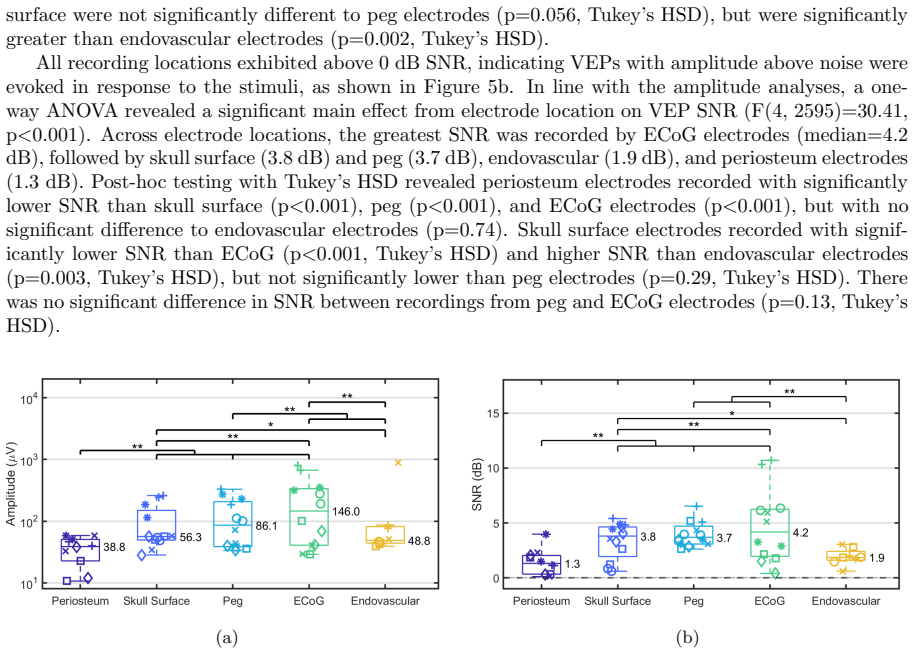
Across six sheep, electrodes were placed successively at five depths: endovascular in the transverse sinus, on the periosteum, on the skull surface, partially embedded in the skull as peg electrodes, and subdurally as ECoG. Median VEP SNR was ordered by depth for the sub-scalp sites (periosteum 1.3 dB, skull surface 3.8 dB, peg 3.7 dB), with ECoG highest at 4.2 dB; the peg-ECoG difference was not statistically significant, whereas endovascular electrodes (1.9 dB) were statistically indistinguishable from the periosteum. Maximum bandwidth showed a similar depth trend among sub-scalp sites (120, 140, and 180 Hz for periosteum, skull surface, and peg), with ECoG at 200 Hz and endovascular at 195 Hz, meaning all sub-scalp depths captured high-gamma activity. A finite-element volume-conduction model with equivalent cortical dipoles reproduced the amplitude ordering, predicting ECoG highest and no significant difference between the three sub-scalp depths and the endovascular electrode, and attributed the ranking largely to tissue attenuation and dipole-electrode distance.
Load-bearing premise
The peg-versus-ECoG parity assumes the ECoG recordings, made last roughly six to eight hours into the session, were not weakened by eye dryness or cumulative anaesthesia, which would inflate how close peg electrodes appear to the ECoG baseline.
Editorial extensions
If this is right
- Since VEP SNR from peg electrodes was statistically indistinguishable from ECoG, a BCI could obtain comparable evoked-response quality without a craniotomy.
- Skull-surface electrodes may offer a safer compromise: their SNR and bandwidth were not significantly different from peg electrodes, so removing the periosteum alone may be enough.
- Endovascular arrays showed no SNR advantage over periosteum electrodes in this acute setting, weakening the case for their higher procedural risk and limited spatial coverage in VEP-based BCIs.
- All sub-scalp depths captured high-gamma activity, with maximum bandwidth of 120-180 Hz, supporting the use of sub-scalp EEG for BCI features beyond evoked potentials.
- The finite-element model offers a quantitative way to predict relative signal amplitudes across candidate depths and electrode designs before implantation.
Reading between the lines
- If peg-ECoG parity survives a randomized or simultaneous recording design, the practical bottleneck for sub-scalp BCI shifts from raw signal quality to spatial resolution, which this acute study did not measure.
- Because electrode surface area and interface impedance were not modeled, a testable design hypothesis is that larger-area sub-scalp electrodes could close the remaining gap to ECoG without going deeper.
- Chronic conditions could move the ranking in either direction: endothelialisation may strengthen endovascular recordings, while tissue growth around sub-scalp electrodes may weaken them, making the acute ordering a baseline rather than a lifetime guarantee.
- Anatomical differences between sheep and human skull mean the exact depth-versus-SNR curve should be re-estimated from human intraoperative or cadaveric recordings before choosing a target sub-scalp depth for clinical trials.
Editorial analysis
A structured set of objections, weighed in public.
Referee Report
Summary. This manuscript reports an acute sheep study comparing visual evoked potential (VEP) SNR and maximum bandwidth across five electrode configurations: endovascular, periosteum, skull surface, peg, and subdural ECoG. It supplements the in vivo data with an FEM forward model of the ovine head, predicting ECoG to have the highest amplitude and the sub-scalp and endovascular configurations to be broadly similar. The authors' headline claim is that peg electrodes achieve VEP SNR approaching ECoG, while endovascular arrays are comparable to periosteal electrodes, and that sub-scalp electrodes record high-gamma activity with maximum bandwidth 120-180 Hz.
Significance. If validated, the peg-versus-ECoG SNR result would be an important step toward less invasive chronic BCI recording. The study's strengths include direct multi-modality comparison in the same animals, a forward model built from independent conductivity data rather than fitted to the experimental amplitudes (low circularity), and an unusually explicit discussion of simulation assumptions and experimental confounds in Sections 4.2.1 and 4.5. The model also makes testable predictions about distance/angle dependence and electrode orientation effects. However, the central experimental claim currently rests on statistics that do not support it, and on a recording-order confound that the authors themselves identify.
major comments (4)
- [Section 3.2 / Table 2] The ANOVAs reported as F(4,2595) treat individual epochs as independent observations even though epochs are nested within sheep and within the two selected channels per electrode type. This pseudoreplication inflates degrees of freedom and makes the Tukey HSD p-values, including the load-bearing peg-versus-ECoG SNR comparison (p=0.1261), uninterpretable as evidence of equivalence or of a real difference. Please reanalyze the data with a mixed-effects model (random intercepts for sheep and channel) or with per-sheep summary statistics, and report confidence intervals for the peg-versus-ECoG SNR difference rather than relying on a non-significant pairwise test.
- [Sections 2.1 and 4.2.1] ECoG was always recorded last, 6-8 hours after the endovascular and periosteum sessions, and Section 4.2.1 acknowledges that eye dryness and prolonged anaesthesia may have reduced late VEP responses. Because the abstract's central claim is that peg SNR approaches ECoG SNR, this fixed recording order is a major confound: even a small suppression of the late ECoG baseline (on the order of 0.5-1 dB) would fully explain the observed median gap of 3.7 dB versus 4.2 dB. The authors should randomize or interleave the recording order, include time-matched control recordings, or substantially soften the abstract and conclusion claims to reflect the tentative nature of the comparison.
- [Sections 2.3.1 and 3.2] The analysis selects the two channels with the highest VEP SNR for each recording method after SNR is computed. This outcome-dependent selection biases every median upward, and the bias likely differs across methods because the numbers of available channels differ (five for sub-scalp arrays, six for ECoG, four for endovascular). The bandwidth analysis in Section 3.3 inherits the same selected channels. Please report results for all channels as a sensitivity analysis, or motivate the selection with a pre-registered or independent criterion.
- [Section 2.3.1] The SNR definition is ambiguous: it is not clear whether the ratio of post-stimulus to pre-stimulus variance is computed on single epochs or on the trial-averaged VEP, and it is not stated how the random sample of 50 trials interacts with this computation. Because the SNR values are the paper's primary outcome, the exact formula and unit of analysis should be stated explicitly, and the sensitivity of the median SNRs to the 300 ms window choice should be reported.
minor comments (4)
- [Sections 3.3 and 4.1.2] The bandwidth ANOVA is not significant (F(4,49)=0.47, p=0.76), but Section 4.1.2 describes an increase in median bandwidth with depth; please mark this explicitly as a non-significant trend.
- [Section 2.1] The experimental timeline is described only verbally; a table or figure showing the time of each recording session per sheep would make the confound transparent.
- [Section 2.4] There are several terminology and typographical issues, including 'electromagentic' and 'accomodate' in Section 2.4, 'ANOV A' in Section 3.2, and the description of the 'spherical peg electrode (Skull surface electrode...)' that appears to conflate peg and skull-surface labels.
- [Availability] No data or code availability statement is provided; given the custom MATLAB pipeline and FEM model, a repository would substantially aid reproducibility.
Circularity Check
No significant circularity: the peg-vs-ECoG SNR claim rests on directly measured trial data, and the FEM forward model is parameter-free with respect to experimental amplitudes.
full rationale
This paper's derivation chain is self-contained, so no circular step is identified. The central experimental claims — peg VEP SNR not significantly different from ECoG (p=0.1261), endovascular SNR comparable to periosteum (p=0.74), and maximum bandwidth of 120-180 Hz for sub-scalp depths — are computed directly from the recorded data under fully stated analysis rules (5-40 Hz band-pass, 1 mV epoch rejection, fixed 300 ms pre/post-stimulus windows, and a Q3+1.5·IQR noise-floor threshold for bandwidth). The two outcome-informed analysis choices, namely the post-stimulus window (Section 2.3.1: 'These windows were used because the VEP waveform was observed to span 300 ms post-stimulus') and the best-channel selection ('The two channels that exhibited the highest SNR were included in the analysis for each recording method'), are applied identically to every electrode type, so they cannot by construction force the peg-ECoG comparison; the same pipeline found the peg-ECoG amplitude difference significant (p<0.001) and the endovascular-periosteum difference non-significant, showing the metric is sensitive to genuine distinctions. The FEM simulation is a genuine forward model rather than a fit: tissue conductivities are taken from the independent IT'IS database, the dipole moment is arbitrary with only relative amplitudes used, dipole locations are randomly sampled, and no parameter is tuned to the experimental amplitudes. Its prediction (ECoG highest, sub-scalp not significantly different from endovascular) is falsifiable and, in fact, partially fails: simulated endovascular amplitudes exceed skull-surface amplitudes contrary to the experiment, a discrepancy the authors transparently attribute to electrode surface area effects. Self-citations with overlapping authors ([4] Benovitski et al., [13] John et al., [19] Mahoney et al., [30,31,33,34] Opie/Oxley et al.) are used for context, for a fully disclosed noise-floor method, or for prior results that this study re-derives independently; none carries the load of the peg-vs-ECoG claim, so they are not load-bearing under the review rules. The acknowledged 6-8 hour recording-order confound (Section 4.2.1) is a validity and correctness risk, not a circular dependency, and the paper discloses it explicitly. Overall circularity score: 0.
Assumptions & free parameters
free parameters (9)
- SNR post-stimulus window =
300 ms post-stimulus
- Channels analyzed per method =
2 highest-SNR channels
- Epoch rejection threshold =
1 mV range
- Random trial sample =
50 trials
- Periosteum conductivity and thickness =
0.06 S/m and 0.23 mm (dura values)
- EPSP time constant tau =
10 ms
- Cortical propagation speed =
0.3 m/s
- Noise floor threshold =
third quartile + 1.5*IQR of 400-500 Hz band
- Dipole moment =
1e-6 A m (arbitrary)
assumptions (6)
- domain assumption Quasi-static approximation of Maxwell's equations holds at EEG frequencies in the ovine head
- domain assumption Tissue conductivities are isotropic, homogeneous, and frequency-independent within each layer
- domain assumption Cortical sources can be represented by equivalent current dipoles oriented normal to the grey-white matter boundary
- domain assumption Sheep head geometry and tissue properties approximate human conditions sufficiently for BCI signal-quality conclusions
- ad hoc to paper The two highest-SNR channels provide an unbiased measure of each electrode type's performance
- domain assumption VEP SNR can be quantified as the ratio of post-stimulus to pre-stimulus variance in the 5-40 Hz band
Cite this review
Pith. "Pith review of Minimally Invasive Brain Computer Interfaces: Evaluating the Impact of Tissue Layers on Signal Quality of Sub-Scalp EEG." pith.science (2026). https://pith.science/paper/MBODDYRA
@misc{pith2026250603452,
  author       = {Pith},
  title        = {Pith review of: Minimally Invasive Brain Computer Interfaces: Evaluating the Impact of Tissue Layers on Signal Quality of Sub-Scalp EEG},
  year         = {2026},
  howpublished = {\url{https://pith.science/paper/MBODDYRA}},
  note         = {Machine review of arXiv:2506.03452}
}
read the original abstract
Individuals with severe physical disabilities often experience diminished quality of life stemming from limited ability to engage with their surroundings. Brain-Computer Interface (BCI) technology aims to bridge this gap by enabling direct technology interaction. However, current BCI systems require invasive procedures, such as craniotomy or implantation of electrodes through blood vessels, posing significant risks to patients. Sub-scalp electroencephalography (EEG) offers a lower risk alternative. This study investigates the signal quality of sub-scalp EEG recordings from various depths in a sheep model, and compares results with other methods: ECoG and endovascular arrays. A computational model was also constructed to investigate the factors underlying variations in electrode performance. We demonstrate that peg electrodes placed within the sub-scalp space can achieve visual evoked potential signal-to-noise ratios (SNRs) approaching that of ECoG. Endovascular arrays exhibited SNR comparable to electrodes positioned on the periosteum. Furthermore, sub-scalp recordings captured high gamma neural activity, with maximum bandwidth ranging from 120 Hz to 180 Hz depending on electrode depth. These findings support the use of sub-scalp EEG for BCI applications, and provide valuable insights for future sub-scalp electrode design. This data lays the groundwork for human trials, ultimately paving the way for chronic, in-home BCIs that empower individuals with physical disabilities.
Figures


Figures from the paper (5 more)



Reference graph
Works this paper leans on
-
[1]
T. Ball, E. Demandt, I. Mutschler, E. Neitzel, C. Mehring, K. Vogt, A. Aertsen, and A. Schulze- Bonhage. Movement related activity in the high gamma range of the human EEG. NeuroImage, 41(2):302–310, June 2008. ISSN 10538119. doi: 10.1016/j.neuroimage.2008.02.032. URL https: //linkinghub.elsevier.com/retrieve/pii/S1053811908001717. 15
-
[2]
T. Ball, M. Kern, I. Mutschler, A. Aertsen, and A. Schulze-Bonhage. Signal quality of simultaneously recorded invasive and non-invasive EEG.NeuroImage, 46(3):708–716, 2009. ISSN 1053-8119. doi: 10. 1016/j.neuroimage.2009.02.028. URL https://www.sciencedirect.com/science/article/pii/ S1053811909001827
work page 2009
-
[3]
A. L. Benabid, T. Costecalde, A. Eliseyev, G. Charvet, A. Verney, S. Karakas, M. Foerster, A. Lambert, B. Morini` ere, N. Abroug, M.-C. Schaeffer, A. Moly, F. Sauter-Starace, D. Ratel, C. Moro, N. Torres-Martinez, L. Langar, M. Oddoux, M. Polosan, S. Pezzani, V. Auboiroux, T. Aksenova, C. Mestais, and S. Chabardes. An exoskeleton controlled by an epidural...
work page 2019
-
[4]
Y. B. Benovitski, A. Lai, C. C. McGowan, O. Burns, V. Maxim, D. A. X. Nayagam, R. Millard, G. D. Rathbone, M. A. le Chevoir, R. A. Williams, D. B. Grayden, C. N. May, M. Murphy, W. J. D’Souza, M. J. Cook, and C. E. Williams. Ring and peg electrodes for minimally-Invasive and long-term sub-scalp EEG recordings. Epilepsy Research, 135:29–37, 2017. ISSN 0920...
-
[5]
U. Chaudhary, N. Birbaumer, and A. Ramos-Murguialday. Chapter 5 - Brain-computer interfaces in the completely locked-in state and chronic stroke. In D. Coyle, editor, Progress in Brain Research, volume 228, pages 131–161. Elsevier, 2016. ISBN 0079-6123. doi: https://doi.org/10.1016/bs.pbr. 2016.04.019. Type: Book Section
-
[6]
P. G. Clarke and D. Whitteridge. The cortical visual areas of the sheep. The Journal of Physiology , 256(3):497–508, Apr. 1976. ISSN 0022-3751, 1469-7793. doi: 10.1113/jphysiol.1976.sp011335. URL https://physoc.onlinelibrary.wiley.com/doi/10.1113/jphysiol.1976.sp011335
-
[7]
J. Duun-Henriksen, M. Baud, M. P. Richardson, M. Cook, G. Kouvas, J. M. Heasman, D. Friedman, J. Peltola, I. C. Zibrandtsen, and T. W. Kjaer. A new era in electroencephalographic monitoring? Subscalp devices for ultra-long-term recordings. EPILEPSIA, 2020. ISSN 00139580. doi: 10.1111/ epi.16630. Type: Journal Article
work page 2020
-
[8]
V. L. Feigin, T. Vos, E. Nichols, M. O. Owolabi, W. M. Carroll, M. Dichgans, G. Deuschl, P. Parmar, M. Brainin, and C. Murray. The global burden of neurological disorders: trans- lating evidence into policy. The Lancet Neurology , 19(3):255–265, Mar. 2020. ISSN 14744422. doi: 10.1016/S1474-4422(19)30411-9. URL https://linkinghub.elsevier.com/retrieve/pii/...
Show all 62 references
-
[9]
Haneef, K
Z. Haneef, K. Yang, S. A. Sheth, F. Z. Aloor, B. Aazhang, V. Krishnan, and C. Karakas. Sub-scalp electroencephalography: A next-generation technique to study human neurophysiology. Clinical Neurophysiology, 141:77–87, 2022. ISSN 1388-2457. doi: 10.1016/j.clinph.2022.07.003. UR...
2022 doi
-
[10]
L. R. Hochberg, D. Bacher, B. Jarosiewicz, N. Y. Masse, J. D. Simeral, J. Vogel, S. Haddadin, J. Liu, S. S. Cash, P. Van Der Smagt, and J. P. Donoghue. Reach and grasp by people with tetraplegia using a neurally controlled robotic arm. Nature, 485(7398):372–375, May 2012. ISSN...
2012 doi
-
[11]
Tissue Properties Database V4.2, 2024
IT’IS Foundation. Tissue Properties Database V4.2, 2024. URL https://itis.swiss/ virtual-population/tissue-properties/downloads/database-v4-2/
2024
-
[12]
Jiang, P
Y. Jiang, P. Gu, B. Li, X. Gao, B. Sun, Y. Song, G. Wang, Y. Yuan, C. Wang, M. Liu, D. Han, and P. Dai. Analysis and Management of Complications in a Cohort of 1,065 Mini- mally Invasive Cochlear Implantations. Otology & Neurotology , 38(3):347–351, Mar. 2017. ISSN 1531-7129, ...
2017 doi
-
[13]
S. E. John, N. L. Opie, Y. T. Wong, G. S. Rind, S. M. Ronayne, G. Gerboni, S. H. Bauquier, T. J. O’Brien, C. N. May, D. B. Grayden, and T. J. Oxley. Signal quality of simultaneously recorded endovascular, subdural and epidural signals are comparable. Scientific Reports , 8(1):...
2018 doi
-
[14]
J. M. Johnston, F. T. Mangano, J. G. Ojemann, T. S. Park, E. Trevathan, and M. D. Smyth. Complications of invasive subdural electrode monitoring at St. Louis Children’s Hospital, 1994–2005. Journal of Neurosurgery: Pediatrics PED , 105(5):343–347, 2006. doi: 10.3171/ped.2006.1...
1994 doi
-
[15]
M. A. Khan, R. Das, H. K. Iversen, and S. Puthusserypady. Review on motor imagery based BCI systems for upper limb post-stroke neurorehabilitation: From designing to application.Computers in Biology and Medicine, 123:103843, 2020. ISSN 0010-4825. doi: 10.1016/j.compbiomed.2020...
2020
-
[16]
Kinaci, W
A. Kinaci, W. Bergmann, R. L. Bleys, A. Van Der Zwan, and T. P. Van Doormaal. Histologic Comparison of the Dura Mater among Species. Comparative Medicine, 70(2):170–175, Apr. 2020. ISSN 1532-0820. doi: 10.30802/AALAS-CM-19-000022. URL https://aalas.kglmeridian.com/ view/journa...
2020
-
[17]
Kubler, E
A. Kubler, E. M. Holz, A. Riccio, C. Zickler, T. Kaufmann, S. C. Kleih, P. Staiger-Salzer, L. Desideri, E. J. Hoogerwerf, and D. Mattia. The user-centered design as novel perspective for evaluating the usability of BCI-controlled applications. PLoS One , 9(12):e112392, 2014. I...
2014 doi
-
[18]
Logroscino, M
G. Logroscino, M. Piccininni, B. Marin, E. Nichols, F. Abd-Allah, A. Abdelalim, F. Alahdab, S. W. Asgedom, A. Awasthi, Y. Chaiah, A. Daryani, H. P. Do, M. Dubey, A. Elbaz, S. Eskan- darieh, F. Farhadi, F. Farzadfar, S.-M. Fereshtehnejad, E. Fernandes, I. Filip, K. J. Foreman, ...
1990
-
[19]
T. B. Mahoney, P.-C. Liu, D. B. Grayden, and S. E. John. Comparison of Sub-Scalp EEG and Endovascular Stent-Electrode Array for Visual Evoked Potential Brain-Computer Interface. In 2023 45th Annual International Conference of the IEEE Engineering in Medicine & Biology Society ...
2023
-
[20]
D. J. McFarland. Brain-computer interfaces for amyotrophic lateral sclerosis. Muscle & nerve , 61 (6):702–707, 2020. ISSN 1097-4598 0148-639X. doi: 10.1002/mus.26828. Type: Journal Article
2020 doi
-
[21]
D. J. McFarland, W. A. Sarnacki, and J. R. Wolpaw. Electroencephalographic (EEG) control of three-dimensional movement. Journal of Neural Engineering , 7(3):036007, June 2010. ISSN 1741-2560, 1741-2552. doi: 10.1088/1741-2560/7/3/036007. URL https://iopscience.iop.org/ article...
2010 doi
-
[22]
K. J. Miller, L. B. Sorensen, J. G. Ojemann, and M. den Nijs. Power-Law Scaling in the Brain Surface Electric Potential. PLOS Computational Biology , 5(12):1–10, Dec. 2009. doi: 10.1371/journal.pcbi. 1000609. URL https://doi.org/10.1371/journal.pcbi.1000609. Publisher: Public ...
2009 doi
-
[23]
Miralles, E
F. Miralles, E. Vargiu, X. Rafael-Palou, M. Sol` a, S. Dauwalder, C. Guger, C. Hinterm¨ uller, A. Es- pinosa, H. Lowish, S. Martin, E. Armstrong, and J. Daly. Brain–Computer Interfaces on Track to Home: Results of the Evaluation at Disabled End-Users’ Homes and Lessons Learnt....
2015
-
[24]
K. Modi, M. P. Soos, and K. Mahajan. Stent Thrombosis. In StatPearls. StatPearls Publishing, Treasure Island (FL), 2024. URL http://www.ncbi.nlm.nih.gov/books/NBK441908/
2024
-
[25]
Nagahama, A
Y. Nagahama, A. J. Schmitt, D. Nakagawa, A. S. Vesole, J. Kamm, C. K. Kovach, D. Hasan, M. Granner, B. J. Dlouhy, M. A. Howard, and H. Kawasaki. Intracranial EEG for seizure focus 17 localization: evolving techniques, outcomes, complications, and utility of combining surface a...
2019 doi
-
[26]
F. S. d. A. Neto and J. L. G. Rosa. Depression biomarkers using non-invasive EEG: A review.Neuro- science & Biobehavioral Reviews , 105:83–93, 2019. ISSN 0149-7634. doi: 10.1016/j.neubiorev.2019. 07.021. URL https://www.sciencedirect.com/science/article/pii/S0149763419303823
2019 doi
-
[27]
Neufeld, A
E. Neufeld, A. M. Cassar´ a, H. Montanaro, N. Kuster, and W. Kainz. Functionalized anatomical models for EM-neuron Interaction modeling. Physics in Medicine and Biology , 61(12):4390–4401, June 2016. ISSN 0031-9155, 1361-6560. doi: 10.1088/0031-9155/61/12/4390. URL https:// io...
2016 doi
-
[28]
Nitzsche, S
B. Nitzsche, S. Frey, L. D. Collins, J. Seeger, D. Lobsien, A. Dreyer, H. Kirsten, M. H. Stoffel, V. S. Fonov, and J. Boltze. A stereotaxic, population-averaged T1w ovine brain atlas includ- ing cerebral morphology and tissue volumes. Frontiers in Neuroanatomy , 9, June 2015. ...
2015
-
[29]
J. D. Olson, J. D. Wander, L. Johnson, D. Sarma, K. Weaver, E. J. Novotny, J. G. Ojemann, and F. Darvas. Comparison of subdural and subgaleal recordings of cortical high-gamma activity in humans. Clinical neurophysiology : official journal of the International Federation of Cl...
2016 doi
-
[30]
Type: Journal Article
-
[31]
N. L. Opie, N. R. van der Nagel, S. E. John, K. Vessey, G. S. Rind, S. M. Ronayne, E. L. Fletcher, C. N. May, O. B. TJ, and T. J. Oxley. Micro-CT and Histological Evaluation of an Neural Interface Implanted Within a Blood Vessel. IEEE Trans Biomed Eng, 64(4):928–934, 2017. ISS...
2017
-
[32]
N. L. Opie, S. E. John, G. S. Rind, S. M. Ronayne, C. N. May, D. B. Grayden, and T. J. Oxley. Effect of Implant Duration, Anatomical Location and Electrode Orientation on Bandwidth Recorded with a Chronically Implanted Endovascular Stent-Electrode Array. In 2018 40th Annual In...
2018
-
[33]
T. J. Oxley, N. L. Opie, S. E. John, G. S. Rind, S. M. Ronayne, T. L. Wheeler, J. W. Judy, A. J. McDonald, A. Dornom, T. J. H. Lovell, C. Steward, D. J. Garrett, B. A. Moffat, E. H. Lui, N. Yassi, B. C. V. Campbell, Y. T. Wong, K. E. Fox, E. S. Nurse, I. E. Bennett, S. H. Bauq...
-
[34]
Ormachea, B
E. Ormachea, B. Calsin, E. Aguilar, B. Ormachea, H. Gonzales, and Y. Masias. Principal Compo- nent Analysis of Morphological Characteristics in Creole Sheep (Ovis aries). Advances in Animal and Veterinary Sciences , 11(6):903–909, Dec. 2022. ISSN 2307-8316. doi: https://dx.doi...
2022
-
[35]
Perez-Valero, M
E. Perez-Valero, M. A. Vaquero-Blasco, M. A. Lopez-Gordo, and C. Morillas. Quantitative As- sessment of Stress Through EEG During a Virtual Reality Stress-Relax Session. Frontiers in Computational Neuroscience, 15, 2021. ISSN 1662-5188. doi: 10.3389/fncom.2021.684423. URL http...
2021
-
[36]
Pieber and S
M. Pieber and S. DeSaSouza. Cochlear Implant Reliability. In S. DeSaSouza, editor, Cochlear Implants, pages 473–499. Springer Nature Singapore, Singapore, 2022. ISBN 978-981-19045-1-6 978-981-19045-2-3. doi: 10.1007/978-981-19-0452-3 23. URL https://link.springer.com/10. 1007/...
2022 doi
-
[37]
T. J. Oxley, P. E. Yoo, G. S. Rind, S. M. Ronayne, C. M. S. Lee, C. Bird, V. Hampshire, R. P. Sharma, A. Morokoff, D. L. Williams, C. MacIsaac, M. E. Howard, L. Irving, I. Vrljic, C. Williams, S. E. John, F. Weissenborn, M. Dazenko, A. H. Balabanski, D. Friedenberg, A. N. Burk...
2021
-
[38]
P. Qi, H. Ru, L. Gao, X. Zhang, T. Zhou, Y. Tian, N. Thakor, A. Bezerianos, J. Li, and Y. Sun. Neural Mechanisms of Mental Fatigue Revisited: New Insights from the Brain Connec- tome. Engineering, 5(2):276–286, 2019. ISSN 2095-8099. doi: 10.1016/j.eng.2018.11.025. URL https://...
2019 doi
-
[39]
Rashid, N
M. Rashid, N. Sulaiman, A. P. P. Abdul Majeed, R. M. Musa, A. F. Ab. Nasir, B. S. Bari, and S. Khatun. Current Status, Challenges, and Possible Solutions of EEG-Based Brain-Computer Interface: A Comprehensive Review. Frontiers in Neurorobotics, 14(25), 2020. ISSN 1662-5218. do...
2020
-
[40]
Plourde, S
G. Plourde, S. J. Reed, and C. A. Chapman. Attenuation of High-Frequency (50–200 Hz) Thalamocortical Electroencephalographic Rhythms by Isoflurane in Rats Is More Pronounced for the Thalamus Than for the Cortex. Anesthesia & Analgesia , 122(6):1818–1825, June 2016. ISSN 0003-2...
2016 doi
-
[41]
K. S. Scheuer, J. M. Judge, X. Zhao, and M. B. Jackson. Velocity of conduction between columns and layers in barrel cortex reported by parvalbumin interneurons. Cerebral Cortex (New York, NY), 33(17):9917–9926, July 2023. ISSN 1047-3211. doi: 10.1093/cercor/bhad254. URL https:...
2023 doi
-
[42]
Schimpf, C
P. Schimpf, C. Ramon, and J. Haueisen. Dipole models for the EEG and MEG. IEEE Transactions on Biomedical Engineering , 49(5):409–418, May 2002. ISSN 00189294. doi: 10.1109/10.995679. URL http://ieeexplore.ieee.org/document/995679/
2002 doi
-
[43]
J. D. Rolston, D. Ouyang, D. J. Englot, D. D. Wang, and E. F. Chang. National trends and complication rates for invasive extraoperative electrocorticography in the USA. Journal of clinical neuroscience : official journal of the Neurosurgical Society of Australasia, 22(5):823–8...
2015 doi
-
[44]
M. M. Smith, K. E. Weaver, T. J. Grabowski, R. P. N. Rao, and F. Darvas. Non-invasive detection of high gamma band activity during motor imagery. Frontiers in Human Neuroscience , 8, Oct
-
[45]
R. M. Starke, T. Wang, D. Ding, C. R. Durst, R. W. Crowley, N. Chalouhi, D. M. Hasan, A. S. Dumont, P. Jabbour, and K. C. Liu. Endovascular Treatment of Venous Sinus Stenosis in Idiopathic Intracranial Hypertension: Complications, Neurological Outcomes, and Radiographic Result...
2015 doi
-
[46]
Smith, K
M. Smith, K. Weaver, T. Grabowski, and F. Darvas. Utilizing High Gamma (HG) Band Power Changes as a Control Signal for Non-Invasive BCI. In C. Guger, B. Z. Allison, and G. Edlinger, editors, Brain-Computer Interface Research, pages 83–91. Springer Berlin Heidelberg, Berlin, He...
2013 doi
-
[47]
Taussig, G
D. Taussig, G. Dorfm¨ uller, M. Fohlen, C. Jalin, C. Bulteau, S. Ferrand-Sorbets, M. Chipaux, and O. Delalande. Invasive explorations in children younger than 3years. Seizure, 21(8):631–638, 2012. ISSN 1059-1311. doi: 10.1016/j.seizure.2012.07.004. Type: Journal Article
2012 doi
-
[48]
H. J. Theunisse, R. J. E. Pennings, H. P. M. Kunst, J. J. Mulder, and E. A. M. Mylanus. Risk factors for complications in cochlear implant surgery. European Archives of Oto-Rhino-Laryngology, 275(4):895–903, 2018. ISSN 1434-4726. doi: 10.1007/s00405-018-4901-z. URL https://doi...
2018 doi
-
[49]
Weder, C
S. Weder, C. Shaul, A. Wong, S. O’Leary, and R. J. Briggs. Management of Severe Cochlear Implant Infections—35 Years Clinical Experience. Otology & Neurotology , 41(10):1341–1349, Dec
-
[50]
R. E. Stirling, M. I. Maturana, P. J. Karoly, E. S. Nurse, K. McCutcheon, D. B. Grayden, S. G. Ringo, J. M. Heasman, R. J. Hoare, A. Lai, W. D’Souza, U. Seneviratne, L. Seiderer, K. J. McLean, K. J. Bulluss, M. Murphy, B. H. Brinkmann, M. P. Richardson, D. R. Freestone, and M....
2021
-
[51]
B. D. Winslow and P. A. Tresco. Quantitative analysis of the tissue response to chronically implanted microwire electrodes in rat cortex. Biomaterials, 31(7):1558–1567, 2010. ISSN 0142-9612. doi: 10.1016/j.biomaterials.2009.11.049. Type: Journal Article
2010 doi
-
[52]
B. D. Winslow, M. B. Christensen, W.-K. Yang, F. Solzbacher, and P. A. Tresco. A compari- son of the tissue response to chronically implanted Parylene-C-coated and uncoated planar silicon microelectrode arrays in rat cortex. Biomaterials, 31(35):9163–9172, 2010. ISSN 0142-9612...
2010 doi
-
[53]
Wittevrongel, E
B. Wittevrongel, E. Khachatryan, M. Fahimi Hnazaee, F. Camarrone, E. Carrette, L. De Taeye, A. Meurs, P. Boon, D. Van Roost, and M. Van Hulle. Decoding Steady-State Visual Evoked Potentials From Electrocorticography. Frontiers in Neuroinformatics , 12:65, Sept. 2018. doi: 10. ...
2018
-
[54]
X. Zhao, X. Wang, T. Yang, S. Ji, H. Wang, J. Wang, Y. Wang, and Q. Wu. Classification of sleep apnea based on EEG sub-band signal characteristics. Scientific Reports , 11(1):5824, Mar
-
[55]
Weisdorf, J
S. Weisdorf, J. Duun-Henriksen, M. J. Kjeldsen, F. R. Poulsen, S. W. Gangstad, and T. W. Kjær. Ultra-long-term subcutaneous home monitoring of epilepsy—490 days of EEG from nine patients. Epilepsia, 60(11):2204–2214, 2019. ISSN 0013-9580. doi: 10.1111/epi.16360. Type: Journal Article
2019 doi
-
[56]
¨Onal, H
C. ¨Onal, H. Otsubo, T. Araki, S. Chitoku, A. Ochi, S. Weiss, W. Logan, I. Elliott, O. C. Snead, and J. T. Rutka. Complications of invasive subdural grid monitoring in children with epilepsy. Journal of Neurosurgery, 98(5):1017–1026, 2003. doi: 10.3171/jns.2003.98.5.1017. Type...
2003 doi
-
[61]
P. Zis, A. Liampas, A. Artemiadis, G. Tsalamandris, P. Neophytou, Z. Unwin, V. K. Kimiskidis, G. M. Hadjigeorgiou, G. Varrassi, Y. Zhao, and P. G. Sarrigiannis. EEG Recordings as Biomarkers of Pain Perception: Where Do We Stand and Where to Go? Pain and Therapy , 11(2):369–380...
2022 doi
-
[2014]
doi: 10.3389/fnhum.2014.00817
ISSN 1662-5161. doi: 10.3389/fnhum.2014.00817. URL http://journal.frontiersin.org/ article/10.3389/fnhum.2014.00817/abstract
2014
-
[2016]
doi: 10.1038/nbt.3428
ISSN 1546-1696. doi: 10.1038/nbt.3428. URL https://doi.org/10.1038/nbt.3428. Type: Journal Article
-
[2018]
doi: 10.1109/EMBC.2018.8512385
2018
-
[2020]
doi: 10.1097/MAO.0000000000002783
ISSN 1531-7129, 1537-4505. doi: 10.1097/MAO.0000000000002783. URL https://journals. lww.com/10.1097/MAO.0000000000002783
-
[2021]
doi: 10.1038/s41598-021-85138-0
ISSN 2045-2322. doi: 10.1038/s41598-021-85138-0. URL https://doi.org/10.1038/ s41598-021-85138-0
Reviewed August 7, 2026 · model on record in the stance chip above.
Discussion (0). Sign in to comment.