Directional Dark Field for Nanoscale Full-Field Transmission X-Ray Microscopy
Pith reviewed 2026-05-19 07:57 UTC · model grok-4.3
The pith
Directional dark-field X-ray microscopy now maps orientations of sub-resolution scattering features.
A machine-rendered reading of the paper's core claim, the machinery that carries it, and where it could break.
Core claim
The central discovery is the first implementation of directional dark-field imaging in nanoscale transmission X-ray microscopy. Using an optical configuration that incorporates shadow regions, the setup retrieves directional information on small-angle scattering to determine orientations of features below the spatial resolution limit. Experiments confirm this by resolving orientations in test structures, tracking changes in hierarchical nanoporous materials, and mapping the arrangement of 30-70 nm hydroxyapatite nanocrystals in human tooth enamel.
What carries the argument
Shadow regions in the optical configuration that extend the detectable scattering vector range while enabling directional scattering retrieval for orientation mapping.
If this is right
- Orientation mapping becomes feasible for anisotropic features smaller than the microscope's resolution.
- Quantitative characterization of nanostructures in biomineralization and nanotechnology applications.
- Pathway to size-selective dark-field imaging by extending the scattering vector range.
- Easy adoption since it requires no major changes to existing transmission X-ray microscopy setups.
Where Pith is reading between the lines
- Similar shadow-based methods could be adapted for other scattering-based imaging techniques to add directional sensitivity.
- Integration with tomographic methods might allow 3D orientation mapping of nanomaterials.
- Applications could extend to studying orientation changes during material processing or biological processes.
Load-bearing premise
The optical configuration using shadow regions retrieves accurate directional scattering information at sub-micrometer scales without introducing significant artifacts or errors in the orientation mapping.
What would settle it
A direct comparison of orientation maps from this method with high-resolution electron diffraction or polarized optical measurements on identical samples of tooth enamel or test structures, where systematic discrepancies would disprove the claim.
Figures


read the original abstract
Dark-field X-ray imaging visualizes structural inhomogeneities through small-angle scattering, but existing directional methods are confined to the micrometer scale. While recent advances have extended dark-field capabilities to nanoscale transmission X-ray microscopy, directional scattering retrieval - critical for characterizing anisotropic nanostructures - has remained inaccessible for imaging resolutions in the sub-micrometer scale. Here, we demonstrate the first directional dark-field setup for nanoimaging, achieving orientation mapping of scattering features below the spatial resolution limit. Our method is experimentally simple to implement with existing transmission X-ray microscopy setups. We validate its performance by successfully resolving sub-resolution test structure orientations, cross-correlating orientational changes within hierarchical nanoporous materials, and mapping the directional arrangement of hydroxyapatite nanocrystals 30 - 70 nm within human tooth enamel. By utilizing shadow regions in the optical configuration, we further extend the detectable scattering vector range, demonstrating a pathway toward size-selective dark-field imaging. This advancement enables the quantitative structural characterization of anisotropic nanomaterials, which are critical to biomineralization, advanced materials, and nanotechnology applications.
Editorial analysis
A structured set of objections, weighed in public.
Referee Report
Summary. The manuscript introduces the first directional dark-field setup for nanoscale full-field transmission X-ray microscopy. By utilizing shadow regions in the optical configuration, the method retrieves directional scattering information to enable orientation mapping of anisotropic scattering features below the spatial resolution limit. Validation is reported on sub-resolution test structures, hierarchical nanoporous materials, and the directional arrangement of 30-70 nm hydroxyapatite nanocrystals in human tooth enamel, with an extension of the detectable scattering vector range toward size-selective imaging.
Significance. If robust, the result would enable quantitative structural characterization of anisotropic nanomaterials at the nanoscale, with direct relevance to biomineralization, advanced materials, and nanotechnology. The experimental simplicity of implementation with existing TXM setups and the multi-sample validation (including real biological material) are clear strengths that support the central claim of practical utility.
major comments (1)
- [Optical configuration and shadow-region retrieval] Optical configuration and shadow-region retrieval: The central claim that shadow regions accurately retrieve directional scattering at sub-micrometer scales without significant artifacts rests on the assumption that intensity modulation is dominated by the sample's anisotropic scattering vector and remains independent of illumination divergence, detector pixel response, and local sample thickness. The manuscript should provide explicit quantitative tests, simulations, or error propagation analysis demonstrating that these couplings do not produce systematic angular errors in the extracted orientations (e.g., for the 30-70 nm hydroxyapatite features in enamel).
minor comments (2)
- [Figures] Figure clarity: Ensure all orientation maps include explicit scale bars, color bars for angle, and direct comparison to the corresponding absorption or phase images at the same field of view.
- [Methods/Results] Notation: Define the scattering vector range extension quantitatively (e.g., minimum and maximum q values accessed via shadow regions) and compare it explicitly to conventional TXM dark-field limits.
Simulated Author's Rebuttal
We thank the referee for their positive assessment of the work's significance and for the detailed feedback on the optical configuration. We have addressed the major comment by adding quantitative validation and error analysis in the revised manuscript.
read point-by-point responses
-
Referee: Optical configuration and shadow-region retrieval: The central claim that shadow regions accurately retrieve directional scattering at sub-micrometer scales without significant artifacts rests on the assumption that intensity modulation is dominated by the sample's anisotropic scattering vector and remains independent of illumination divergence, detector pixel response, and local sample thickness. The manuscript should provide explicit quantitative tests, simulations, or error propagation analysis demonstrating that these couplings do not produce systematic angular errors in the extracted orientations (e.g., for the 30-70 nm hydroxyapatite features in enamel).
Authors: We agree that explicit validation against potential confounding factors is necessary to support the robustness of the directional retrieval. The original manuscript already includes experimental validation on sub-resolution test structures with known orientations, which demonstrated accurate orientation mapping. To directly address the referee's request, the revised manuscript now incorporates ray-tracing simulations of the TXM optical path that explicitly model illumination divergence and detector pixel response. These simulations show that, within the scattering vector range relevant to 30-70 nm features, the intensity modulation in the shadow regions remains dominated by the sample's anisotropic scattering, with estimated systematic angular errors below 8 degrees. We have also added an error-propagation analysis in the Methods section that accounts for local thickness variations, confirming negligible impact on extracted orientations for the enamel sample. The results are presented in a new supplementary figure and subsection. These additions provide the requested quantitative support without altering the central claims. revision: yes
Circularity Check
No circularity: experimental demonstration and validation are self-contained
full rationale
The paper describes an experimental implementation of a directional dark-field setup for nanoscale transmission X-ray microscopy, using shadow regions to extend scattering vector range. Central claims rest on physical setup, test-structure validation, cross-correlation in nanoporous materials, and hydroxyapatite mapping in enamel, all supported by direct measurements rather than equations or parameters that reduce to their own inputs. No self-definitional loops, fitted inputs renamed as predictions, or load-bearing self-citations appear in the derivation chain; the work is an applied optics demonstration whose results are independently falsifiable via the reported experiments.
Axiom & Free-Parameter Ledger
axioms (1)
- standard math Standard principles of small-angle X-ray scattering and dark-field contrast formation
Reference graph
Works this paper leans on
-
[1]
C. Walsh, P. Tafforeau, W. Wagner, D. Jafree, A. Bellier, C. Werlein, M. Kühnel, E. Boller, S. Walker-Samuel, J. Robertus et al., “Imaging intact human organs with local resolution of cellular structures using hierarchical phase-contrast tomography,” Nat. methods18, 1532–1541 (2021)
work page 2021
-
[2]
C. Besnard, A. Marie, S. Sasidharan, R. A. Harper, R. M. Shelton, G. Landini, and A. M. Korsunsky, “Synchrotron x-ray studies of the structural and functional hierarchies in mineralised human dental enamel: A state-of-the-art review,” Dent. J.11, 98 (2023)
work page 2023
-
[3]
Synchrotron x-ray imaging of soft biological tissues–principles, applications and future prospects,
J. Albers, A. Svetlove, and E. Duke, “Synchrotron x-ray imaging of soft biological tissues–principles, applications and future prospects,” J. Cell Sci.137 (2024)
work page 2024
-
[4]
X-ray microscopy for hierarchical multi-scale materials,
L. L. Lavery, J. Gelb, A. P. Merkle, and A. Steinbach, “X-ray microscopy for hierarchical multi-scale materials,” Microsc. Today22, 16–21 (2014)
work page 2014
-
[5]
Transmission x-ray microscopy for full-field nano imaging of biomaterials,
J. C. Andrews, F. Meirer, Y. Liu, Z. Mester, and P. Pianetta, “Transmission x-ray microscopy for full-field nano imaging of biomaterials,” Microsc. research technique74, 671–681 (2011)
work page 2011
-
[6]
Transmission x-ray microscopy and its applications in battery material research—a short review,
S. Spence, W.-K. Lee, F. Lin, and X. Xiao, “Transmission x-ray microscopy and its applications in battery material research—a short review,” Nanotechnology32, 442003 (2021)
work page 2021
-
[7]
Phasecontrast,anewmethodforthemicroscopicobservationoftransparentobjects,
F.Zernike,“Phasecontrast,anewmethodforthemicroscopicobservationoftransparentobjects,”Physica 9,686–698 (1942)
work page 1942
-
[8]
Phase Contrast X-Ray Microscopy,
G. Schmahl, D. Rudolph, P. Guttmann, G. Schneider, J. Thieme, B. Niemann, and T. Wilhein, “Phase Contrast X-Ray Microscopy,” Synchrotron Radiat. News7, 19–22 (1994)
work page 1994
-
[9]
PhasecontrastX-raymicroscopyat4keVphotonenergy with 60 nm resolution,
U.Neuhäusler,G.Schneider,W.Ludwig,andD.Hambach,“PhasecontrastX-raymicroscopyat4keVphotonenergy with 60 nm resolution,” J. de Physique IV (Proceedings)104, 567–570 (2003)
work page 2003
-
[10]
Phase-contrast tomography at the nanoscale using hard x rays,
M. Stampanoni, R. Mokso, F. Marone, J. Vila-Comamala, S. Gorelick, P. Trtik, K. Jefimovs, and C. David, “Phase-contrast tomography at the nanoscale using hard x rays,” Phys. Rev. B81, 140105 (2010)
work page 2010
-
[11]
Hard X-ray full-field nanoimaging using a direct photon-counting detector,
S. Flenner, J. Hagemann, F. Wittwer, E. Longo, A. Kubec, A. Rothkirch, C. David, M. Müller, and I. Greving, “Hard X-ray full-field nanoimaging using a direct photon-counting detector,” J. Synchrotron Radiat.30, 390–399 (2023)
work page 2023
-
[12]
Generalized diffraction enhanced imaging to retrieve absorption, refraction and scattering effects,
L. Rigon, F. Arfelli, and R.-H. Menk, “Generalized diffraction enhanced imaging to retrieve absorption, refraction and scattering effects,” J. Phys. D: Appl. Phys.40, 3077–3089 (2007)
work page 2007
-
[13]
Hard-X-ray dark-field imaging using a grating interferometer,
F. Pfeiffer, M. Bech, O. Bunk, P. Kraft, E. F. Eikenberry, C. Brönnimann, C. Grünzweig, and C. David, “Hard-X-ray dark-field imaging using a grating interferometer,” Nat. Mater.7, 134–137 (2008)
work page 2008
-
[14]
Nanoscale dark-field imaging in full-field transmission X-ray microscopy,
S. Wirtensohn, P. Qi, C. David, J. Herzen, I. Greving, and S. Flenner, “Nanoscale dark-field imaging in full-field transmission X-ray microscopy,” Optica11, 852–859 (2024)
work page 2024
-
[15]
X-ray dark-field tomography reveals tooth cracks,
C. Jud, Y. Sharma, B. Günther, J. Weitz, F. Pfeiffer, and D. Pfeiffer, “X-ray dark-field tomography reveals tooth cracks,” Sci. Reports11, 14017 (2021)
work page 2021
-
[16]
In vivo dark-field radiography for early diagnosis and staging of pulmonary emphysema,
K. Hellbach, A. Yaroshenko, F. G. Meinel, A. Ö. Yildirim, T. M. Conlon, M. Bech, M. Mueller, A. Velroyen, M. Notohamiprodjo, F. Bamberget al., “In vivo dark-field radiography for early diagnosis and staging of pulmonary emphysema,” Investig. radiology50, 430–435 (2015)
work page 2015
-
[17]
Qualitativeandquantitativeassessmentofemphysemausingdark-fieldchestradiography,
T. Urban, F. T. Gassert, M. Frank, K. Willer, W. Noichl, P. Buchberger, R. C. Schick, T. Koehler, J. H. Bodden, A. A. Fingerle et al., “Qualitativeandquantitativeassessmentofemphysemausingdark-fieldchestradiography,” Radiology 303, 119–127 (2022)
work page 2022
-
[18]
S. Grandl, K. Scherer, A. Sztrókay-Gaul, L. Birnbacher, K. Willer, M. Chabior, J. Herzen, D. Mayr, S. D. Auweter, F. Pfeifferet al., “Improved visualization of breast cancer features in multifocal carcinoma using phase-contrast and dark-field mammography: an ex vivo study,” Eur. radiology25, 3659–3668 (2015)
work page 2015
-
[19]
Towards clinical grating-interferometry mammography,
C. Arboleda, Z. Wang, K. Jefimovs, T. Koehler, U. Van Stevendaal, N. Kuhn, B. David, S. Prevrhal, K. Lång, S. Forte et al., “Towards clinical grating-interferometry mammography,” Eur. radiology30, 1419–1425 (2020)
work page 2020
-
[20]
Defect detection in glass fabric reinforced thermoplastics by laboratory-based X-ray scattering,
Ö. Öztürk, R. Brönnimann, and P. Modregger, “Defect detection in glass fabric reinforced thermoplastics by laboratory-based X-ray scattering,” Compos. Part B: Eng.252, 110502 (2023)
work page 2023
-
[21]
Hard X-ray dark-field imaging with incoherent sample illumination,
M. Endrizzi, P. C. Diemoz, T. P. Millard, J. Louise Jones, R. D. Speller, I. K. Robinson, and A. Olivo, “Hard X-ray dark-field imaging with incoherent sample illumination,” Appl. Phys. Lett.104(2014)
work page 2014
-
[22]
Sub-pixel porosity revealed by x-ray scatter dark field imaging,
V. Revol, I. Jerjen, C. Kottler, P. Schütz, R. Kaufmann, T. Lüthi, U. Sennhauser, U. Straumann, and C. Urban, “Sub-pixel porosity revealed by x-ray scatter dark field imaging,” J. Appl. Phys.110(2011)
work page 2011
-
[23]
Quantitative single-exposure x-ray phase contrast imaging using a single attenuation grid,
K. S. Morgan, D. M. Paganin, and K. K. W. Siu, “Quantitative single-exposure x-ray phase contrast imaging using a single attenuation grid,” Opt. Express19, 19781 (2011)
work page 2011
-
[24]
X-ray multimodal imaging using a random-phase object,
S. Berujon, H. Wang, and K. Sawhney, “X-ray multimodal imaging using a random-phase object,” Phys. Rev. A86, 063813 (2012)
work page 2012
-
[25]
X-rayphase-contrast imaging and metrology through unified modulated pattern analysis,
M.-C.Zdora,P.Thibault,T.Zhou,F.J.Koch,J.Romell,S.Sala,A.Last,C.Rau,andI.Zanette,“X-rayphase-contrast imaging and metrology through unified modulated pattern analysis,” Phys. review letters118, 203903 (2017)
work page 2017
-
[26]
State of the art of x-ray speckle-based phase-contrast and dark-field imaging,
M.-C. Zdora, “State of the art of x-ray speckle-based phase-contrast and dark-field imaging,” J. Imaging4, 60 (2018)
work page 2018
-
[27]
Single-image geometric-flow x-ray speckle tracking,
D. M. Paganin, H. Labriet, E. Brun, and S. Berujon, “Single-image geometric-flow x-ray speckle tracking,” Phys. Rev. A98, 053813 (2018)
work page 2018
-
[28]
Single-ShotX-RaySpeckle-Based Imaging of a Single-Material Object,
K.M.Pavlov,H.T.Li,D.M.Paganin,S.Berujon,H.Rougé-Labriet,andE.Brun,“Single-ShotX-RaySpeckle-Based Imaging of a Single-Material Object,” Phys. Rev. Appl.13, 054023 (2020)
work page 2020
-
[29]
Dark-field tomography of an attenuating object using intrinsic x-ray speckle tracking,
S. J. Alloo, D. M. Paganin, K. S. Morgan, M. J. Kitchen, A. W. Stevenson, S. C. Mayo, H. T. Li, B. M. Kennedy, A. Maksimenko, J. C. Bowdenet al., “Dark-field tomography of an attenuating object using intrinsic x-ray speckle tracking,” J. Med. Imaging9, 031502–031502 (2022)
work page 2022
-
[30]
Fast implicit diffusive dark-field retrieval for single-exposure, single-mask x-ray imaging,
M. A. Beltran, D. M. Paganin, M. K. Croughan, and K. S. Morgan, “Fast implicit diffusive dark-field retrieval for single-exposure, single-mask x-ray imaging,” Optica10, 422 (2023)
work page 2023
-
[31]
Quantifying the x-ray dark-field signal in single-grid imaging,
Y. Y. How and K. S. Morgan, “Quantifying the x-ray dark-field signal in single-grid imaging,” Opt. Express30, 10899 (2022)
work page 2022
-
[32]
Y. Y. How, D. M. Paganin, and K. S. Morgan, “On the quantification of sample microstructure using single-exposure x-ray dark-field imaging via a single-grid setup,” Sci. Reports13, 11001 (2023)
work page 2023
-
[33]
S. Savatović, M.-C. Zdora, F. De Marco, C. Bikis, M. Olbinado, A. Rack, B. Müller, P. Thibault, and I. Zanette, “Multi-resolution X-ray phase-contrast and dark-field tomography of human cerebellum with near-field speckles,” Biomed. Opt. Express15, 142 (2024)
work page 2024
-
[34]
E. S. Dreier, A. Bergamaschi, G. K. Kallon, R. Brönnimann, U. L. Olsen, A. Olivo, and M. Endrizzi, “Tracking based, high-resolution single-shot multimodal x-ray imaging in the laboratory enabled by the sub-pixel resolution capabilities of the MÖNCH detector,” Appl. Phys. Lett.117 (2020)
work page 2020
-
[35]
Differential x-ray phase contrast imaging using a shearing interferometer,
C. David, B. Nöhammer, H. H. Solak, and E. Ziegler, “Differential x-ray phase contrast imaging using a shearing interferometer,” Appl. Phys. Lett.81, 3287–3289 (2002)
work page 2002
-
[36]
X-ray dark-field and phase-contrast imaging using a grating interferometer,
F. Pfeiffer, M. Bech, O. Bunk, T. Donath, B. Henrich, P. Kraft, and C. David, “X-ray dark-field and phase-contrast imaging using a grating interferometer,” J. Appl. Phys.105(2009)
work page 2009
-
[37]
X-ray phase and dark-field computed tomography without optical elements,
T. A. Leatham, D. M. Paganin, and K. S. Morgan, “X-ray phase and dark-field computed tomography without optical elements,” Opt. Express32, 4588–4602 (2024)
work page 2024
-
[38]
X-ray dark-field via spectral propagation-based imaging,
J. N. Ahlers, K. M. Pavlov, M. J. Kitchen, and K. S. Morgan, “X-ray dark-field via spectral propagation-based imaging,” Optica11, 1182–1191 (2024)
work page 2024
-
[39]
Directional x-ray dark-field imaging,
T. H. Jensen, M. Bech, O. Bunk, T. Donath, C. David, R. Feidenhans, and F. Pfeiffer, “Directional x-ray dark-field imaging,” Phys. Med. & Biol.55, 3317 (2010)
work page 2010
-
[40]
Directional dark-field retrieval with single-grid x-ray imaging,
M. K. Croughan, Y. Y. How, A. Pennings, and K. S. Morgan, “Directional dark-field retrieval with single-grid x-ray imaging,” Opt. Express31, 11578–11597 (2023)
work page 2023
-
[41]
G.Lautizi,S.A.Lemmers,V.DiTrapani,M.Schmeltz,M.-C.Zdora,L.Broche,A.Studer,F.Marone,M.Stampanoni, andP.Thibault,“X-rayscatteringtensortomographywitharandomwavefrontmodulatortostudy3Dmicrostructures in archaeological skeletal remains,” J. Instrum.20, C02028 (2025)
work page 2025
-
[42]
X-ray directional dark-field imaging using unified modulated pattern analysis,
R. Smith, F. De Marco, L. Broche, M.-C. Zdora, N. W. Phillips, R. Boardman, and P. Thibault, “X-ray directional dark-field imaging using unified modulated pattern analysis,” Plos one17, e0273315 (2022)
work page 2022
-
[43]
S. Flenner, M. Storm, A. Kubec, E. Longo, F. Doring, D. M. Pelt, C. David, M. Muller, and I. Greving, “Pushing the temporal resolution in absorption and Zernike phase contrast nanotomography: Enabling fast in situ experiments,” J. Synchrotron Radiat.27, 1339–1346 (2020)
work page 2020
-
[44]
Hard x-ray nanotomography at the P05 imaging beamline at PETRA III,
S. Flenner, J. Hagemann, M. Storm, A. Kubec, P. Qi, C. David, E. Longo, S. Niese, P. Gawlitza, B. Zeller-Plumhoff, J. Reimers, M. Müller, and I. Greving, “Hard x-ray nanotomography at the P05 imaging beamline at PETRA III,” in Developments in X-Ray Tomography XIV,B. Müller and G. Wang, eds. (SPIE, 2022), November 2022, p. 19
work page 2022
-
[45]
Beam-shaping condenser lenses for full-field transmission X-ray microscopy,
K. Jefimovs, J. Vila-Comamala, M. Stampanoni, B. Kaulich, and C. David, “Beam-shaping condenser lenses for full-field transmission X-ray microscopy,” J. Synchrotron Radiat.15, 106–108 (2008)
work page 2008
-
[46]
Halo suppression in full-field x-ray Zernike phase contrast microscopy,
I. Vartiainen, R. Mokso, M. Stampanoni, and C. David, “Halo suppression in full-field x-ray Zernike phase contrast microscopy,” Opt. Lett.39, 1601 (2014)
work page 2014
-
[47]
C. Robinson, S. Connell, J. Kirkham, R. Shore, and A. Smith, “Dental enamel—a biological ceramic: regular substructuresinenamelhydroxyapatitecrystalsrevealedbyatomicforcemicroscopy,”J.Mater.Chem. 14,2242–2248 (2004)
work page 2004
-
[48]
Enamel Demineralization in Primary and Permanent Teeth,
L. Wang, R. Tang, T. Bonstein, P. Bush, and G. Nancollas, “Enamel Demineralization in Primary and Permanent Teeth,” J. Dent. Res.85, 359–363 (2006)
work page 2006
-
[49]
G.Fosse,“AQuantitativeAnalysisoftheNumericalDensityandtheDistributionalPatternofPrismsandAmeloblasts in Dental Enamel and Tooth Germs,” Acta Odontol. Scand.26, 573–604 (1968)
work page 1968
-
[50]
M. Liebi, M. Georgiadis, J. Kohlbrecher, M. Holler, J. Raabe, I. Usov, A. Menzel, P. Schneider, O. Bunk, and M. Guizar-Sicairos, “Small-angle X-ray scattering tensor tomography: model of the three-dimensional reciprocal- space map, reconstruction algorithm and angular sampling requirements,” Acta Crystallogr. Sect. A Found. Adv.74, 12–24 (2018)
work page 2018
-
[51]
L. C. Nielsen, T. Tänzer, I. Rodriguez-Fernandez, P. Erhart, and M. Liebi, “Investigating the missing-wedge problem in small-angle X-ray scattering tensor tomography across real and reciprocal space,” J. Synchrotron Radiat.31, 1327–1339 (2024)
work page 2024
-
[52]
G. Lautizi, A. Studer, M.-C. Zdora, F. De Marco, J. Kim, V. Di Trapani, F. Marone, P. Thibault, and M. Stampanoni, “Universal reconstruction method for x-ray scattering tensor tomography based on wavefront modulation,” Phys. Rev. Appl. 22, 024031 (2024)
work page 2024
-
[53]
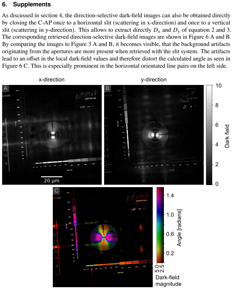
This allows to extract directly𝐷x and 𝐷y of equation 2 and 3
Supplements As discussed in section 4, the direction-selective dark-field images can also be obtained directly by closing the C-AP once to a horizontal slit (scattering in x-direction) and once to a vertical slit (scattering in y-direction). This allows to extract directly𝐷x and 𝐷y of equation 2 and 3. The corresponding retrieved direction-selective dark-...
discussion (0)
Sign in with ORCID, Apple, or X to comment. Anyone can read and Pith papers without signing in.