REVIEW 4 major objections 5 minor 1 cited by
GUSL: A Novel and Efficient Machine Learning Model for Prostate Segmentation on MRI
T0 review · 4 major / 5 minor · reviewed 2026-08-06 · deepseek-v4-flash
Pith's one-line read GUSL, a feed-forward model that learns without backpropagation, rivals or beats deep-learning baselines on prostate gland and zonal segmentation across three MRI datasets while using a fraction of their parameters and computation.
desk verdict Feed-forward Green Learning segmentation architecture worth a look; the efficiency results are strong but the public-benchmark SOTA claim is not yet supported and the abstract overstates the DSC results. read the letter →
The pith
A machine-rendered reading of the paper's core claim, the machinery that carries it, and where it could break.
The reading
What carries the argument
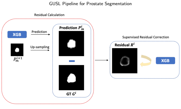
The central mechanism is the multi-level residual-correction regression: at each scale a second regressor learns the residual between the current upsampled prediction and a continuously downsampled ground-truth map, restricted to a boundary ROI, so the model focuses attention on error-prone boundaries. Feature extraction is performed by cascaded VoxelHop units using the channel-wise Saab transform, a data-driven linear transform based on principal component analysis, which the paper argues makes the features inspectable. The predictors are XGBoost regressors, and the overall architecture is a U-shaped cascade with four scales, deployed inside a two-stage pipeline that first localizes the gland at low resolution and then refines the cropped ROI at full resolution.
What would settle it
Retrain the compared deep baselines (such as V-Net, U-Net, and nnU-Net) on exactly the same 5-fold splits and preprocessing as GUSL on ISBI-2013 and PROMISE12, using identical metric computations; if any baseline then matches or exceeds GUSL's reported DSC, HD95, or ABD, the state-of-the-art claim fails. A simpler check is to verify whether the cited baseline papers ([57], [60], [63]) used the same training partitions; if they did not, the comparison is invalid.
Extended reading notes
Core claim
On the paper's own terms, the central discovery is that an all-regression, U-shaped architecture can perform coarse-to-fine 3D segmentation without training a neural network by backpropagation. The encoder builds voxel features through cascaded VoxelHop units that apply the channel-wise Saab transform, a PCA-derived linear transform, in a fully unsupervised way, then refines those features with supervised least-squares normal transforms and feature selection. The decoder is a chain of XGBoost regressors: the deepest one emits an initial low-resolution probability map, and each subsequent level regresses the residual between the upsampled prediction and a continuously downsampled ground truth, while a boundary-focused ROI sampler directs the residual regressor toward error-prone regions. The final probability map is thresholded to produce the segmentation. The authors report that this design reaches DSC above 0.9 for gland segmentation on all three datasets, outperforming reported baselines on ISBI-2013 gland and transition zone, matching or slightly trailing nnU-Net on PROMISE12, and consistently best on a private institutional cohort, while using roughly 16 to 40 times fewer parameters than the compared U-Net variants.
Load-bearing premise
The claim of state-of-the-art performance assumes that the performance numbers reported for the compared deep-learning models in earlier papers are directly comparable to GUSL's own 5-fold cross-validation runs, meaning the data splits, preprocessing, and evaluation metrics are matched across studies.
Editorial extensions
If this is right
- Prostate segmentation on T2-weighted MRI can be performed competitively without a neural network or backpropagation.
- A model with about 1.1 million parameters and 7 GFLOPs can reach DSC above 0.9 for whole-gland segmentation across datasets, making it usable on hardware without high-end GPUs.
- Because the feature transform is linear and the regressors are tree-based, intermediate features and correction maps can be inspected, opening an interpretability route for clinicians.
- The same two-stage, coarse-to-fine residual-correction scheme is presented as a general method extendable to other organs and modalities, with CT and X-ray named in the paper as targets.
- GUSL's fold-to-fold standard deviations are competitive with or lower than those of the deep baselines, indicating stable behavior across training folds.
Reading between the lines
- The residual-correction chain behaves like gradient boosting on masks: each scale fits a regressor to the previous scale's error, so adding more scales or more regressors per scale would likely improve accuracy further at modest parameter cost.
- If the interpretability claim holds, the linear Saab features could be projected back into voxel space to identify which local textures drive boundary decisions—a testable extension the paper does not carry out.
- The energy advantage is robust to protocol mismatch: even if baselines matched GUSL's accuracy under identical splits, GUSL's parameter and FLOPS advantage would still favor it for low-resource deployment.
Editorial analysis
A structured set of objections, weighed in public.
Referee Report
Summary. The paper introduces GUSL, a feed-forward, backpropagation-free 3D segmentation model for prostate MRI, built on the Green Learning paradigm. GUSL combines cascaded VoxelHop units with channel-wise Saab transforms, supervised LNT feature learning, RFT feature selection, and XGBoost regressors that perform coarse-to-fine residual correction. The method is evaluated on ISBI-2013, PROMISE12, and a private USC-Keck dataset for whole-gland, transition-zone, and peripheral-zone segmentation, and is compared with deep learning baselines in terms of DSC, ABD, HD95, model size, and FLOPS.
Significance. If the reported performance holds under matched evaluation protocols, GUSL would be a meaningful contribution to efficient and interpretable medical image segmentation, with an order-of-magnitude reduction in model size and complexity relative to deep learning baselines. The paper provides a novel all-regression, multi-scale residual-correction design and extensive experiments across three datasets. However, the central state-of-the-art claim is weakened by protocol-mismatched comparisons on the public benchmarks, and the abstract overstates the results. The work is of interest to the green learning and medical imaging communities, but the evaluation needs strengthening.
major comments (4)
- [Section 4.4, Tables 5–8] The comparison of GUSL's 5-fold cross-validation results with published baselines from [57], [60], and [63] is not protocol-matched. The paper does not establish that data splits, preprocessing (Lanczos resampling, CLAHE, ROI cropping), and metric computation are identical. Baselines in Tables 5–7 are quoted without standard deviations. For instance, in Table 8, GUSL's HD95 is 1.607 versus nnU-Net's 9.735, a discrepancy that suggests differing metric definitions or evaluation protocols. Consequently, the state-of-the-art claim on ISBI-2013 and PROMISE12 is not supported. Please retrain baselines under the same protocol, or clearly confine the SOTA claim to the Keck cohort and label public-dataset numbers as independent, non-matched comparisons.
- [Abstract, Table 4] The abstract states that 'In all datasets, GUSL achieved a DSC performance greater than 0.9 for gland segmentation.' This is contradicted by Table 4, where the T2-w Keck cohort gland DSC is 0.891 ± 0.070. Also, the abstract's claim of 'state-of-the-art performance among other DL-based models' is not supported by Table 7 (ISBI PZ: 3D Attention U-Net 0.790 vs GUSL 0.774) or Table 8 (PROMISE12: nnU-Net 0.910 vs GUSL 0.907). Please correct these claims in the abstract and Section 5.1.
- [Sections 3.2.1–3.2.5] The description of GUSL omits key hyperparameters needed for reproduction. Specifically, the number of AC anchor vectors per VoxelHop layer, the PCA energy retention ratio, the ROI boundary sampling strategy, the final probability threshold, and the XGBoost hyperparameters (number of estimators, max depth, learning rate) are not reported; only the LNT subset size K=400 is given. Without these values, the experiments are not reproducible and the sensitivity of the results to these choices is unknown. Please provide a complete hyperparameter table and consider releasing code.
- [Table 9] The model size and FLOPS comparison is not apples-to-apples. GUSL's parameter count (1,132,176) includes PCA anchor vectors and XGBoost components, whereas the deep learning baselines are neural networks. The inference cost of XGBoost ensembles (number of trees, depth, sparsity) is not captured by the quoted FLOPS, and the two-stage ROI processing is apparently not included in the GUSL FLOPS estimate. Please provide a per-component breakdown of parameters and complexity, and, if possible, end-to-end inference time and memory usage on the same hardware.
minor comments (5)
- [Section 3.2.6] The probability threshold used for the final segmentation is not specified; please state how it is chosen and whether it is fixed across datasets.
- [Section 4.1] There is a typo: '3.0 mmmm to 4.0mm' should read '3.0 mm to 4.0 mm.'
- [References] References [23] and [42] appear to cite the same Green Learning overview with different publication years; please consolidate to avoid duplicate citations.
- [Section 4.4 and Tables 3–8] The text states that 'we conduct the same experiment setting in both datasets and train V-Net and U-Net-based architectures,' but it is unclear which baseline results were produced by the authors' own training (e.g., Tables 3–4) and which were quoted from prior papers (Tables 5–8). Please clarify the provenance of each baseline result.
- [Section 5.1] The text claims that GUSL 'outperforms by large margins in certain cases,' but some of the reported margins are small (e.g., Table 8: DSC 0.907 vs 0.910). Please make the wording consistent with the magnitude of the differences.
Circularity Check
No significant circularity; GUSL is trained and validated on held-out data, with self-citations to Green Learning components that are not load-bearing.
full rationale
GUSL is a feed-forward regressor trained on labeled prostate MRI volumes and validated with 5-fold cross-validation (Section 4.4). The core quantities—initial coarse masks, residual maps R_i = G_i - P_i^m (Eq. 3), and corrected masks P_i^r = P_i^m + R_i (Eq. 4)—are computed from training-fold ground truths and applied to validation folds; no test label enters training, and the evaluation metrics (DSC, ABD, HD95) are measured against held-out ground truths. The paper cites prior Green Learning work (Saab/VoxelHop/LNT/RFT/PSHop, Refs. [42]-[50]) with overlapping authors, but these citations supply concrete algorithmic components (PCA-based transforms and feature selectors) that are then empirically retrained and evaluated here; no uniqueness theorem is invoked to forbid alternatives, and no fitted parameter is renamed as a prediction. The main risk is an evaluation-protocol mismatch: Tables 5-8 compare GUSL's 5-fold CV numbers to published baseline values without evidence of identical splits, preprocessing (Lanczos, CLAHE, ROI cropping), or metric definitions, and Table 4's DSC 0.891 contradicts the abstract's claim of >0.9 in all datasets. These are correctness and validity concerns, not circularity. Accordingly, no circular step is identified.
Assumptions & free parameters
free parameters (6)
- PCA energy retention / number of AC anchor vectors per VoxelHop layer =
not specified
- LNT number of feature subsets K =
400
- ROI selection boundary sampling strategy =
not specified
- Final probability threshold =
not specified
- XGBoost hyperparameters =
not specified
- Two-stage resolution sizes and CLAHE parameters =
see Table 1
assumptions (4)
- domain assumption PCA-based linear Saab features are sufficient for voxel-wise regression of prostate presence and residual errors
- domain assumption Average-pooled ground truth masks are valid continuous regression targets at each scale
- domain assumption Residual correction learned on ROI-selected samples improves segmentation across all scales
- domain assumption Published baseline results are directly comparable to GUSL's 5-fold cross-validation numbers
Cite this review
Pith. "Pith review of GUSL: A Novel and Efficient Machine Learning Model for Prostate Segmentation on MRI." pith.science (2026). https://pith.science/paper/SJQBX2F7
@misc{pith2026250623688,
  author       = {Pith},
  title        = {Pith review of: GUSL: A Novel and Efficient Machine Learning Model for Prostate Segmentation on MRI},
  year         = {2026},
  howpublished = {\url{https://pith.science/paper/SJQBX2F7}},
  note         = {Machine review of arXiv:2506.23688}
}
abstract
Prostate and zonal segmentation is a crucial step for clinical diagnosis of prostate cancer (PCa). Computer-aided diagnosis tools for prostate segmentation are based on the deep learning (DL) paradigm. However, deep neural networks are perceived as "black-box" solutions by physicians, thus making them less practical for deployment in the clinical setting. In this paper, we introduce a feed-forward machine learning model, named Green U-shaped Learning (GUSL), suitable for medical image segmentation without backpropagation. GUSL introduces a multi-layer regression scheme for coarse-to-fine segmentation. Its feature extraction is based on a linear model, which enables seamless interpretability during feature extraction. Also, GUSL introduces a mechanism for attention on the prostate boundaries, which is an error-prone region, by employing regression to refine the predictions through residue correction. In addition, a two-step pipeline approach is used to mitigate the class imbalance, an issue inherent in medical imaging problems. After conducting experiments on two publicly available datasets and one private dataset, in both prostate gland and zonal segmentation tasks, GUSL achieves state-of-the-art performance among other DL-based models. Notably, GUSL features a very energy-efficient pipeline, since it has a model size several times smaller and less complexity than the rest of the solutions. In all datasets, GUSL achieved a Dice Similarity Coefficient (DSC) performance greater than $0.9$ for gland segmentation. Considering also its lightweight model size and transparency in feature extraction, it offers a competitive and practical package for medical imaging applications.
Figures


Figures from the paper (7 more)




Forward citations
Cited by 1 Pith paper
-
Interpretable and backpropagation-free Green Learning for efficient multi-task echocardiographic segmentation and classification
A backprop-free Green Learning pipeline with a PCA-based VoxelHop encoder and XGBoost decoders achieves 94.3% three-class LVEF accuracy and 0.912 Dice on EchoNet-Dynamic.
Reference graph
Works this paper leans on
-
[57]
A novel prostate segmentation method:triplefusionmodelwithhybridloss
Hakan Ocal and Necaattin Barisci. A novel prostate segmentation method:triplefusionmodelwithhybridloss. NeuralComputingand Applications, 34(16):13559–13574, 2022
work page 2022
-
[60]
Xiangxiang Qin, Yu Zhu, Wei Wang, Shaojun Gui, Bingbing Zheng, and Peijun Wang. 3d multi-scale discriminative network with multi- directionaledgelossforprostatezonalsegmentationinbi-parametric mr images.Neurocomputing, 418:148–161, 2020
work page 2020
-
[63]
Investigation and benchmarking of u-nets on prostate segmentation tasks
Shrajan Bhandary, Dejan Kuhn, Zahra Babaiee, Tobias Fechter, MatthiasBenndorf,ConstantinosZamboglou,Anca-LigiaGrosu,and Radu Grosu. Investigation and benchmarking of u-nets on prostate segmentation tasks. Computerized Medical Imaging and Graphics, 107:102241, 2023
work page 2023
-
[1]
Global cancer statistics 2022: the trends projection analysis
Bhupender S Chhikara, Keykavous Parang, et al. Global cancer statistics 2022: the trends projection analysis. Chemical Biology Letters, 10(1):451–451, 2023
work page 2022
-
[2]
Cancer statistics, 2023.CA: a cancer journal for clinicians, 73(1):17–48, 2023
Rebecca L Siegel, Kimberly D Miller, Nikita Sandeep Wagle, and Ahmedin Jemal. Cancer statistics, 2023.CA: a cancer journal for clinicians, 73(1):17–48, 2023. Jiaxin Yang et al.:Preprint Page 12 of 14 GUSL Pipeline for Prostate Segmentation
work page 2023
-
[3]
AndiKCani,KevinHu,Chia-JenLiu,JavedSiddiqui,YingyeZheng, SuminHan,SrinivasNallandhighal,DanielHHovelson,LanboXiao, Trinh Pham, et al. Development of a whole-urine, multiplexed, next-generationrna-sequencingassayforearlydetectionofaggressive prostate cancer.European urology oncology, 5(4):430–439, 2022
work page 2022
-
[4]
Prostate specific antigen densitycorrelateswithfeaturesofprostatecanceraggressiveness
ShilajitDKundu,KimberlyARoehl,XiaoyingYu,JoAnnVAntenor, Brian K Suarez, and William J Catalona. Prostate specific antigen densitycorrelateswithfeaturesofprostatecanceraggressiveness. The Journal of urology, 177(2):505–509, 2007
work page 2007
-
[5]
Derek W Cool, Jeff Bax, Cesare Romagnoli, Aaron D Ward, Lori Gardi, Vaishali Karnik, Jonathan Izawa, Joseph Chin, and Aaron Fenster. Fusion of mri to 3d trus for mechanically-assisted targeted prostate biopsy: system design and initial clinical experience. In Prostate Cancer Imaging. Image Analysis and Image-Guided Inter- ventions:InternationalWorkshop,He...
work page 2011
Show all 65 references
-
[6]
Prostate zones and cancer: lost in transition?Nature Reviews Urology, 19(2):101–115, 2022
Amin Ali, Alexander Du Feu, Pedro Oliveira, Ananya Choudhury, Robert G Bristow, and Esther Baena. Prostate zones and cancer: lost in transition?Nature Reviews Urology, 19(2):101–115, 2022
2022
-
[7]
Normal central zone of the prostate and central zone in- volvement by prostate cancer: clinical and mr imaging implications
Hebert Alberto Vargas, Oguz Akin, Tobias Franiel, Debra A Gold- man, Kazuma Udo, Karim A Touijer, Victor E Reuter, and Hedvig Hricak. Normal central zone of the prostate and central zone in- volvement by prostate cancer: clinical and mr imaging implications. Radiology, 262(3):...
2012
-
[8]
Prostate tumor delineation using multiparametric magnetic resonance imag- ing:Inter-observervariabilityandpathologyvalidation
Peter Steenbergen, Karin Haustermans, Evelyne Lerut, Raymond Oyen,LiesbethDeWever,LauraVandenBergh,LindaGWKerkmei- jer,FrankAPameijer,WouterBVeldhuis,FlorisJPos,etal. Prostate tumor delineation using multiparametric magnetic resonance imag- ing:Inter-observervariabilityandpath...
2015
-
[9]
A survey of prostate segmentation methodologies in ultrasound, magnetic resonance and computed tomography images
Soumya Ghose, Arnau Oliver, Robert Martí, Xavier Lladó, Joan C Vilanova, Jordi Freixenet, Jhimli Mitra, Désiré Sidibé, and Fabrice Meriaudeau. A survey of prostate segmentation methodologies in ultrasound, magnetic resonance and computed tomography images. Computer methods and...
2012
-
[10]
Challenge of prostate mri segmentation on t2-weighted images: inter-observer variabilityandimpactofprostatemorphology
Sarah Montagne, Dimitri Hamzaoui, Alexandre Allera, Malek Ezziane, Anna Luzurier, Raphaelle Quint, Mehdi Kalai, Nicholas Ayache, Hervé Delingette, and Raphaële Renard-Penna. Challenge of prostate mri segmentation on t2-weighted images: inter-observer variabilityandimpactofpros...
2021
-
[11]
Survey of denoising, segmentation and classification of magnetic resonance imaging for prostate cancer.Multimedia Tools and Ap- plications, 80(19):29199–29249, 2021
Mamta Juneja, Sumindar Kaur Saini, Jatin Gupta, Poojita Garg, Niharika Thakur, Aviral Sharma, Manan Mehta, and Prashant Jindal. Survey of denoising, segmentation and classification of magnetic resonance imaging for prostate cancer.Multimedia Tools and Ap- plications, 80(19):29...
2021
-
[12]
A survey on cancer detection via convolutional neural networks: Current challenges and future directions.Neural Networks, 169:637–659, 2024
Pallabi Sharma, Deepak Ranjan Nayak, Bunil Kumar Balabantaray, Muhammad Tanveer, and Rajashree Nayak. A survey on cancer detection via convolutional neural networks: Current challenges and future directions.Neural Networks, 169:637–659, 2024
2024
-
[13]
U-net:Convo- lutional networks for biomedical image segmentation, 2015
OlafRonneberger,PhilippFischer,andThomasBrox. U-net:Convo- lutional networks for biomedical image segmentation, 2015
2015
-
[14]
Saunet: Shape attentive u-net for interpretable medical image segmentation, 2020
Jesse Sun, Fatemeh Darbehani, Mark Zaidi, and Bo Wang. Saunet: Shape attentive u-net for interpretable medical image segmentation, 2020
2020
-
[15]
nnu-net: Self-adapting frame- work for u-net-based medical image segmentation.arXiv preprint arXiv:1809.10486, 2018
Fabian Isensee, Jens Petersen, Andre Klein, David Zimmerer, Paul F Jaeger, Simon Kohl, Jakob Wasserthal, Gregor Koehler, Tobias No- rajitra, Sebastian Wirkert, et al. nnu-net: Self-adapting frame- work for u-net-based medical image segmentation.arXiv preprint arXiv:1809.10486, 2018
2018 arXiv
-
[16]
Medical image segmentation network based on multi-scale frequency domain filter.Neural Networks, 175:106280, 2024
Yufeng Chen, Xiaoqian Zhang, Lifan Peng, Youdong He, Feng Sun, and Huaijiang Sun. Medical image segmentation network based on multi-scale frequency domain filter.Neural Networks, 175:106280, 2024
2024
-
[17]
Evolution of image segmentation using deep convolutional neural network: A survey
Farhana Sultana, Abu Sufian, and Paramartha Dutta. Evolution of image segmentation using deep convolutional neural network: A survey. Knowledge-Based Systems, 201:106062, 2020
2020
-
[18]
Deeply-supervised cnn for prostate segmentation
Qikui Zhu, Bo Du, Baris Turkbey, Peter L Choyke, and Pingkun Yan. Deeply-supervised cnn for prostate segmentation. In 2017 international joint conference on neural networks (IJCNN), pages 178–184. IEEE, 2017
2017
-
[19]
Attention u-net: Learning where to look for the pancreas.arXiv preprint arXiv:1804.03999, 2018
Ozan Oktay, Jo Schlemper, Loic Le Folgoc, Matthew Lee, Mat- tias Heinrich, Kazunari Misawa, Kensaku Mori, Steven McDonagh, Nils Y Hammerla, Bernhard Kainz, et al. Attention u-net: Learning where to look for the pancreas.arXiv preprint arXiv:1804.03999, 2018
2018 arXiv
-
[20]
A multi-scale channel attention network for prostate segmentation
Meiwen Ding, Zhiping Lin, Chau Hung Lee, Cher Heng Tan, and Weimin Huang. A multi-scale channel attention network for prostate segmentation. IEEETransactionsonCircuitsandSystemsII:Express Briefs, 2023
2023
-
[21]
Explainableaiforhealthcare: from black box to interpretable models
AminaAdadiandMohammedBerrada. Explainableaiforhealthcare: from black box to interpretable models. InEmbedded systems and artificialintelligence:proceedingsofESAI2019,Fez,Morocco ,pages 327–337. Springer, 2020
2020
-
[22]
Energy andpolicyconsiderationsformoderndeeplearningresearch
Emma Strubell, Ananya Ganesh, and Andrew McCallum. Energy andpolicyconsiderationsformoderndeeplearningresearch. In Pro- ceedingsoftheAAAIconferenceonartificialintelligence ,volume34, pages 13693–13696, 2020
2020
-
[23]
Green learning: Introduction, examples and outlook.Journal of Visual Communication and Image Representation, page 103685, 2022
C-C Jay Kuo and Azad M Madni. Green learning: Introduction, examples and outlook.Journal of Visual Communication and Image Representation, page 103685, 2022
2022
-
[24]
Automatic segmentationoftheprostatein3dmrimagesbyatlasmatchingusing localized mutual information
Stefan Klein, Uulke A Van Der Heide, Irene M Lips, Marco Van Vulpen, Marius Staring, and Josien PW Pluim. Automatic segmentationoftheprostatein3dmrimagesbyatlasmatchingusing localized mutual information. Medical physics, 35(4):1407–1417, 2008
2008
-
[25]
A multi- atlas approach for prostate segmentation in mr images
Geert Litjens, Nico Karssemeijer, and Henkjan Huisman. A multi- atlas approach for prostate segmentation in mr images. MICCAI Grand Challenge: Prostate MR Image Segmentation, 2012, 2012
2012
-
[26]
Fast automatic multi-atlas segmentation of the prostate from 3d mr images
JasonADowling,JurgenFripp,ShekharChandra,JosienPWPluim, Jonathan Lambert, Joel Parker, James Denham, Peter B Greer, and Olivier Salvado. Fast automatic multi-atlas segmentation of the prostate from 3d mr images. InInternational Workshop on Prostate Cancer Imaging, pages 10–21....
2011
-
[27]
Prostate mri segmentationusinglearnedsemanticknowledgeandgraphcuts
Dwarikanath Mahapatra and Joachim M Buhmann. Prostate mri segmentationusinglearnedsemanticknowledgeandgraphcuts. IEEE Transactions on Biomedical Engineering, 61(3):756–764, 2013
2013
-
[28]
Medical image segmentation by combining graph cuts and oriented active appearance models.IEEE transactions on image processing, 21(4):2035–2046, 2012
Xinjian Chen, Jayaram K Udupa, Ulas Bagci, Ying Zhuge, and Jianhua Yao. Medical image segmentation by combining graph cuts and oriented active appearance models.IEEE transactions on image processing, 21(4):2035–2046, 2012
2012
-
[29]
Dual optimization based prostate zonal segmentation in 3d mr images.Medical image analysis, 18(4):660–673, 2014
Wu Qiu, Jing Yuan, Eranga Ukwatta, Yue Sun, Martin Rajchl, and Aaron Fenster. Dual optimization based prostate zonal segmentation in 3d mr images.Medical image analysis, 18(4):660–673, 2014
2014
-
[30]
Medical physics, 44(2):558–569, 2017
Zhiqiang Tian, Lizhi Liu, Zhenfeng Zhang, Jianru Xue, and Baowei Fei.Asupervoxel-basedsegmentationmethodforprostatemrimages. Medical physics, 44(2):558–569, 2017
2017
-
[31]
V-net: Fully convolutional neural networks for volumetric medical image segmentation
Fausto Milletari, Nassir Navab, and Seyed-Ahmad Ahmadi. V-net: Fully convolutional neural networks for volumetric medical image segmentation. In 2016 fourth international conference on 3D vision (3DV), pages 565–571. Ieee, 2016
2016
-
[32]
Unet++: Redesigning skip connections to exploitmultiscalefeaturesinimagesegmentation
Zongwei Zhou, Md Mahfuzur Rahman Siddiquee, Nima Tajbakhsh, and Jianming Liang. Unet++: Redesigning skip connections to exploitmultiscalefeaturesinimagesegmentation. IEEEtransactions on medical imaging, 39(6):1856–1867, 2019
2019
-
[33]
3dpbv-net:anautomatedprostatemridataseg- mentationmethod
Yao Jin, Guang Yang, Ying Fang, Ruipeng Li, Xiaomei Xu, Yongkai Liu,andXiaoboLai. 3dpbv-net:anautomatedprostatemridataseg- mentationmethod. Computersinbiologyandmedicine ,128:104160, 2021
2021
-
[34]
Attention-guided multi-scale learning network for automatic prostate and tumor segmentation on mri.Computers in Biology and Medicine, 165:107374, 2023
YuchunLi,YuanyuanWu,MengxingHuang,YuZhang,andZhiming Bai. Attention-guided multi-scale learning network for automatic prostate and tumor segmentation on mri.Computers in Biology and Medicine, 165:107374, 2023. Jiaxin Yang et al.:Preprint Page 13 of 14 GUSL Pipeline for Prostat...
2023
-
[35]
Multiclass ensemble framework for enhanced prostate gland segmentation: Integrating self-onn decoders with efficientnet
Md Shaheenur Islam Sumon, Muhammad EH Chowdhury, Ena- mul Hoque Bhuiyan, Md Sohanur Rahman, Muntakim Mahmud Khan, Israa Al-Hashimi, Adam Mushtak, and Sohaib Bassam Zoghoul. Multiclass ensemble framework for enhanced prostate gland segmentation: Integrating self-onn decoders wi...
2025
-
[36]
A two-stage cnn method for mri image segmentation of prostate with lesion
Zixuan Wang, Ruofan Wu, Yanran Xu, Yi Liu, Ruimei Chai, and He Ma. A two-stage cnn method for mri image segmentation of prostate with lesion. Biomedical Signal Processing and Control, 82:104610, 2023
2023
-
[37]
A dual attention-guided 3d convolution network for automatic segmentation of prostate and tumor.Biomedical Signal Processing and Control, 85:104755, 2023
YuchunLi,MengxingHuang,YuZhang,SilingFeng,JingChen,and Zhiming Bai. A dual attention-guided 3d convolution network for automatic segmentation of prostate and tumor.Biomedical Signal Processing and Control, 85:104755, 2023
2023
-
[38]
3dapa-net:3dadversarialpyramid anisotropic convolutional network for prostate segmentation in mr images
Haozhe Jia, Yong Xia, Yang Song, Donghao Zhang, Heng Huang, YanningZhang,andWeidongCai. 3dapa-net:3dadversarialpyramid anisotropic convolutional network for prostate segmentation in mr images. IEEE transactions on medical imaging, 39(2):447–457, 2019
2019
-
[39]
Cat-net: A cross-slice attention transformer model for prostate zonal segmentation in mri
Alex Ling Yu Hung, Haoxin Zheng, Qi Miao, Steven S Raman, Demetri Terzopoulos, and Kyunghyun Sung. Cat-net: A cross-slice attention transformer model for prostate zonal segmentation in mri. IEEE transactions on medical imaging, 42(1):291–303, 2022
2022
-
[40]
Semi-supervised representation learning for segmentation on medical volumes and sequences.IEEE Transactions on Medical Imaging, 2023
Zejian Chen, Wei Zhuo, Tianfu Wang, Jun Cheng, Wufeng Xue, and Dong Ni. Semi-supervised representation learning for segmentation on medical volumes and sequences.IEEE Transactions on Medical Imaging, 2023
2023
-
[41]
Hd-net:hybriddiscriminativenetworkforprostatesegmentationinmr images
Haozhe Jia, Yang Song, Heng Huang, Weidong Cai, and Yong Xia. Hd-net:hybriddiscriminativenetworkforprostatesegmentationinmr images. In International Conference on Medical Image Computing andComputer-AssistedIntervention ,pages110–118.Springer,2019
2019
-
[42]
Green learning: Introduction, examples and outlook.Journal of Visual Communication and Image Representation, 90:103685, 2023
C-C Jay Kuo and Azad M Madni. Green learning: Introduction, examples and outlook.Journal of Visual Communication and Image Representation, 90:103685, 2023
2023
-
[43]
Interpretable convolutional neural networks via feedforward design
C-C Jay Kuo, Min Zhang, Siyang Li, Jiali Duan, and Yueru Chen. Interpretable convolutional neural networks via feedforward design. Journal of Visual Communication and Image Representation, 2019
2019
-
[44]
In 2023 IEEE 20th International Symposium on Biomedical Imaging (ISBI), pages 1–5
XiaofengLiu,FangxuXing,HannaKGaggin,C-CJayKuo,Georges ElFakhri,andJonghyeWoo.Successivesubspacelearningforcardiac disease classification with two-phase deformation fields from cine mri. In 2023 IEEE 20th International Symposium on Biomedical Imaging (ISBI), pages 1–5. IEEE, 2023
2023
-
[45]
Voxel- hop: Successive subspace learning for als disease classification using structural mri
XiaofengLiu,FangxuXing,ChaoYang,Chung-ChiehJayKuo,Suma Babu,GeorgesElFakhri,ThomasJenkins,andJonghyeWoo. Voxel- hop: Successive subspace learning for als disease classification using structural mri. IEEE journal of biomedical and health informatics, 26(3):1128–1139, 2021
2021
-
[46]
Pca-radhop:Atransparent andlightweightfeed-forwardmethodforclinicallysignificantprostate cancer segmentation.Computerized Medical Imaging and Graphics, 116:102408, 2024
Vasileios Magoulianitis, Jiaxin Yang, Yijing Yang, Jintang Xue, MasatomoKaneko,GiovanniCacciamani,AndreAbreu,VinayDud- dalwar,C-CJayKuo,InderbirSGill,etal. Pca-radhop:Atransparent andlightweightfeed-forwardmethodforclinicallysignificantprostate cancer segmentation.Computerized...
2024
-
[47]
Pshop:Alightweightfeed- forward method for 3d prostate gland segmentation.arXiv preprint arXiv:2403.15971, 2024
Yijing Yang, Vasileios Magoulianitis, Jiaxin Yang, Jintang Xue, MasatomoKaneko,GiovanniCacciamani,AndreAbreu,VinayDud- dalwar,C-CJayKuo,InderbirSGill,etal. Pshop:Alightweightfeed- forward method for 3d prostate gland segmentation.arXiv preprint arXiv:2403.15971, 2024
2024 arXiv
-
[48]
Pixelhop++: A small successive-subspace-learning- based (ssl-based) model for image classification
YueruChen,MozhdehRouhsedaghat,SuyaYou,RaghuveerRao,and C-C Jay Kuo. Pixelhop++: A small successive-subspace-learning- based (ssl-based) model for image classification. In 2020 IEEE International Conference on Image Processing (ICIP), pages 3294–
2020
-
[49]
Enhancing edge intelligence with highly discriminant lnt features
Xinyu Wang, Vinod K Mishra, and C-C Jay Kuo. Enhancing edge intelligence with highly discriminant lnt features. In 2023 IEEE International Conference on Big Data (BigData), pages 3880–3887. IEEE, 2023
2023
-
[50]
On supervised feature selection from high dimensional feature spaces
Yijing Yang, Wei Wang, Hongyu Fu, C-C Jay Kuo, et al. On supervised feature selection from high dimensional feature spaces. APSIPA Transactions on Signal and Information Processing, 11(1), 2022
2022
-
[51]
Xgboost: A scalable tree boosting system
Tianqi Chen and Carlos Guestrin. Xgboost: A scalable tree boosting system. InProceedings of the 22nd acm sigkdd international confer- enceonknowledgediscoveryanddatamining ,pages785–794,2016
2016
-
[52]
NBloch,AMadabhushi,HHuisman,JFreymann,JKirby,MGrauer, AEnquobahrie,CJaffe,LClarke,andKFarahani.Nci-isbi2013chal- lenge: Automated segmentation of prostate structures.The Cancer Imaging Archive, 2015
2015
-
[53]
Evaluation of prostate segmentation algorithms for mri: the promise12 challenge.Medical image analysis, 18(2):359–373, 2014
Geert Litjens, Robert Toth, Wendy Van De Ven, Caroline Hoeks, Sjoerd Kerkstra, Bram Van Ginneken, Graham Vincent, Gwenael Guillard, Neil Birbeck, Jindang Zhang, et al. Evaluation of prostate segmentation algorithms for mri: the promise12 challenge.Medical image analysis, 18(2)...
2014
-
[54]
In Graphics Gems, 1994
KarelJ.Zuiderveld.Contrastlimitedadaptivehistogramequalization. In Graphics Gems, 1994
1994
-
[55]
Comparison and evaluation of methods for liver segmentation from ct datasets.IEEE transactions on medical imaging, 28(8):1251–1265, 2009
Tobias Heimann, Bram Van Ginneken, Martin A Styner, Yulia Arzhaeva, Volker Aurich, Christian Bauer, Andreas Beck, Christoph Becker, Reinhard Beichel, György Bekes, et al. Comparison and evaluation of methods for liver segmentation from ct datasets.IEEE transactions on medical ...
2009
-
[56]
Comparing images using the hausdorff distance.IEEE Trans- actionsonpatternanalysisandmachineintelligence ,15(9):850–863, 1993
DanielPHuttenlocher,GregoryA.Klanderman,andWilliamJRuck- lidge. Comparing images using the hausdorff distance.IEEE Trans- actionsonpatternanalysisandmachineintelligence ,15(9):850–863, 1993
1993
-
[58]
Large kernel matters–improve semantic segmentation by global con- volutional network
Chao Peng, Xiangyu Zhang, Gang Yu, Guiming Luo, and Jian Sun. Large kernel matters–improve semantic segmentation by global con- volutional network. In Proceedings of the IEEE conference on computer vision and pattern recognition, pages 4353–4361, 2017
2017
-
[59]
Voxresnet: Deep voxelwise residual networks for brain segmentation from 3d mr images.NeuroImage, 170:446–455, 2018
Hao Chen, Qi Dou, Lequan Yu, Jing Qin, and Pheng-Ann Heng. Voxresnet: Deep voxelwise residual networks for brain segmentation from 3d mr images.NeuroImage, 170:446–455, 2018
2018
-
[61]
Journal of medical imaging, 6(1):014006–014006, 2019
Md Zahangir Alom, Chris Yakopcic, Mahmudul Hasan, Tarek M Taha,andVijayanKAsari.Recurrentresidualu-netformedicalimage segmentation. Journal of medical imaging, 6(1):014006–014006, 2019
2019
-
[62]
Attention gated networks: Learning to leverage salient regions in medical images
Jo Schlemper, Ozan Oktay, Michiel Schaap, Mattias Heinrich, Bern- hard Kainz, Ben Glocker, and Daniel Rueckert. Attention gated networks: Learning to leverage salient regions in medical images. Medical image analysis, 53:197–207, 2019
2019
-
[64]
3d mri brain tumor segmentation using autoen- coderregularization
Andriy Myronenko. 3d mri brain tumor segmentation using autoen- coderregularization. InInternationalMICCAIbrainlesionworkshop , pages 311–320. Springer, 2018
2018
-
[65]
Prostate segmentation using z-net
Yue Zhang, Jiong Wu, Wanli Chen, Yifan Chen, and Xiaoying Tang. Prostate segmentation using z-net. In2019 IEEE 16th International Symposium on Biomedical Imaging (ISBI 2019), pages 11–14. IEEE, 2019. Jiaxin Yang et al.:Preprint Page 14 of 14
2019
Reviewed August 6, 2026 · model on record in the stance chip above.
Discussion (0). Continue with ORCID to comment.