REVIEW 2 major objections 4 minor 13 references
The fast X-ray detector system of the FAMU experiment at RAL
T0 review · 2 major / 4 minor · reviewed 2026-08-06 · deepseek-v4-flash
Pith's one-line read The FAMU fast X-ray detector system, built on LaBr3:Ce crystals with SiPM array readout, achieves energy resolution comparable to photomultiplier readout and detects muonic oxygen X-ray lines after background subtraction, supporting the…
desk verdict A useful, honest progress report on the FAMU LaBr3:Ce detector system; the lab measurements carry the paper, the in-beam oxygen-line claim is the soft spot. read the letter →
The pith
A machine-rendered reading of the paper's core claim, the machinery that carries it, and where it could break.
The reading
What carries the argument
The central mechanism is the LaBr3:Ce scintillator read out by an array of silicon photomultipliers, with the 1-inch detectors using a '4-1' circuit that groups the outputs of four nearby $6\times6$ mm$^2$ SiPM cells, applies per-group pole-zero compensation and amplification with OPA695 operational amplifiers, and then sums the four sub-array signals and inverts them. This circuit is what compresses the signal fall time by up to a factor of four (used at a conservative factor of two in the final setup) while keeping the energy resolution near 3% at 662 keV, and it is the piece that allows delayed muonic-oxygen X-rays to be separated from the prompt beam background. The system also includes an online gain-drift correction that stabilizes the SiPM response against temperature changes, reducing the variation of the $^{137}$Cs photopeak position from 41% to 5% over a 10-30 C scan.
What would settle it
Re-analyse the December 2023 H$_2$+O$_2$ beam data using a different background estimate, such as interpolating the H$_2$ continuum from time sidebands before the delayed X-ray window or fitting the continuum shape analytically, and check whether the peaks near 133, 158, and 167 keV persist. Alternatively, examine the same target mixture with the HPGe detector, whose $1.26\%$ resolution at 142 keV would resolve the lines cleanly if they are real; absence of the lines in the HPGe spectrum would indicate the LaBr3 signal is a subtraction artefact.
Extended reading notes
Core claim
The paper establishes, on the basis of laboratory and beam tests, that LaBr3:Ce detectors read by SiPM arrays are a viable replacement for photomultiplier readout in the FAMU fast X-ray system. The 1-inch SiPM detectors show a linear response from 100 to 1200 keV, FWHM energy resolution of $2.94 \pm 0.14\%$ at 662 keV and $8.03 \pm 0.39\%$ at 120 keV in the laboratory, and $7.0 \pm 0.3\%$ at the 142 keV muonic silver peak in beam data, compared with $12.3 \pm 1.2\%$ for the PMT-read detectors at that same line. The 4-1 SiPM readout circuit, which groups four $6\times6$ mm$^2$ SiPMs with individual pole-zero compensation and amplification before summing, reduces the 10-90% signal fall time to $147 \pm 13$ ns from $372 \pm 17$ ns for the 1/2-inch parallel-ganged version, a speed-up used conservatively as a factor of two in the experiment. In December 2023 beam data with a H$_2$+O$_2$ (1.5% wt.) gas mixture, the detectors show clear K$\alpha$ and K$\beta$/ $\gamma$ muonic oxygen lines after H$_2$ background subtraction, which is the signature the experiment needs to see the laser-excited hyperfine transition.
Load-bearing premise
The claim of seeing muonic oxygen signal lines in beam data rests on a background subtraction that the authors explicitly note is imperfect, so the lines could in principle be artifacts of that subtraction rather than genuine X-rays.
Editorial extensions
If this is right
- With the demonstrated energy resolution and reduced fall time, the FAMU detector system is capable of recording the delayed muonic-oxygen X-rays that encode the laser-driven hyperfine transition, so the 2023-2024 data can be used to extract the proton Zemach radius.
- The 4-1 SiPM readout design can be applied to other large-area LaBr3:Ce detectors where PMTs are undesirable, giving comparable resolution with a shorter fall time.
- The comparable performance of SiPM and PMT readout means that future detector rings can be built entirely with SiPM readout, simplifying mechanics and lowering cost at equal physics output.
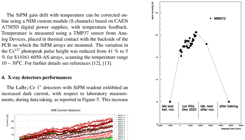
- The recovery of neutron-damaged SiPM arrays by a 24-hour bake at 125 C provides a maintenance procedure that keeps the detector system operational over multiple beam campaigns.
Reading between the lines
- If the residual lines survive a re-analysis with an independent background model (e.g., time-sideband subtraction), the muonic-oxygen detection would be on much firmer ground; the authors' own footnote about imperfect subtraction leaves this as the main open check.
- The fall-time reduction achieved by circuit-level grouping of SiPM cells may generalize to other fast scintillator readouts that need to distinguish prompt and delayed signals in pulsed beams.
- The neutron-damage/baking recovery behaviour suggests that SiPM-based detectors operating near muon targets should budget for periodic annealing runs, a practical design consideration for future experiments.
- Because the HPGe detector has far better energy resolution, correlating its spectra with the LaBr3:Ce spectra for the same target gas could provide an independent cross-check of the oxygen lines without relying on the LaBr3 background subtraction.
Editorial analysis
A structured set of objections, weighed in public.
Referee Report
Summary. The paper describes the fast X-ray detection system built for the FAMU experiment at RAL, which aims to measure the proton Zemach radius via the hyperfine splitting of muonic hydrogen. The system consists of 34 LaBr3:Ce crystals (six with PMT readout, 28 with SiPM array readout) plus one HPGe detector for inter-calibration. The paper reports laboratory measurements of energy resolution and linearity for the SiPM-readout detectors, a dedicated 4-1 readout circuit that reduces signal fall time, a temperature feedback system that controls gain drift, and in-beam data showing muonic-oxygen K-alpha and K-beta/gamma lines after background subtraction. The central conclusion is that the SiPM array readout performs comparably to PMT readout in energy resolution while offering lower cost and flexible geometry.
Significance. If the reported performance is sustained, the FAMU detector system meets the experimental requirements: FWHM energy resolutions of about 3% at 662 keV and 7-8% at 120-142 keV are sufficient to separate the oxygen X-ray lines, and the fall times are compatible with the delayed-signal requirement. The paper contains credible, directly measured laboratory data (linear response over 100-1200 keV, resolution values, and gain-drift reduction from 41% to 5% with temperature feedback) and identifies an operational issue (neutron-induced dark-current increase) together with a recovery procedure. The 4-1 SiPM readout circuit is a useful technical development. These are positive, concrete contributions to detector development for muonic X-ray spectroscopy.
major comments (2)
- [Section 4, Figure 8] The in-beam demonstration that the system detects the muonic-oxygen signal lines rests entirely on a background-subtracted spectrum with no statistical uncertainties, and the footnote in Figure 8 explicitly states that the background subtraction is imperfect, as evidenced by negative values at low energies. The authors do not provide error bars on the subtracted spectrum, a systematic variation of the background normalization, an alternative background model, or a comparison with the HPGe inter-calibration detector for the same runs. Because a small gain shift or normalization mismatch between the H2-only and H2+O2 runs could in principle produce positive residuals near 133, 158, and 167 keV, the claim of 'clear' detection of the oxygen lines is not yet quantitatively supported. Please provide a robustness study (e.g., varying the normalization by ±1–2%), fit the residuals with known line positions to extract significances, or show the same lines in the HPGe data.
- [Table 1] The PMT readout entries (fall time ~60 ns, R 662 keV 3.5–4.6, R 120 keV 7.2–8.1) are quoted as single values or ranges without uncertainties, while the SiPM entries are given as mean ± RMS. Since the paper concludes that SiPM readout 'performs comparably' to PMT readout, the PMT numbers should carry the same statistical treatment (sample mean and spread, number of detectors) or be explicitly referenced to the earlier publication [7]. As written, the comparison between the two readout schemes is not statistically assessable.
minor comments (4)
- [Section 3] The sentence 'scanning the temperature range 10−300C' appears to contain a typo; it should read '10–30 °C'.
- [Figure 4] The bottom panel's y-axis label 'FWHM Energy Resolution [%]' appears to plot fractional values between 0.02 and 0.2, not percentages; either change the label to a fraction or multiply the plotted values by 100.
- [Sections 2 and 3] The SiPM model numbers are inconsistent: Section 2 lists Hamamatsu S4161-6050-04-AS arrays, while Section 3 refers to S14161-6050-AS arrays; please unify the notation.
- [Table 1] For the PMT row, the ranges for R 662 keV and R 120 keV should specify whether they represent the spread over individual detectors, and the number of detectors used for the average should be stated.
Circularity Check
No circularity: the paper reports direct measurements; self-citations provide background details and do not carry the central claims.
full rationale
The paper is an instrumentation and performance report. Its central claims—energy resolution, linearity, fall time, and the in-beam observation of muonic-oxygen lines—are based on laboratory measurements and beam data described in the paper, not on a derivation that assumes its conclusion. The energy resolutions in Table 1 (e.g., 2.94 ± 0.14% at 662 keV for 1-inch SiPM detectors) are measured quantities, not predictions fitted from a model. The in-situ calibration uses known peaks from C, Pb, and Ag to convert ADC to energy; this is standard calibration, not a circular fit of the oxygen lines. Figure 8 shows oxygen K-alpha and K-beta/gamma lines after subtracting an H2-only background; the paper's own footnote admits imperfect background subtraction, which is a data-quality caveat relevant to correctness risk, but it is not a circular reduction. The background subtraction does not assume the presence of the oxygen lines; it subtracts a separately measured spectrum. Self-citations (refs. [8]–[12]) are used for detector construction details and prior characterization, but the performance values in the present paper are independently measured. No load-bearing step reduces to its own input by construction. Score 0: no significant circularity found.
Assumptions & free parameters
free parameters (1)
- Parabolic calibration coefficients per detector =
Not listed in the paper; fitted from Am241, Ba133, Cs137, C, Pb, Ag peaks
assumptions (3)
- domain assumption Muonic oxygen X-ray lines (K-alpha ~133 keV, K-beta ~158 keV, K-gamma ~167 keV) are produced when muons transfer from muonic hydrogen to oxygen.
- domain assumption The SiPM gain drift with temperature can be corrected online using a TMP37 sensor and CAEN power supplies, restoring stable operation.
- domain assumption The increased dark current in SiPM arrays during the 2024 run is due to neutron damage from mu-Au interactions in the silver moderator, and baking restores performance.
Cite this review
Pith. "Pith review of The fast X-ray detector system of the FAMU experiment at RAL." pith.science (2026). https://pith.science/paper/XRWKN7SZ
@misc{pith2026250701819,
  author       = {Pith},
  title        = {Pith review of: The fast X-ray detector system of the FAMU experiment at RAL},
  year         = {2026},
  howpublished = {\url{https://pith.science/paper/XRWKN7SZ}},
  note         = {Machine review of arXiv:2507.01819}
}
abstract
The FAMU experiment at RAL has been designed to study the hyperfine splitting (HFS) of muonic hydrogen and thus measure the Zemach radius of the proton, with a precision better than 1 %. The HFS transition is excited by a tunable MIR laser at ~ 6790 nm and is recognized by delayed ($\mu$O) X-ray emission around 130-170 keV. The fast X-ray detection system is based on 34 scintillating LaBr3:Ce crystals and one HPGe detector for inter-calibration.
Figures


Figures from the paper (3 more)


Reference graph
Works this paper leans on
-
[10]
M. Bonesini et al., Large area LaBr3:Ce crystals read by SiPM arrays with improved timing and temperature drift control, Nucl. Instr. Meth. A1046 (2023) 167677
work page 2023
-
[11]
M. Bonesini et al. , Improving the Time Resolution of Large-Area LaBr3:Ce Detectors with SiPM Array Read- out, Condens. Matter 8 (2023) 99
work page 2023
-
[7]
Baldazzi et al., The LaBr3:Ce based detection system for the FAMU experiment, JINST 12 (2017) C03067
G. Baldazzi et al., The LaBr3:Ce based detection system for the FAMU experiment, JINST 12 (2017) C03067
work page 2017
-
[1]
A. Vacchi et al., Measuring the size of the proton , SPIE Newsroom (2012), DOI:10.1117/2/1201207.004274; A. Adamczack et al., The FAMU experiment at RIKEN- RAL to study the muon transfer rate , JINST 13 (2018) P12033; M. Bonesini, The FAMU experiment at RIKEN RAL for a precise measurement of the proton radius , PoS EPS- HEP2019 (2019) 132. C. Pizzolotto e...
-
[2]
Zemach, Proton structure and the hyperfine shift of hy- drogen, Phys
A. Zemach, Proton structure and the hyperfine shift of hy- drogen, Phys. ReV . 104 (1956) 1771
work page 1956
-
[3]
Pohl et al., The size of the proton , Nature 466 (2010) 213; A
R. Pohl et al., The size of the proton , Nature 466 (2010) 213; A. Antognini et al., Proton Structure from the Measure- ment of 2S − 2P Transition Frequencies of Muonic Hy- drogen,Science 339 (2013) 417
work page 2010
-
[4]
Matsuzaki et al., The RIKEN RAL pulsed muon facility, Nucl
T. Matsuzaki et al., The RIKEN RAL pulsed muon facility, Nucl. Instr. Meth. A465 (2001) 365
work page 2001
-
[5]
Baruzzo et al., A mid-IR laser source for muonic hy- drogen spectroscopy: The FAMU laser system, Opt
M. Baruzzo et al., A mid-IR laser source for muonic hy- drogen spectroscopy: The FAMU laser system, Opt. Laser Techn. 179 (2024) 111375
work page 2024
Show all 13 references
-
[6]
A.Adamczazck et al., Hyperfine spectroscopy of muonic hydrogen and the PSI Lamb shift experiment, Nucl. Instr. Meth. B281 (2012) 72
2012
-
[8]
M. Bonesini et al., 1" LaBr3:Ce dettectors, with tempera- ture control and improved time resolution for low-energy X-rays spectroscopy, Pos(EPS-HEP2023), 547; M.Bonesini et al., Large area LaBr3:Ce crystals read by SiPM arrays with improved timing and gain drift control, Nucl....
2022
-
[9]
Bonesini et al., Detection of low-energy X-rays with 1/2 and 1 inch LaBr 3:Ce crystals read by SiPM arrays , PoS (EPS-HEP2021) 770; M
M. Bonesini et al., Detection of low-energy X-rays with 1/2 and 1 inch LaBr 3:Ce crystals read by SiPM arrays , PoS (EPS-HEP2021) 770; M. Bonesini et al. , Ce:LaBr3 crystals with SiPM array readout and temperature control for the FAMU experi- ment at RAL, JINST 15 (2020) 05, C05065
2020
-
[12]
Bonesini, R
M. Bonesini, R. Bertoni, M. Prata, M. Rossella, Online control of the gain drift with temperature of SiPM ar- rays used for the readout of LaBr3:Ce crystals, JINST 17 (2022) C10004
2022
-
[13]
Otte, Characterization of Three High E fficiency and Blue Sensitive Silicon Photomultipliers Nucl
N. Otte, Characterization of Three High E fficiency and Blue Sensitive Silicon Photomultipliers Nucl. Instr. Meth. A846 (2017) 106. 4
2017
Reviewed August 6, 2026 · model on record in the stance chip above.
Discussion (0). Sign in to comment.