REVIEW 3 major objections 5 minor 40 references
Surface scattering of atoms for high-sensitivity spectroscopy
T0 review · 3 major / 5 minor · reviewed 2026-08-06 · deepseek-v4-flash
Pith's one-line read A single scattering event from a polydimethylsiloxane-coated surface can cool iron atoms from roughly 1400 K to room temperature, and the same surface scatters ytterbium atoms with negligible adsorption down to about 200 K.
desk verdict Useful new measurements of Fe and Yb scattering on PDMS show real cooling, but the single-bounce claim outruns the evidence. read the letter →
The pith
A machine-rendered reading of the paper's core claim, the machinery that carries it, and where it could break.
The reading
What carries the argument
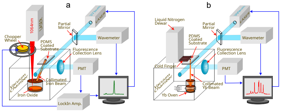
The load-bearing object is the spin-coated, cured PDMS surface (a roughly 1 µm film of Dow Sylgard 184), which combines two properties: a low sticking probability of order $10^{-5}$ (consistent with rubidium measurements on PDMS) so that nearly all incident atoms return to the vacuum, and rapid thermal accommodation so that the returning atoms leave at the surface temperature. The argument's operative event is a single adsorption–desorption cycle; the authors infer this because the collimated beam interacts with the surface only once and the observed fluorescence comes from straight-line paths, with no build-up of atoms in the chamber.
What would settle it
Measure the full velocity distribution of the scattered atoms, for example by isotope-resolved Doppler spectroscopy or time-of-flight detection: if the scattered-iron spectrum shows a non-thermal component at velocities corresponding to 1400 K rather than a single distribution at about 293 K, the single-scattering equilibration claim fails. Alternatively, a quartz-crystal microbalance at 200 K should show measurable ytterbium adsorption; if instead no adsorption is observed at 150 K, the claimed low-temperature cutoff would need revision.
Extended reading notes
Core claim
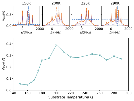
The central discovery is that a thin cured PDMS film acts as a thermalizing, nearly non-sticking surface: hot atoms impinging on it desorb at the surface temperature after a single scattering event. For iron, the measured temperature of the scattered atoms is 285 ± 22 K (mean ± standard deviation over 44 spectra), indistinguishable from the laboratory-temperature substrate at 293 K and a factor of about five below the 1418 ± 130 K estimated for the incoming beam. For ytterbium, fluorescence backgrounds show that scattering persists with roughly constant yield as the substrate is cooled from 290 K to about 200 K, then drops sharply below 190 K, which the authors attribute to embrittlement of PDMS.
Load-bearing premise
The scattered-atom temperature is inferred by fitting one Voigt profile to a broad fluorescence background that actually contains Doppler-broadened contributions from several iron isotopes with different isotope shifts, so the temperature estimate presumes the scattered atoms form a single thermal Maxwell–Boltzmann distribution that one Voigt can represent.
Editorial extensions
If this is right
- A single scattering event can cut an atom's most probable velocity roughly in half (758 m/s at 1970 K oven to about 298 m/s at 298 K), doubling the transit time across an excitation laser and increasing the number of excitation–emission cycles per atom.
- Because the surface preserves flux while cooling, Doppler broadening can be reduced without the flux loss that slit collimation alone would cause.
- The simulated geometry places the slit out of line-of-sight of the ablation point and still delivers about 5.2% of the original flux at 50 MHz Doppler width, a roughly 50% improvement over a slit alone at the same width, but at room temperature rather than 1400 K.
- PDMS-coated surfaces remain effective scattering agents for ytterbium down to roughly 200 K, which extends the useful range of this cooling approach toward cryogenic operation.
- The 'non-stick' property means a coated surface can be placed in an atom beam for extended periods without accumulating a coating that would spoil the surface (no visible ytterbium coating after 200 minutes, versus 162 nm on a bare crystal in 100 minutes).
Reading between the lines
- If single-scattering thermal accommodation is generic, then anti-relaxation-coated vapor cells—where atoms undergo many wall collisions—likely contain atoms already at the wall temperature; this could simplify models of spin relaxation and transit-time broadening in such cells.
- The same mechanism may extend to other refractory elements with high oven temperatures, since the cooling is purely mechanical and does not depend on the atom's internal level structure.
- Tetracontane coatings, which scatter rubidium at temperatures down to 123 K, are a natural testbed for pushing surface-scattering cooling below the 200 K limit observed here for PDMS.
- A direct test of the single-scattering claim would be to compare the temperature of atoms scattered once versus atoms that have undergone multiple bounces; the paper's geometry isolates a single bounce, but a two-surface 'corner' reflector could test whether additional collisions perturb the distribution.
Signed reviews
Editorial analysis
A structured set of objections, weighed in public.
Referee Report
Summary. The paper reports that a polydimethylsiloxane (PDMS)-coated surface scatters hot iron atoms (from laser ablation, initially ~1400 K) and ytterbium atoms (from an effusion oven) and that the scattered atoms appear thermally equilibrated to roughly room temperature (mean 285 K for iron, against a 293 K surface). The authors also report that ytterbium scattering persists down to substrate temperatures near 200 K, that a quartz crystal microbalance shows negligible net ytterbium deposition on PDMS, and that a numerical simulation of a scattering-based atom source can provide collimated flux with reduced velocity and modest flux enhancement compared with a slit alone.
Significance. If the observations hold, the work offers a simple, general method to reduce Doppler broadening and increase transit time in atomic-beam spectroscopy, potentially serving as a first cooling stage for high-temperature atoms. The non-stick property of PDMS for ytterbium down to ~200 K is notable and extends prior work on anti-relaxation coatings. The numerical simulation is a useful proof-of-principle, and the experimental data are direct measurements with a clear spectroscopic approach. However, the strength of the central claims currently exceeds the evidence in three respects: the single-scattering inference, the iron adsorption claim, and the unquantified systematic uncertainty in the Voigt temperature extraction. These issues are addressable by rephrasing or by additional analysis/measurements.
major comments (3)
- [III A, Abstract] The claim that 'a single scattering event is enough for atoms to equilibrate with the PDMS surface temperature' is not supported by the time-integrated fluorescence data. The lock-in detected spectrum measures steady-state fluorescence, so a thermal velocity distribution could result from direct inelastic scattering, from adsorption-desorption with a finite dwell time, or from multiple surface collisions before detection. The argument that 'there is no build-up' rules out a persistent trapped vapor but does not exclude trapping-desorption or multi-bounce trajectories. Since the single-event statement is a headline claim, the authors should either add time-resolved or velocity-selective evidence, or rephrase to state that atoms equilibrate to the surface temperature after scattering without specifying the number of events.
- [III A, Figs. 6 and 7] The temperature of scattered iron atoms is extracted by fitting a single Voigt profile to the background, but the background is a Doppler-broadened superposition of at least four iron isotopes (54Fe, 56Fe, 57Fe, 58Fe), with hyperfine structure for 57Fe. The systematic bias in the fitted Gaussian width from this simplification is not quantified. The reported 285 ± 22 K is the spread over 44 fits, not the total uncertainty. The authors should fit a sum of Voigt functions with known isotope shifts and abundances, or otherwise estimate the model bias, to support the stated temperature and its uncertainty.
- [Abstract, III B] The claim that PDMS shows 'very low adsorption of iron and ytterbium atoms' is only directly measured for ytterbium via the quartz crystal monitor. For iron, the fluorescence data show that a scattered signal exists, but they do not quantify the sticking probability. The abstract and conclusions should restrict the low-adsorption claim to ytterbium unless an iron adsorption measurement is provided.
minor comments (5)
- [III A] "Uncertainity" should be "Uncertainty", and the Fig. 6 caption contains "scatted atoms" instead of "scattered atoms".
- [II B] "15 0 from the normal" should read "15° from the normal".
- [II D] The simulation parameters (surface height, tilt, slit width) are described as chosen for ease of comparison, but the sensitivity of the flux enhancement to these parameters is not discussed; a brief parameter study would strengthen the proof-of-principle claim.
- [III C, Fig. 9] The text says the background intensity maxima are marked with dashed blue lines, but the bottom row shows green triangles and a red dot-dash reference line; the caption and text should be made consistent.
- [Fig. 2] The Grotrian diagram labels a 501.2 nm transition for iron, but the text only uses the 372 nm transition; please verify whether the additional labels are intended.
Circularity Check
No significant circularity: the temperatures are measured directly and the simulation explicitly assumes thermalization rather than deriving it.
full rationale
The paper's derivation chain contains no step in which an output is equivalent to an input by construction. Scattered-atom temperatures (Section III A) are extracted by fitting Voigt profiles to fluorescence backgrounds; the fitted width is compared with the independently known surface temperature, so the reported 285 K is a measurement rather than a parameter recycled into the claim. The numerical source design (Section II D) explicitly states that atoms 'are further assumed to thermally equilibrate with the surface, and scatter at room temperature'; because the simulation adopts, rather than purports to prove, the thermalization result, its flux and Doppler-width outputs are a design calculation and not a disguised prediction of the same physics. The single-scattering inference ('The experiments suggest that the temperature equilibration occurs through a single scattering event...') is geometrically motivated and is not an equation whose conclusion is embedded in its premise; if steady-state fluorescence cannot distinguish single-bounce from trapping-desorption or multi-bounce histories, that is an evidence limitation, not circularity. The only near-self citation, Ref. [39] for the Yb oven, is not load-bearing. The paper also candidly notes that Yb temperature could not be fitted with a single Voigt, using background intensity instead. No self-definitional, fitted-input-as-prediction, imported-uniqueness, ansatz-by-citation, or renaming pattern is present. Hence no circularity is found.
Assumptions & free parameters
free parameters (2)
- Voigt Gaussian width for scattered-atom background =
285 K equivalent (mean over 44 fits, std dev 22 K)
- Simulation geometry parameters (surface height, tilt, slit width) =
4 mm above ablation point, 45 degrees tilt, 1.2 mm slit width
assumptions (4)
- domain assumption Atoms in the collimated beam have a Maxwell-Boltzmann velocity distribution at a single temperature.
- domain assumption Ablation flux follows a cosine angular distribution relative to the surface normal.
- domain assumption Scattered atoms thermally equilibrate with the PDMS surface before leaving.
- domain assumption Each point on the scattering surface acts as an independent diffuse point source.
Cite this review
Pith. "Pith review of Surface scattering of atoms for high-sensitivity spectroscopy." pith.science (2026). https://pith.science/paper/DE4KDBIS
@misc{pith2026250709951,
  author       = {Pith},
  title        = {Pith review of: Surface scattering of atoms for high-sensitivity spectroscopy},
  year         = {2026},
  howpublished = {\url{https://pith.science/paper/DE4KDBIS}},
  note         = {Machine review of arXiv:2507.09951}
}
read the original abstract
High-sensitivity laser spectroscopy is integral to applications like atomic clocks, quantum computers, and chemical sensing. Lowering atomic temperature decreases spectral Doppler broadening and increases transit time across the excitation laser. We find that a polydimethylsiloxane (PDMS) coated surface can cool iron atoms from ~1400 K to room temperature by surface scattering. It is also apparent that a single scattering event is enough for atoms to equilibrate with the PDMS surface temperature. Further, we show the very low adsorption of iron and ytterbium atoms on PDMS, an effect that persists to surface temperatures of 200 K. Through numerical simulation, we demonstrate the potential use of surface scattering in making a room temperature source of collimated atoms with enhanced flux and reduced velocity compared to that without surface scattering.
Figures


Figures from the paper (6 more)


Reference graph
Works this paper leans on
-
[1]
W. Demtr¨ oder, Applications of Laser Spectroscopy, in Laser Spectroscopy 2: Experimental Techniques (Springer Berlin Heidelberg, 2015) pp. 589–650
work page 2015
-
[2]
A. D. Ludlow, M. M. Boyd, J. Ye, E. Peik, and P. Schmidt, Optical atomic clocks, Rev. Mod. Phys. 87, 637 (2015)
work page 2015
-
[3]
Derevianko and H
A. Derevianko and H. Katori, Colloquium: Physics of optical lattice clocks, Rev. Mod. Phys. 83, 331 (2011)
2011
-
[4]
T. D. Ladd, F. Jelezko, R. Laflamme, Y. Nakamura, C. Monroe, and J. L. O’Brien, Quantum computers, Nature 464, 45 (2010). 16
work page 2010
-
[5]
T. Walker and P. Feng, Measurements of Collisions Between Laser-Cooled Atoms, inAdvances In Atomic, Molecular, and Optical Physics, Vol. 34, edited by B. Bederson and H. Walther (Academic Press, 1994) pp. 125–170
work page 1994
-
[6]
P. R. Berman, Study of Collisions by Laser Spectroscopy, inAdvances in Atomic and Molecular Physics, Vol. 13, edited by D. R. Bates and B. Bederson (Academic Press, 1978) pp. 57–112
work page 1978
-
[7]
R. G. Brewer, Study of atom—wall collisions by optical pumping, The Journal of Chemical Physics 38, 3015 (1963)
work page 1963
-
[8]
E. Kerstel and L. Gianfrani, Advances in laser-based isotope ratio measurements: selected applications, Appl. Phys. B 92, 439 (2008)
work page 2008
Show all 40 references
-
[9]
R. S. Harmon, F. C. DeLucia, C. E. McManus, N. J. McMillan, T. F. Jenkins, M. E. Walsh, and A. Miziolek, Laser-induced breakdown spectroscopy – An emerging chemical sensor technol- ogy for real-time field-portable, geochemical, mineralogical, and environmental applications, Ap...
2006
-
[10]
DeLucia, A
F. DeLucia, A. Samuels, R. Harmon, R. Walters, K. McNesby, A. LaPointe, R. Winkel, and A. Miziolek, Laser-induced breakdown spectroscopy (LIBS): a promising versatile chemical sensor technology for hazardous material detection, IEEE Sensors Journal 5, 681 (2005)
2005
-
[11]
Wysocki and D
G. Wysocki and D. Weidmann, Molecular dispersion spectroscopy for chemical sensing using chirped mid-infrared quantum cascade laser, Opt. Express, OE 18, 26123 (2010)
2010
-
[12]
N. Huet, M. Pettens, and T. Bastin, Isotope shifts and hyperfine structure of the laser-cooling Fe I 358-nm line, Phys. Rev. A 92, 052507 (2015)
2015
-
[13]
W. D. Phillips, Nobel Lecture: Laser cooling and trapping of neutral atoms, Rev. Mod. Phys. 70, 721 (1998)
1998
-
[14]
Degen, F
C. Degen, F. Reinhard, and P. Cappellaro, Quantum sensing, Rev. Mod. Phys. 89, 035002 (2017)
2017
-
[15]
M. H. Anderson, J. R. Ensher, M. R. Matthews, C. E. Wieman, and E. A. Cornell, Observation of Bose-Einstein Condensation in a Dilute Atomic Vapor, Science 269, 198 (1995)
1995
-
[16]
J. V. Prodan and W. D. Phillips, Chirping the light—fantastic? Recent NBS atom cooling experiments, Progress in Quantum Electronics 8, 231 (1984)
1984
-
[17]
N. R. Hutzler, H.-I. Lu, and J. M. Doyle, The Buffer Gas Beam: An Intense, Cold, and Slow Source for Atoms and Molecules, Chem. Rev. 112, 4803 (2012). 17
2012
-
[18]
O. Kock, W. He, D. ´Swierad, L. Smith, J. Hughes, K. Bongs, and Y. Singh, Laser controlled atom source for optical clocks, Scientific reports 6, 37321 (2016)
2016
-
[19]
Yasuda, T
M. Yasuda, T. Tanabe, T. Kobayashi, D. Akamatsu, T. Sato, and A. Hatakeyama, Laser- controlled cold ytterbium atom source for transportable optical clocks, Journal of the Physical Society of Japan 86, 125001 (2017)
2017
-
[20]
Stephens, R
M. Stephens, R. Rhodes, and C. Wieman, Study of wall coatings for vapor-cell laser traps, Journal of Applied Physics 76, 3479 (1994)
1994
-
[21]
H. M. Goldenberg, D. Kleppner, and N. F. Ramsey, Atomic Beam Resonance Experiments with Stored Beams, Phys. Rev. 123, 530 (1961)
1961
-
[22]
Rahman and H
C. Rahman and H. Robinson, Rb oo hyperfine transition in evacuated wall-coated cell at melting temperature, IEEE journal of quantum electronics 23, 452 (1987)
1987
-
[23]
Y. Yi, H. Robinson, S. Knappe, J. Maclennan, C. Jones, C. Zhu, N. Clark, and J. Kitching, Method for characterizing self-assembled monolayers as antirelaxation wall coatings for alkali vapor cells, Journal of Applied Physics 104 (2008)
2008
-
[24]
H. Chi, W. Quan, J. Zhang, L. Zhao, and J. Fang, Advances in anti-relaxation coatings of alkali-metal vapor cells, Applied Surface Science 501, 143897 (2020)
2020
-
[25]
Sekiguchi, A
N. Sekiguchi, A. Hatakeyama, K. Okuma, and H. Usui, Scattering of an alkali-metal atomic beam on anti-spin-relaxation coatings, Phys. Rev. A 98, 042709 (2018)
2018
-
[26]
Wu, Wall interactions of spin-polarized atoms, Rev
Z. Wu, Wall interactions of spin-polarized atoms, Rev. Mod. Phys. 93, 035006 (2021)
2021
-
[27]
Liberman and R
V. Liberman and R. Knize, Relaxation of optically pumped cs in wall-coated cells, Physical Review A 34, 5115 (1986)
1986
-
[28]
Budker, L
D. Budker, L. Hollberg, D. F. Kimball, J. Kitching, S. Pustelny, and V. V. Yashchuk, Mi- crowave transitions and nonlinear magneto-optical rotation in anti-relaxation-coated cells, Physical Review A—Atomic, Molecular, and Optical Physics 71, 012903 (2005)
2005
-
[29]
S. N. Atutov and A. I. Plekhanov, Accurate measurement of the sticking time and sticking probability of Rb atoms on a polydimethylsiloxane coating, J. Exp. Theor. Phys.120, 1 (2015)
2015
-
[30]
M. A. Bouchiat and J. Brossel, Relaxation of Optically Pumped Rb Atoms on Paraffin-Coated Walls, Phys. Rev. 147, 41 (1966)
1966
-
[31]
K. F. Zhao, M. Schaden, and Z. Wu, Method for Measuring the Dwell Time of Spin-Polarized Rb Atoms on Coated Pyrex Glass Surfaces Using Light Shift, Phys. Rev. Lett. 103, 073201 (2009). 18
2009
-
[32]
Ulanski and Z
E. Ulanski and Z. Wu, Measurement of dwell times of spin polarized rubidium atoms on octadecyltrichlorosilane- and paraffin-coated surfaces, Applied Physics Letters 98, 201115 (2011)
2011
-
[33]
Gozzini, F
A. Gozzini, F. Mango, J. H. Xu, G. Alzetta, F. Maccarrone, and R. A. Bernheim, Light- induced ejection of alkali atoms in polysiloxane coated cells, Il Nuovo Cimento D 15, 709 (1993)
1993
-
[34]
S. N. Atutov, V. Biancalana, P. Bicchi, C. Marinelli, E. Mariotti, M. Meucci, A. Nagel, K. A. Nasyrov, S. Rachini, and L. Moi, Light-induced diffusion and desorption of alkali metals in a siloxane film: Theory and experiment, Phys. Rev. A 60, 4693 (1999)
1999
-
[35]
Asakawa, Y
K. Asakawa, Y. Tanaka, K. Uemura, N. Matsuzaka, K. Nishikawa, Y. Oguma, H. Usui, and A. Hatakeyama, Measurement of the temperature dependence of dwell time and spin relaxation probability of Rb atoms on paraffin surfaces using a beam-scattering method, Phys. Rev. A 104, 063106 (2021)
2021
-
[36]
S 1) and more details of the numerical simulation showing the flux enchancement
See supplemental material at [URL will be inserted by publisher] for an optical image of the spin-coated PDMS surface(Fig. S 1) and more details of the numerical simulation showing the flux enchancement
-
[37]
D. B. Hall, P. Underhill, and J. M. Torkelson, Spin coating of thin and ultrathin polymer films, Polymer Engineering & Science 38, 2039 (1998)
1998
-
[38]
Braˇ ciˇ c, T
M. Braˇ ciˇ c, T. Mohan, R. Kargl, T. Griesser, S. Hribernik, S. K¨ ostler, K. Stana-Kleinschek, and L. Fras-Zemljiˇ c, Preparation of PDMS ultrathin films and patterned surface modification with cellulose, RSC Advances 4, 11955 (2014)
2014
-
[39]
Bucay, Surface Ionization of Metastable Calcium and Ytterbium Atoms, Ph.D., The Uni- versity of Texas at Austin, United States – Texas (2019), iSBN: 9798684608384
I. Bucay, Surface Ionization of Metastable Calcium and Ytterbium Atoms, Ph.D., The Uni- versity of Texas at Austin, United States – Texas (2019), iSBN: 9798684608384
2019
-
[40]
Zhang, Y
G. Zhang, Y. Sun, B. Qian, H. Gao, and D. Zuo, Experimental study on mechanical perfor- mance of polydimethylsiloxane (PDMS) at various temperatures, Polymer Testing 90, 106670 (2020). 19
2020
Reviewed August 6, 2026 · model on record in the stance chip above.
Discussion (0). Continue with ORCID to comment.