REVIEW 3 major objections 4 minor 41 references
Optics for broadband x-ray ptychography
T0 review · 3 major / 4 minor · reviewed 2026-08-15 · deepseek-v4-flash
Pith's one-line read Smaller zone plates widen x-ray ptychography's usable bandwidth, per a new chromatic-error metric.
desk verdict Useful parameter-free relation and honest instrument study, but the 'small FZP is best' claim is conditional on an under-review companion and on a probe-size metric that is thinner than the paper's own coherence requirement. read the letter →
The pith
A machine-rendered reading of the paper's core claim, the machinery that carries it, and where it could break.
The reading
What carries the argument
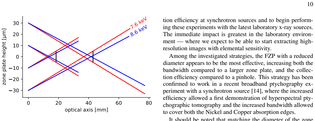
The central object is the Fresnel zone plate, a diffractive lens whose focal length scales inversely with wavelength, $f_\lambda = D_{\mathrm{FZP}}\Delta_{\mathrm{FZP}}/n\lambda$, which is what makes the probe size chromatic. The argument runs through the geometric probe-size formula $D_\lambda(z)=|n z \lambda/\Delta_{\mathrm{FZP}} - D_{\mathrm{FZP}}|$ and the Nyquist sampling bound $D^*_\lambda = Z_{\mathrm{det}}\lambda/(2\Delta_{\mathrm{det}})$. Combining them yields Eq. 4, the probe-size error metric whose linear dependence on $D_{\mathrm{FZP}}$ carries the recommendation to shrink the zone plate.
What would settle it
Set up a small FZP and measure the probe diameter at the sample plane across an energy band broad enough that the highest-energy focus lies upstream of the sample; if the relative probe-size variation grows faster than $|\lambda'-\lambda|/\lambda$ in that regime, the linear scaling of Eq. 4 fails there.
Extended reading notes
Core claim
The central claim is that the chromatic probe-size problem in broadband ptychography is quantified by the error metric $$\varepsilon = \left|\frac{\$\lambda$'-\$\lambda$}{\$\lambda$}\right|\frac{D_{\mathrm{FZP}}}{D^*_\$\lambda$},$$ where $\lambda$ is a reference wavelength, $\lambda'$ another wavelength in the band, $D_{\mathrm{FZP}}$ the zone plate diameter, and $D^*_\lambda$ the largest probe diameter allowed by the Nyquist sampling condition at that wavelength. Because $\varepsilon$ is directly proportional to $D_{\mathrm{FZP}}$, reducing the zone plate diameter is the direct lever for accepting a wider bandwidth without increasing probe-size variation. The paper argues that a small-diameter FZP retains the three useful properties of a larger one, controlled probe size, a diverging disc that fills the detector, and high spatial frequencies in the probe, and that its diffraction efficiency is independent of the number of zones, so the main trade-offs are the smaller collecting area and the fraction blocked by the central stop. Tested against pinhole-based alternatives, the small FZP proved most effective, and the strategy was used in a broadband ptychotomography experiment covering both nickel and copper absorption edges.
Load-bearing premise
The linear error formula in Eq. 4 holds only while every wavelength in the band is still on the converging side of its focus at the chosen sample plane; for very broad bands where the shortest wavelengths have already passed focus, the probe-size error grows differently and the diameter-reduction rule may no longer apply.
Editorial extensions
If this is right
- Reducing the FZP diameter by a given factor raises the tolerable relative bandwidth by the same factor for a fixed probe-size variation, according to Eq. 4.
- A small FZP keeps the converging-then-diverging beam shape and the diffraction efficiency of a larger zone plate, avoiding the pinhole's high dynamic range while retaining efficient detector use.
- The chromatic focal shift combined with an order-sorting aperture can act as a tunable spectral filter, letting a sequence of small FZPs select which energy range reaches the detector.
- The strategy has already been used in a broadband ptychotomography experiment covering both nickel and copper absorption edges in a single acquisition.
- At lower-brilliance laboratory sources, matching the FZP diameter to the coherence length keeps the setup compact while preserving coherent flux.
Reading between the lines
- An implication the authors leave implicit is that Eq. 4's linear scaling holds only while every wavelength in the band is still converging at the chosen sample plane; for ultra-broad bands where short-wavelength probes have already passed focus, the error metric changes and the optimal diameter may differ from the linear rule.
- If hyperspectral detector count-rate limits improve substantially, the small-FZP efficiency trade-off caused by the central stop blocking a larger fraction of the aperture becomes more significant, and the ranking of optics strategies could shift.
- The same diameter-reduction logic could extend to other diffractive optics in visible-light or EUV multispectral ptychography, where chromatic probe spread similarly constrains bandwidth.
- A testable extension would combine a small FZP with multi-beam ptychography to recover the lost coherent flux from the reduced aperture, making the bandwidth gain and throughput gain additive.
Signed reviews
Editorial analysis
A structured set of objections, weighed in public.
Referee Report
Summary. The manuscript analyzes pre-sample focusing optics for broadband x-ray ptychography with hyperspectral detectors. It derives a chromatic probe-size error metric ε = (|λ′−λ|/λ)(D_FZP/D*_λ) for Fresnel zone plates, argues that reducing the zone-plate diameter is the most effective way to broaden the tolerable bandwidth while preserving collection efficiency, and compares this strategy with pinhole-plus-attenuator and pinhole-plus-diffuser schemes. Experimental data and wave-propagation simulations are used to support the comparative assessment, and the conclusion is that a reduced-diameter FZP increases both bandwidth and collection efficiency relative to larger zone plates and pinholes.
Significance. If the central scaling relation and the comparative ranking hold, the paper provides a simple and practical optics rule for broadband ptychography, which is directly relevant to the development of laboratory-scale and synchrotron hyperspectral imaging. The paper is honest about trade-offs and includes wave-optical simulations and experimental diffraction-pattern analysis. However, the quantitative centerpiece, Eq. (4), is derived without stating a necessary branch condition, and the 'most effective' conclusion rests on a geometric size metric rather than a full demonstration of complex-probe stability within energy bins. The paper therefore offers a useful framework and a plausible recommendation, but the support for the headline claim is incomplete as presented.
major comments (3)
- [Section IV D, Eq. (4)] The derivation of ε = (|λ′−λ|/λ)(D_FZP/D*_λ) silently assumes that both λ and λ′ lie on the same branch of the absolute value in Eq. (2). If the reference wavelength is on the converging branch, the equality holds only for r = λ′/λ ≤ D_FZP/(D_FZP − D*_λ); if on the diverging branch, it holds only for r ≥ D_FZP/(D_FZP + D*_λ). Outside these ranges the probe-size error takes a different form and is not proportional to D_FZP. The manuscript never states this bandwidth limit, even though it is load-bearing for the central recommendation; for example, with D_FZP = 200 µm and D*_λ = 10 µm, a wavelength 10% longer than λ is already outside the valid range, and the claimed proportionality to D_FZP does not hold.
- [Section IV D and Section V] The metric ε quantifies only the probe diameter, whereas Section III explicitly states that the complete complex probe—amplitude and phase—must be stable within each energy bin for the ptychographic single-wavefield model to be a good fit. The wavefield simulations in Figure 6 display only magnitude profiles, not phase, and no full ptychographic reconstruction using broadband, energy-binned data with a reduced-diameter FZP is presented in this manuscript. The claim that this strategy is 'the most effective' therefore rests on a geometric proxy and on the under-review companion paper [14]. A full-wavefield test, even for one representative band, is needed to substantiate the causal chain from Eq. (4) to reconstruction fidelity.
- [Section IV C] The conclusion that all tested diffusers are 'antithetical' to the stated aim is drawn from a single set of diffraction patterns that the authors themselves describe as 'not suitable for reconstruction' (page 8). The homogeneity metric mean/max is reported without error bars or repeated measurements, so the observed reductions could be within experimental noise. This is a strong negative conclusion for a whole strategy and needs more quantitative support.
minor comments (4)
- [Section IV D, Eq. (4)] The resolution of the absolute value in Eq. (2) that leads to Eq. (4) should be stated explicitly, including the branch condition, so that the valid bandwidth range is transparent to the reader.
- [Figure 5 caption] The caption states that the 7.6 keV beam creates a 10 µm probe, but it does not specify whether the sample plane is on the converging or diverging branch of the focus. Adding this detail would make the example reproducible.
- [Section IV C] The estimate that a sample-plane autocorrelation FWHM of about 300 nm is needed to fill a 1 cm detector after 10 m propagation is useful, but the calculation is presented without the underlying formula; a one-line derivation would improve clarity.
- [References] Reference [21] contains an unmatched parenthesis in the text where it is cited ('[21])'), which should be corrected.
Circularity Check
Equation 4 is an algebraic consequence of textbook FZP optics with no fitted constants; the only caveat is a supporting self-citation to an under-review companion paper.
full rationale
The central derivation is self-contained. Equation 4 follows from Equation 2 (geometric FZP probe size) and Equation 3 (Nyquist sampling limit) by direct algebra under the stated assumption D_lambda(z_lambda) = D*_lambda and with all wavelengths on the converging branch; no parameter is fitted to data and no conclusion is imported from a prior paper. The reduced-diameter-FZP recommendation is therefore not circular in its main line. The attenuator transmission used in Section IV B is calibrated on the same diffraction data it corrects, an in-loop calibration, but the paper explicitly treats it as a correction rather than as a prediction, and it does not support the central optics recommendation. The only noteworthy self-citation issue is reference [14] (Stolp et al., under review): the conclusion says the small-FZP strategy 'has been confirmed to work' in that paper. Since [14] is not available or independently checkable here, this confirmation is a supporting self-citation, but it is not load-bearing for Equation 4 or for the geometric argument; the central derivation remains independent of it. Accordingly, no equation-to-equation reduction or fitted-parameter-renamed-as-prediction circularity is present, and the paper's core claim is honestly non-circular.
Assumptions & free parameters
free parameters (1)
- Soft attenuator transmission profile =
Not quoted; characterized from diffraction data
assumptions (6)
- standard math FZP focal length f_λ = D Δ/(n λ) for diffraction order n (Eq. 1).
- domain assumption Geometrical probe size D_λ(z) = |n z λ/Δ − D_FZP| (Eq. 2) is valid in the parallel sample plane and beyond focus.
- domain assumption Nyquist sampling condition D*_λ = Z_det λ / (2 Δ_det) (Eq. 3).
- ad hoc to paper At the sample plane z_λ, all wavelengths in the band remain on the converging branch of Eq. 2, so D_λ′(z_λ) = D − n z_λ λ′/Δ.
- domain assumption Diffraction efficiency of an FZP is independent of the number of zones.
- domain assumption The mean/max pixel ratio is a valid proxy for detector saturation-limited flux capture.
Cite this review
Pith. "Pith review of Optics for broadband x-ray ptychography." pith.science (2026). https://pith.science/paper/AXPD3UD4
@misc{pith2026250816953,
  author       = {Pith},
  title        = {Pith review of: Optics for broadband x-ray ptychography},
  year         = {2026},
  howpublished = {\url{https://pith.science/paper/AXPD3UD4}},
  note         = {Machine review of arXiv:2508.16953}
}
read the original abstract
In conventional x-ray ptychography, diffraction data is collected by scanning a sample through a monochromatic, and spatially coherent, x-ray beam. A high-resolution image is then retrieved using an iterative algorithm. Combined with a scan of the incident photon energy, it is also possible to access chemical and elemental information. Although powerful, the high brilliance required currently constrains the method to 3rd and 4th generation synchrotron sources and long scanning times. An alternative approach is to use a broadband illumination in combination with an energy resolving detector. These detectors record the data in a series of energy channels simultaneously, creating a stack of coherent data suitable for a ptychographic reconstruction. This approach promises to unlock the full power of the radiation source and provide spectral imaging at a higher rate and in a single acquisition. However, these detectors currently saturate well below reaching the flux rates produced at synchrotrons, which is preventing the uptake of this approach. Furthermore, current monochromatic synchrotron setups typically employ Fresnel zone plates for pre-sample focusing due to their stability, flexibility, and affordability, but these diffractive optics limit the spectral bandwidth that the setup can accept. In this article, we analyze the problem and consider alternative optics that can both maximize the total photon detection rates and broaden the tolerable bandwidth. Broadband x-ray ptychography has the potential to dramatically reduce data collection times at synchrotron sources, but also to harness the full power of lower brilliance sources and transition x-ray ptychography into a laboratory technique.
Figures


Figures from the paper (3 more)


Reference graph
Works this paper leans on
-
[14]
L. Loetgering, S. Witte, and J. Rothhardt, Advances in laboratory-scale ptychography using high harmonic sources [Invited], Optics Express 30, 4133 (2022)
work page 2022
-
[1]
Probe size control. In the sampling theory of coher- ent diffractive imaging, the diameter of the probe at the sample is an important parameter [13, 16] since larger probes allow for a larger step size in the ptychographic scan, but also cause smaller fringes in the diffraction patterns in the detector plane. These fringes require finer sampling, which co...
-
[2]
Detector plane beam spreading. The FZP produces a converging beam that transitions into a diverging beam beyond focus, forming a relatively uniform, disc- shaped intensity distribution in the far field. When the numerical aperture of the FZP is sufficiently large and the detector is sufficiently far, this uniform disc occu- pies a significant portion of t...
-
[3]
High spatial frequencies. The diverging probe wave- front contains (by its very nature) high spatial frequen- cies, which has been shown to benefit reconstruction quality [17]. This is different from point 1, which con- cerned the spatial frequencies contained in the far field diffraction pattern. III. FRESNEL ZONE PLATE CHROMATICITY IN BROADBAND PTYCHOGR...
-
[4]
A. M. Maiden, G. R. Morrison, B. Kaulich, A. Gianoncelli, and J. M. Rodenburg, Soft X-ray spectromicroscopy using ptychog- raphy with randomly phased illumination, Nature Communica- tions 4, 10.1038/ncomms2640 (2013)
- [5]
-
[6]
D. J. Batey, S. Cipiccia, F. Van Assche, S. Vanheule, J. Van- mechelen, M. N. Boone, and C. Rau, Spectroscopic imaging with single acquisition ptychography and a hyperspectral de- tector, Scientific Reports 9, 1 (2019)
work page 2019
-
[7]
J. M. Rodenburg, A. C. Hurst, A. G. Cullis, B. R. Dobson, F. Pfeiffer, O. Bunk, C. David, K. Jefimovs, and I. Johnson, Hard-X-Ray Lensless Imaging of Extended Objects, Physical Review Letters 98, 034801 (2007)
work page 2007
Show all 41 references
-
[8]
Aidukas, N
T. Aidukas, N. W. Phillips, A. Diaz, E. Poghosyan, E. Müller, A. F. J. Levi, G. Aeppli, M. Guizar-Sicairos, and M. Holler, High-performance 4-nm-resolution X-ray tomography using burst ptychography, Nature 632, 81 (2024)
2024
-
[9]
Beckers, T
M. Beckers, T. Senkbeil, T. Gorniak, M. Reese, K. Gieweke- meyer, S. C. Gleber, T. Salditt, and A. Rosenhahn, Chemical contrast in soft X-ray ptychography, Physical Review Letters 10.1103/PhysRevLett.107.208101 (2011)
2011 doi
-
[10]
P. J. Duke, Synchrotron Radiation Production and Properties (Oxford University Press, 2000)
2000
-
[11]
can mitigate this problem, and has already enabled the first lab based ptychography experiment with a hyperspectral detector [12]. In addition to the spectral imaging capabili- ties introduced by the detector, a larger part of the source flux can be put to use in this configur...
-
[12]
D. J. Batey, F. Van Assche, S. Vanheule, M. N. Boone, A. J. Parnell, O. O. Mykhaylyk, C. Rau, and S. Cipiccia, X-Ray Pty- chography with a Laboratory Source, Physical Review Letters 126, 193902 (2021)
2021
-
[13]
Ordavo, S
I. Ordavo, S. Ihle, V . Arkadiev, O. Scharf, H. Soltau, A. Bjeoumikhov, S. Bjeoumikhova, G. Buzanich, R. Gubzhokov, A. Günther, R. Hartmann, P. Holl, N. Kimmel, M. Kühbacher, M. Lang, N. Langhoff, A. Liebel, M. Radtke, U. Reinholz, H. Riesemeier, G. Schaller, F. Schopper, L. S...
2011
-
[15]
Zhang, D
B. Zhang, D. F. Gardner, M. H. Seaberg, E. R. Shanblatt, C. L. Porter, R. Karl, C. A. Mancuso, H. C. Kapteyn, M. M. Mur- nane, and D. E. Adams, Ptychographic hyperspectral spectro- microscopy with an extreme ultraviolet high harmonic comb, Optics Express 24, 18745 (2016)
2016
-
[16]
T. B. Edo, D. J. Batey, A. M. Maiden, C. Rau, U. Wagner, Z. D. Peši ´c, T. A. Waigh, and J. M. Rodenburg, Sampling in x-ray ptychography, Physical Review A - Atomic, Molecular, and Optical Physics 87, 1 (2013)
2013
-
[17]
D. H. Larsson, P. a. C. Takman, U. Lundström, A. Burvall, and H. M. Hertz, A 24 keV liquid-metal-jet x-ray source for biomedical applications, Review of Scientific Instruments 82, 123701 (2011)
2011
-
[18]
D. J. Batey, T. B. Edo, C. Rau, U. Wagner, Z. D. Peši ´c, T. A. Waigh, and J. M. Rodenburg, Reciprocal-space up-sampling from real-space oversampling in x-ray ptychography, Physi- cal Review A - Atomic, Molecular, and Optical Physics 89, 1 (2014)
2014
-
[19]
Spence, U
J. Spence, U. Weierstall, and M. Howells, Coherence and sam- pling requirements for diffractive imaging, Ultramicroscopy 101, 149 (2004)
2004
-
[20]
Stolp, F
W. Stolp, F. Van Assche, R. Tang, K. Desplenter, C. Organ- ista Castelblanco, R. Ziesche, S. Cipiccia, D. Batey, and M. N. Boone, Broadband ptychotomography with a hyperspectral de- tector, under review (2025)
2025
-
[21]
Paganin, Coherent X-Ray Optics (Oxford University Press, 2006)
D. Paganin, Coherent X-Ray Optics (Oxford University Press, 2006)
2006
-
[22]
A. M. Maiden, M. J. Humphry, F. Zhang, and J. M. Rodenburg, Superresolution imaging via ptychography, Journal of the Opti- cal Society of America A 28, 604 (2011)
2011
-
[23]
The two level attenuator was attached to a Kapton foil a few mm upstream of the detector
and spectral data was acquired using the SLcam hyper- spectral detector [24]. The two level attenuator was attached to a Kapton foil a few mm upstream of the detector. A 10 µm pinhole just upstream of the sample served as the pre-sample beam-defining optics. A Siemens star tes...
-
[24]
Guizar-Sicairos, M
M. Guizar-Sicairos, M. Holler, A. Diaz, J. Vila-Comamala, O. Bunk, and A. Menzel, Role of the illumination spatial- frequency spectrum for ptychography, Physical Review B - Condensed Matter and Materials Physics 86, 10.1103/Phys- RevB.86.100103 (2012)
2012 doi
-
[25]
D. J. Batey, D. Claus, and J. M. Rodenburg, Information multi- plexing in ptychography, Ultramicroscopy 138, 13 (2014)
2014
-
[26]
Kubec, M.-C
A. Kubec, M.-C. Zdora, U. T. Sanli, A. Diaz, J. Vila-Comamala, and C. David, An achromatic X-ray lens, Nature Communica- tions 13, 1305 (2022)
2022
-
[27]
Loetgering, X
L. Loetgering, X. Liu, A. C. C. D. Beurs, M. Du, G. Kui- jper, K. S. E. Eikema, and S. Witte, Tailoring spatial entropy in extreme ultraviolet focused beams for multispectral ptychog- raphy, Optica 8, 130 (2021)
2021
-
[28]
Kieffer, V
J. Kieffer, V . Valls, N. Blanc, and C. Hennig, New tools for calibrating diffraction setups, Journal of Synchrotron Radiation 27, 558 (2020)
2020
-
[29]
D. A. Mossessian and P. A. Heimann, Characterization of ALS undulator radiation—High K, taper, and the near field effect, Review of Scientific Instruments 66, 5153 (1995)
1995
-
[30]
Bjeoumikhov, G
A. Bjeoumikhov, G. Buzanich, N. Langhoff, I. Ordavo, M. Radtke, U. Reinholz, H. Riesemeier, O. Scharf, H. Soltau, and R. Wedell, The SLcam: A full-field energy dispersive X-ray camera, in Journal of Instrumentation, V ol. 7 (2012) pp. 1–5
2012
-
[31]
A. M. Maiden and J. M. Rodenburg, An improved ptychograph- ical phase retrieval algorithm for diffractive imaging, Ultrami- croscopy 109, 1256 (2009)
2009
-
[32]
A. M. Maiden, M. J. Humphry, M. C. Sarahan, B. Kraus, and J. M. Rodenburg, An annealing algorithm to correct positioning errors in ptychography, Ultramicroscopy 120, 64 (2012)
2012
-
[33]
for a tomography reconstruction. Another approach is to incorporate prior knowledge of the refractive index, either through specific information about the elements in the sam- ple or through more general constraints such as the Kramers- Kronig relations [34]. With such spectra...
-
[34]
Odstr ˇcil, M
M. Odstr ˇcil, M. Lebugle, M. Guizar-Sicairos, C. David, and M. Holler, Towards optimized illumination for high-resolution ptychography, Optics Express 27, 14981 (2019)
2019
-
[35]
J. W. Goodman, Introduction to Fourier Optics, fourth edition ed. (W.H. Freeman, Macmillan Learning, New York, 2017)
2017
-
[36]
Guizar-Sicairos and J
M. Guizar-Sicairos and J. C. Gutiérrez-Vega, Computation of quasi-discrete Hankel transforms of integer order for propagat- 13 ing optical wave fields, Journal of the Optical Society of Amer- ica A 21, 53 (2004)
2004
-
[37]
W. Yun, B. Lai, A. A. Krasnoperova, E. Di Fabrizio, Z. Cai, F. Cerrina, Z. Chen, M. Gentili, and E. Gluskin, Development of zone plates with a blazed profile for hard x-ray applications, Review of Scientific Instruments 70, 3537 (1999)
1999
-
[38]
Y . Yao, Y . Jiang, J. A. Klug, M. Wojcik, E. R. Maxey, N. S. Sirica, C. Roehrig, Z. Cai, S. V ogt, B. Lai, and J. Deng, Multi- beam X-ray ptychography for high-throughput coherent diffrac- tion imaging, Scientific Reports 10, 19550 (2020)
2020
-
[39]
Z. Gao, M. Odstrcil, S. Böcklein, D. Palagin, M. Holler, D. Ferreira Sanchez, F. Krumeich, A. Menzel, M. Stam- panoni, G. Mestl, J. A. van Bokhoven, M. Guizar-Sicairos, and J. Ihli, Sparse ab initio x-ray transmission spectrotomography for nanoscopic compositional analysis of ...
2021
-
[40]
Hirose, K
M. Hirose, K. Shimomura, N. Burdet, and Y . Takahashi, Use of Kramers–Kronig relation in phase retrieval calculation in X-ray spectro-ptychography, Optics Express 25, 8593 (2017)
2017
-
[41]
Van Assche, S
F. Van Assche, S. Vanheule, L. Van Hoorebeke, and M. N. Boone, The spectral X-ray imaging data acquisition (Spexidaq) framework, Sensors (Switzerland) 21, 1 (2021)
2021
Reviewed August 15, 2026 · model on record in the stance chip above.
Discussion (0). Continue with ORCID to comment.