REVIEW 3 major objections 6 minor 1 cited by
INR meets Multi-Contrast MRI Reconstruction
T0 review · 3 major / 6 minor · reviewed 2026-08-05 · deepseek-v4-flash
Pith's one-line read A single implicit neural network jointly reconstructs all MPnRAGE contrasts and outperforms classical compressed sensing at acceleration factors up to 12.
desk verdict Useful incremental INR method for accelerated MPnRAGE, but the load-bearing complementary-sampling claim is untested — no identical-mask ablation. read the letter →
The pith
A machine-rendered reading of the paper's core claim, the machinery that carries it, and where it could break.
The reading
What carries the argument
The load-bearing object is the joint INR: a small MLP with hashgrid encoding that maps 2D image coordinates to complex-valued intensities across all N contrasts simultaneously. It is combined with complementary undersampling masks, where each contrast gets a different Poisson disk sampling pattern so that the sparse high-frequency samples from one contrast fill gaps left by another. The loss function weights k-space errors by distance from the k-space center, ensuring both the densely sampled center and sparsely sampled periphery contribute to the gradient. This jointly enforced data consistency is what allows the network to transfer anatomical structure across contrasts without any external
What would settle it
Reconstruct the same fully sampled MPnRAGE data twice: once with complementary Poisson disk masks across contrasts and once with the identical Poisson disk mask applied to every contrast, using the same joint INR. If SSIM and PSNR with identical masks match or exceed the complementary-mask results, the complementary-sampling mechanism is not carrying the claimed benefit.
Extended reading notes
Core claim
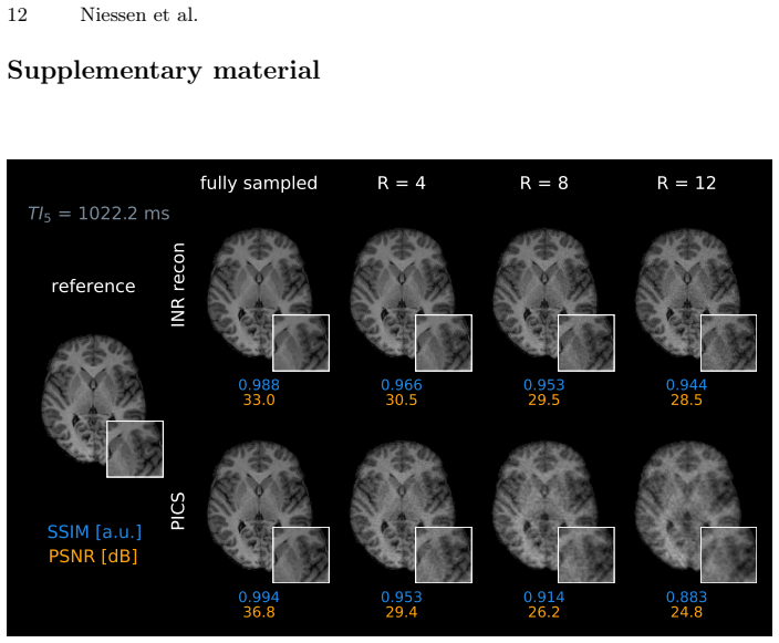
The central claim is that jointly reconstructing all contrast images with a single INR, trained per slice by enforcing data consistency in k-space, lets the network exploit shared anatomy across contrasts and thereby tolerate high undersampling. The INR maps image coordinates (y,z) to the complex signal across all N contrasts, and is trained with a distance-weighted mean-squared-error loss comparing masked learned k-space to acquired k-space. Using complementary variable-density Poisson disk masks per contrast, the method achieves SSIM 0.935 and PSNR 30.0 dB at R=12 for MPnRAGE, compared with PICS at 0.903 and 28.0 dB. The paper also shows that fully sampled INR reconstructions act as a deno
Load-bearing premise
The reported advantage assumes that complementary undersampling patterns across contrasts are what allow the joint INR to exploit shared anatomy; the paper never compares complementary masks with identical masks, so the gain could come from the INR's inherent regularization instead.
Editorial extensions
If this is right
- Multi-contrast acquisitions such as MPnRAGE could be shortened from roughly 13.5 minutes to about 1.1 minutes at R=12 while retaining image quality suitable for structure-preserving reconstruction.
- Because the method is self-supervised and per-subject, it avoids the need for large multi-contrast training datasets and may generalize to other multi-contrast sequences sharing the same underlying anatomy.
- The reported robustness across R=4, 8, and 12 suggests that further acceleration, or alternative undersampling strategies, could be explored instead of relying on random-seed complementarity.
- If the denoising effect is real, the joint INR may also reduce motion-related blurring in multi-contrast scans, potentially improving downstream quantitative T1 mapping.
Reading between the lines
- The paper does not ablate complementary versus identical masks across contrasts, so it remains untested whether the performance gain comes from the sampling design or from the INR's own regularization; a direct same-mask comparison would isolate this mechanism.
- The complementary-sampling idea could be extended to fully 3D Poisson disk sampling or to optimizing masks jointly with the reconstruction, which the authors mention as future work and which would directly test whether even higher accelerations are practical.
- The method's reliance on shared anatomy suggests similar gains might transfer to other joint reconstructions, such as T2-weighted or fluid-attenuated inversion recovery series, but this extrapolation is an inference, not a demonstrated result in the paper.
Editorial analysis
A structured set of objections, weighed in public.
Referee Report
Summary. The paper proposes a scan-specific, self-supervised reconstruction method for multi-contrast MPnRAGE MRI based on an implicit neural representation (INR). A joint INR maps image coordinates to all contrast images simultaneously and is fitted to retrospectively undersampled k-space data using a distance-weighted data-consistency loss. Complementary variable-density Poisson-disk masks are used across contrasts. The method is evaluated on three healthy volunteers at R = 4, 8, and 12 and compared with PICS. The reported results show that the INR is more robust to high acceleration than PICS in SSIM/PSNR, with only a slight decline from fully sampled to R = 12.
Significance. If confirmed, the work offers a training-free, patient-specific reconstruction that does not require large supervised datasets, which is attractive for multi-contrast sequences where training data are scarce. The code is publicly available, and the fully-sampled INR results document a clear denoising property. The main limitation is that the central mechanism, the benefit of complementary undersampling across contrasts, is not experimentally isolated; the comparison with PICS alone cannot attribute the improvement to the sampling pattern or to the INR's inherent regularization. The lack of statistical testing also weakens the strength of the claims. These issues are addressable with additional experiments.
major comments (3)
- [Sec. 3.1, Fig. 1A, Table 1] The complementary-sampling premise is load-bearing for the claimed multi-contrast benefit: the abstract and Sec. 3.1 state that complementary Poisson-disk masks allow the joint INR to exploit shared anatomy. However, no experiment compares complementary masks against identical masks for the same INR. The only comparison is the full INR with complementary masks versus PICS. The fully-sampled result (Table 1: INR SSIM 0.981 vs PICS 0.987, PSNR 33.9 vs 36.0) shows that the INR already acts as a strong regularizer/denoiser. An ablation with identical masks per contrast, and ideally also single-contrast INR reconstructions, is needed to show that the complementary sampling—rather than the INR regularizer alone—is responsible for the robustness at R = 8–12.
- [Sec. 5, Table 1] The Discussion states that 'our joint INR framework significantly outperforms PICS both visually and quantitatively,' but no statistical significance test is reported. The metrics in Table 1 are averaged over all volunteers, contrasts, and slices, with the standard deviation representing within-dataset variability. With only three volunteers, no paired test or confidence interval is provided. To support 'significantly outperforms,' the authors should report per-volunteer metric distributions and perform a paired statistical test (e.g., Wilcoxon signed-rank test across subjects or slices with appropriate multiple-comparison correction).
- [Sec. 4, Table 1] The comparison is against a single classical baseline (PICS), and the implementation details of that baseline are not given (regularization parameter selection, tuning procedure, wavelet/sparsity basis, number of iterations). The claimed 'state-of-the-art' status of the baseline and the fairness of the comparison would be easier to assess if these details were stated. This is not fatal, but it is necessary for reproducibility and for judging the magnitude of the reported improvement.
minor comments (6)
- [Sec. 3.2] The hashgrid encoding parameters (number of levels, base and maximum resolution, feature dimension) are not specified in the paper. The statement that 'we use the same hyperparameters for all datasets' is helpful, but the actual values should be listed for reproducibility; the code availability does not replace a specification in the text.
- [Sec. 3.3] The acquisition details are clear, but the coil sensitivity map estimation procedure is not described. Please state how the coil sensitivities were computed for both the INR and PICS reconstructions.
- [Eq. (2)] There is a minor formatting issue in Equation (2); the norm expression appears as a quotient-like layout. Also, in Eq. (1), the notation D_c ∈ C^{(V_y×V_z)×N} should be clarified to indicate whether this is a product space or a stacked matrix.
- [Sec. 3.2] Please define 'ReLu' as ReLU. Also, the sentence 'The relatively low dimensionality of the MLP is possible thanks to the hashgrid encoding' would benefit from a short explanation of the hashgrid encoding for readers not familiar with Müller et al.
- [Sec. 4, Fig. 2] The caption of Figure 2 reports metrics for a single slice, while Table 1 reports averages. This is fine, but the figure caption should explicitly state the slice index or otherwise make clear that these are example values; currently the numbers in the caption differ from Table 1 and could confuse readers.
- [References] Reference [23] cites Lustig's sparse MRI paper, which is a compressed-sensing method; PICS is a specific parallel-imaging compressed-sensing reconstruction. A more specific citation for the PICS implementation (e.g., the actual parallel-imaging CS method applied) would be useful.
Circularity Check
No significant circularity: the reconstruction is a standard self-supervised data-consistency fit, and the PICS comparison is empirical.
full rationale
The paper's derivation chain is a scan-specific reconstruction: the INR is optimized to minimize the k-space data-consistency loss in Eq. 2, and the optimized network output is the reconstruction. No parameter is fitted to a subset of data and then presented as a prediction of a closely related quantity; the evaluation against the fully sampled reference is an external benchmark not used in the objective. The method is therefore self-contained in the sense that the reported INR-vs-PICS comparison is an empirical experiment, not a consequence of a self-citation. The self-citations [5] and [19] motivate the architecture and the redundancy of MPnRAGE, but the central result (INR achieves competitive or superior metrics at R=12) does not reduce to them. The complementary-sampling design (Sec. 3.1) is asserted as beneficial but not ablated against identical masks; while this is a missing experimental support, it is an assumption about what drives performance, not a circular definition or fitted-input-as-prediction. No step in the paper equates an output to an input by construction, and no load-bearing uniqueness claim is imported from prior author work. Thus no significant circularity.
Assumptions & free parameters
free parameters (3)
- INR training hyperparameters (learning rate, number of iterations, optimizer)
- Hashgrid encoding parameters (number of levels, resolution range, feature size)
- Distance weighting offset in W(k_y,k_z) (the '+1' term) =
1
assumptions (4)
- domain assumption Forward model D_c = M F S_c d + e_c with additive Gaussian noise and known coil sensitivities (Eq. 1).
- domain assumption The anatomy of the object is constant across all N contrasts (Section 1: 'the anatomy of the object being scanned remains constant').
- domain assumption The INR architecture with hashgrid encoding can faithfully represent the multi-contrast signal evolution d = G_theta(y,z) (Section 3.2).
- domain assumption Variable-density Poisson disk sampling with different random seeds per contrast yields incoherent, learnable aliasing and sufficient k-space center coverage (Section 3.1).
Cite this review
Pith. "Pith review of INR meets Multi-Contrast MRI Reconstruction." pith.science (2026). https://pith.science/paper/KS3VWZKY
@misc{pith2026250904888,
  author       = {Pith},
  title        = {Pith review of: INR meets Multi-Contrast MRI Reconstruction},
  year         = {2026},
  howpublished = {\url{https://pith.science/paper/KS3VWZKY}},
  note         = {Machine review of arXiv:2509.04888}
}
read the original abstract
Multi-contrast MRI sequences allow for the acquisition of images with varying tissue contrast within a single scan. The resulting multi-contrast images can be used to extract quantitative information on tissue microstructure. To make such multi-contrast sequences feasible for clinical routine, the usually very long scan times need to be shortened e.g. through undersampling in k-space. However, this comes with challenges for the reconstruction. In general, advanced reconstruction techniques such as compressed sensing or deep learning-based approaches can enable the acquisition of high-quality images despite the acceleration. In this work, we leverage redundant anatomical information of multi-contrast sequences to achieve even higher acceleration rates. We use undersampling patterns that capture the contrast information located at the k-space center, while performing complementary undersampling across contrasts for high frequencies. To reconstruct this highly sparse k-space data, we propose an implicit neural representation (INR) network that is ideal for using the complementary information acquired across contrasts as it jointly reconstructs all contrast images. We demonstrate the benefits of our proposed INR method by applying it to multi-contrast MRI using the MPnRAGE sequence, where it outperforms the state-of-the-art parallel imaging compressed sensing (PICS) reconstruction method, even at higher acceleration factors.
Figures


Figures from the paper (2 more)

Forward citations
Cited by 1 Pith paper
-
PRIME-SVR: Physics-infoRmed Implicit Multi-Echo Slice-to-Volume Reconstruction for Fetal T2 mapping
A self-supervised, physics-regularized neural reconstruction produces high-resolution fetal brain T2 maps at 0.55 T and 1.5 T from multi-echo MRI, with reduced acquisition time.
Reference graph
Works this paper leans on
-
[1]
Quantitative Magnetic Resonance Imaging, 1st Edition, Volume 1,
Seiberlich, N., Gulani, V., Campbell-Washburn, A., Sourbron, S., Doneva, M. I., Calamante, F., Hu, H. H. "Quantitative Magnetic Resonance Imaging, 1st Edition, Volume 1," Elsevier, November 18, 2020
work page 2020
-
[2]
Hammernik, K., Küstner, T., Yaman, B., Huang, Z., Rückert, D., Knoll, F., and Akçakaya, M., “Physics-driven deep learning for computational magnetic resonance imaging: Combining physics and machine learning for improved medical imaging,” IEEE Signal Process. Mag., vol. 40, no. 1, pp. 98–114, 2023. 10 Niessen et al
work page 2023
-
[3]
Deep learning for accelerated and robust MRI reconstruction,
Heckel, R., Jacob, M., Chaudhari, A., Perlman, O., and Shimron, E., “Deep learning for accelerated and robust MRI reconstruction,” Magn. Reson. Mater. Phy., 2024
work page 2024
-
[4]
Mildenhall, B., Srinivasan, P. P., Tancik, M., Barron, J. T., Ramamoorthi, R., & Ng, R. (2021). NeRF: Representing scenes as neural radiance fields for view synthesis. Communications of the ACM, 65(1), 99-106
work page 2021
-
[5]
Subspace Implicit Neural Representations for Real-Time Cardiac Cine MR Imaging
Huang, W., Spieker, V., Xu, S., Cruz, G., Prieto, C., Schnabel, J. A., Hammernik, K., Kuestner, T., and Rueckert, D., “Subspace Implicit Neural Representations for Real-Time Cardiac Cine MR Imaging,” arXiv preprint arXiv:2412.12742, 2024
work page Pith review arXiv 2024
-
[6]
Spatiotemporal implicit neural representation for unsupervised dynamic MRI reconstruction,
Feng,J.,Feng,R.,Wu,Q.,Shen,X.,Chen,L.,Li,X.,Feng,L.etal.,“Spatiotemporal implicit neural representation for unsupervised dynamic MRI reconstruction,” IEEE Trans. Med. Imaging, p. 1, 2025
work page 2025
-
[7]
Kecskemeti, S., Samsonov, A., Hurley, S. A., Dean, D. C., Field, A., and Alexander, A. L., “MPnRAGE: A technique to simultaneously acquire hundreds of differently contrasted MPRAGE images with applications to quantitative T1 mapping,” Mag- netic resonance in medicine, vol. 75, no. 3, pp. 1040–1053, 2016
work page 2016
-
[8]
Use of fluid attenuated inversion recovery (FLAIR) pulse sequences in MRI of the brain,
Hajnal, J. V., Bryant, D. J., Kasuboski, L., Pattany, P. M., De Coene, B., Lewis, P. D., Pennock, J. M., Oatridge, A., Young, I. R., and Bydder, G. M., “Use of fluid attenuated inversion recovery (FLAIR) pulse sequences in MRI of the brain,” *Journal of Computer Assisted Tomography*, vol. 16, no. 6, pp. 841–844, 1992
work page 1992
Show all 23 references
-
[9]
Neural Implicit k-Space for Binning-Free Non-Cartesian Cardiac MR Imaging,
Huang, W., Li, H. B., Pan, J., Cruz, G., Rueckert, D., and Hammernik, K., “Neural Implicit k-Space for Binning-Free Non-Cartesian Cardiac MR Imaging,” in *Infor- mation Processing in Medical Imaging (IPMI 2023)*, Lecture Notes in Computer Science, vol. 13939, pp. 548–560, Spri...
2023
-
[10]
ICoNIK: Gener- ating Respiratory-Resolved Abdominal MR Reconstructions Using Neural Implicit Representations in k-Space,
Spieker, V., Huang, W., Eichhorn, H., Stelter, J., Weiss, K., Zimmer, V. A., Braren, R. F., Karampinos, D. C., Hammernik, K., and Schnabel, J. A., “ICoNIK: Gener- ating Respiratory-Resolved Abdominal MR Reconstructions Using Neural Implicit Representations in k-Space,” in *Lec...
2024
-
[11]
Unsupervised reconstruction of accelerated cardiac cine MRI using Neural Fields,
Catalán, T., Courdurier, M., Osses, A., Botnar, R., Sahli Costabal, F., & Prieto, C. "Unsupervised reconstruction of accelerated cardiac cine MRI using Neural Fields," Computers in Biology and Medicine, Volume 185, 2025
2025
-
[12]
Implicit Neural Networks With Fourier- Feature Inputs for Free-Breathing Cardiac MRI Reconstruction,
Kunz, J. F., Ruschke, S., and Heckel, R., “Implicit Neural Networks With Fourier- Feature Inputs for Free-Breathing Cardiac MRI Reconstruction,” IEEE Trans. Com- put. Imaging, vol. 10, pp. 1280–1289, 2024
2024
-
[13]
IMJENSE: Scan-Specific Implicit Representation for Joint Coil Sensitivity and Image Estima- tion in Parallel MRI,
Feng, R., Wu, Q., Feng, J., She, H., Liu, C., Zhang, Y., and Wei, H., “IMJENSE: Scan-Specific Implicit Representation for Joint Coil Sensitivity and Image Estima- tion in Parallel MRI,” IEEE transactions on medical imaging, vol. 43, no. 4, pp. 1539–1553, 2024
2024
-
[14]
Coordinate-based neural representation enabling zero-shot learning for fast 3D multiparametric quantitative MRI,
Lao, G., Feng, R., Qi, H., Lv, Z., Liu, Q., Liu, C., et al. "Coordinate-based neural representation enabling zero-shot learning for fast 3D multiparametric quantitative MRI," Medical Image Analysis, Volume 102, 2025, Article 103530
2025
-
[15]
NeRP: Implicit Neural Representation Learning With Prior Embedding for Sparsely Sampled Image Reconstruction,
Shen, L., Pauly, J., and Xing, L., “NeRP: Implicit Neural Representation Learning With Prior Embedding for Sparsely Sampled Image Reconstruction,” IEEE trans- actions on neural networks and learning systems, vol. PP, 2022
2022
-
[16]
Joint multi-contrast variational network reconstruction (jVN) with application to rapid 2D and 3D imaging,
Polak, D., Cauley, S., Bilgic, B., Gong, E., Bachert, P., Adalsteinsson, E., and Setsompop, K., “Joint multi-contrast variational network reconstruction (jVN) with application to rapid 2D and 3D imaging,” Magnetic resonance in medicine, vol. 84, no. 3, pp. 1456–1469, 2020
2020
-
[17]
Decomposition-Based Variational Net- work for Multi-Contrast MRI Super-Resolution and Reconstruction,
Lei, P., Fang, F., Zhang, G., and Zeng, T., “Decomposition-Based Variational Net- work for Multi-Contrast MRI Super-Resolution and Reconstruction,” 2023. In 2023 IEEE/CVF International Conference on Computer Vision (ICCV), 21239–49: IEEE. INR meets Multi-Contrast MRI Reconstruction 11
2023
-
[18]
Müller, T., Evans, A., Schied, C., & Keller, A. (2022). Instant neural graphics primitives with a multiresolution hash encoding. ACM Transactions on Graphics (TOG), 41(4), 102
2022
-
[19]
Probing the sparsity of the MPn- RAGE sequence through subspace compression,
Niessen, N., Sprenger, T., Pirkl, C. M., Solana, A. B., Cencini, M., Avventi, E., Tosetti, M., Menzel, M. I., and Schnabel, J. A., “Probing the sparsity of the MPn- RAGE sequence through subspace compression,” International Society for Magnetic Resonance in Medicine conference...
2025
-
[20]
Skare, S., Avventi, E., Norbeck, O., & Ryden, H. (2017). An abstraction layer for simpler EPIC pulse programming on GE MR systems in a clinical environment. In Proceedings of the 25th Annual Meeting of ISMRM, p. 3813
2017
-
[21]
"FSL," NeuroImage, 62(2):782–790, 2012
Jenkinson, M., Beckmann, C.F., Behrens, T.E.J., Woolrich, M.W., Smith, S.M. "FSL," NeuroImage, 62(2):782–790, 2012
2012
-
[22]
Agreement of image quality metrics with radiological evaluation in the presence of motion artifacts,
Marchetto, E., Eichhorn, H., Gallichan, D., et al. "Agreement of image quality metrics with radiological evaluation in the presence of motion artifacts," Magnetic Resonance Materials in Physics, Biology and Medicine (MAGMA), 2025
2025
-
[23]
Sparse MRI: The application of com- pressed sensing for rapid MR imaging,
Lustig, M., Donoho, D., and Pauly, J. M. "Sparse MRI: The application of com- pressed sensing for rapid MR imaging," Magnetic Resonance in Medicine, Volume 58, Issue 6, pp. 1182–1195, December 2007. Wiley. DOI: 10.1002/mrm.21391. 12 Niessen et al. Supplementary material SSIM [...
2007 doi
Reviewed August 5, 2026 · model on record in the stance chip above.
Discussion (0). Sign in to comment.