Development of a Modular Optically Detected Magnetic Resonance Setup for Optical Experiments in a Variable Temperature Insert
Pith reviewed 2026-05-17 00:36 UTC · model grok-4.3
The pith
A modular ODMR apparatus enables nitrogen-vacancy magnetometry inside commercial variable-temperature cryostats by extending the optical path while preserving alignment and beam quality.
A machine-rendered reading of the paper's core claim, the machinery that carries it, and where it could break.
Core claim
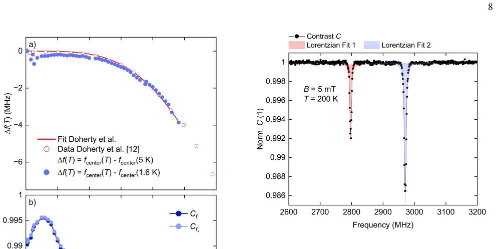
We demonstrate the setup's performance by measuring the temperature dependence of the resonance signal and its behavior under small applied magnetic fields, as well as the magnetic transition of a SrRuO3 sample, thereby showcasing the feasibility of NV magnetometry on a sample in constrained cryogenic environments.
Load-bearing premise
The assumption that an optical path spanning nearly two meters through the cryostat insert preserves sufficient alignment and beam quality to enable effective excitation of NV centers and detection of fluorescence from outside the cryostat.
Figures





read the original abstract
We developed an optically detected magnetic resonance (ODMR) setup designed for compatibility with a widely used, commercially available helium bath cryostat equipped with a variable temperature insert. The optical path extends nearly two meters, spanning the full length of the cryostat insert, enabling excitation of the nitrogen-vacancy (NV) centers and detection of the resulting fluorescence from outside the cryostat. The setup preserves optical alignment and beam quality along this extended path allowing integration into existing cryogenic systems without significant modifications. We demonstrate the setup's performance by measuring the temperature dependence of the resonance signal and its behavior under small applied magnetic fields, as well as the magnetic transition of a SrRuO$_3$ sample, thereby showcasing the feasibility of NV magnetometry on a sample in constrained cryogenic environments.
Editorial analysis
A structured set of objections, weighed in public.
Referee Report
Summary. The manuscript describes the development of a modular optically detected magnetic resonance (ODMR) setup compatible with a commercial helium bath cryostat equipped with a variable temperature insert (VTI). The optical path extends nearly two meters through the insert, enabling NV-center excitation and fluorescence detection from outside the cryostat while claiming to preserve alignment and beam quality without major modifications to existing hardware. Performance is demonstrated via temperature-dependent resonance measurements, response to small applied magnetic fields, and observation of the magnetic transition in a SrRuO3 sample, establishing feasibility of NV magnetometry in constrained cryogenic environments.
Significance. If the demonstrations hold, the work provides a practical, modular solution for integrating NV-based magnetometry into standard commercial cryogenic systems. This lowers the barrier for low-temperature studies of magnetic materials and quantum sensing applications without requiring custom cryostat designs, potentially enabling broader adoption in condensed-matter and quantum-materials research. The emphasis on compatibility with existing VTIs and the direct experimental validations (temperature sweeps, field response, and sample transition) are notable strengths for an instrumentation-focused contribution.
major comments (2)
- [Setup description] Setup description (optical path integration): The central feasibility claim rests on the ~2 m optical path preserving sufficient alignment and beam quality for effective NV excitation and fluorescence collection. However, the manuscript provides no quantitative metrics (e.g., measured beam waist, transmission efficiency through cryostat windows, or alignment drift over temperature cycles), which is load-bearing because degradation along this path would directly undermine the ability to perform ODMR from outside the cryostat.
- [Results] Results on SrRuO3 magnetic transition: The observed feature is presented as evidence of successful NV magnetometry, but the manuscript does not report the transition temperature, compare it to the known ~160 K value for SrRuO3, or include error analysis on resonance shifts. This weakens validation that the signal originates from the sample's magnetism rather than setup artifacts or temperature effects.
minor comments (3)
- [Abstract and results] The abstract and text should consistently specify whether the SrRuO3 is a thin film or bulk crystal, as this affects expected transition behavior and NV-sample coupling.
- [Figures] Figure captions for the temperature-dependence and field-response data lack details on scan parameters, microwave power, or averaging, reducing clarity for readers attempting to reproduce the measurements.
- [Introduction] A brief comparison to prior cryogenic ODMR implementations (e.g., fiber-based or custom-insert approaches) would better contextualize the modularity advantage.
Simulated Author's Rebuttal
We thank the referee for their positive assessment and constructive comments on our manuscript. We address each major comment point by point below and have revised the manuscript to incorporate additional details where this strengthens the presentation without altering the core claims.
read point-by-point responses
-
Referee: Setup description (optical path integration): The central feasibility claim rests on the ~2 m optical path preserving sufficient alignment and beam quality for effective NV excitation and fluorescence collection. However, the manuscript provides no quantitative metrics (e.g., measured beam waist, transmission efficiency through cryostat windows, or alignment drift over temperature cycles), which is load-bearing because degradation along this path would directly undermine the ability to perform ODMR from outside the cryostat.
Authors: We agree that explicit quantitative metrics would provide stronger support for the optical path performance. Although the successful acquisition of ODMR spectra through the extended path demonstrates practical functionality, we have revised the manuscript to include measured beam waist values at the sample position, transmission efficiency estimates through the full optical train including cryostat windows, and observations of alignment stability across temperature cycles. These additions directly address the concern and confirm that beam quality remains adequate for NV excitation and fluorescence collection. revision: yes
-
Referee: Results on SrRuO3 magnetic transition: The observed feature is presented as evidence of successful NV magnetometry, but the manuscript does not report the transition temperature, compare it to the known ~160 K value for SrRuO3, or include error analysis on resonance shifts. This weakens validation that the signal originates from the sample's magnetism rather than setup artifacts or temperature effects.
Authors: We concur that a direct comparison and error analysis would improve validation of the magnetic transition signal. In the revised manuscript we now report the transition temperature extracted from our NV resonance data, compare it explicitly to the established literature value of approximately 160 K for SrRuO3, and include error analysis on the observed resonance shifts. These revisions strengthen the attribution of the feature to the sample magnetism rather than artifacts. revision: yes
Circularity Check
No significant circularity
full rationale
This is an experimental instrumentation paper describing hardware development and performance demonstrations for an ODMR setup in a cryogenic environment. No mathematical derivations, equations, fitted parameters, or theoretical claims appear in the abstract or described content. All assertions rest on physical construction details and direct experimental measurements (temperature dependence, field response, SrRuO3 transition), which are externally falsifiable via the reported hardware and data. The work is self-contained with no self-citation load-bearing steps or reductions by construction.
Axiom & Free-Parameter Ledger
axioms (1)
- domain assumption Optical beam propagation and alignment can be maintained over distances of nearly two meters in the described cryostat geometry
Lean theorems connected to this paper
-
IndisputableMonolith/Foundation/RealityFromDistinction.leanreality_from_one_distinction unclear?
unclearRelation between the paper passage and the cited Recognition theorem.
We developed an optically detected magnetic resonance (ODMR) setup designed for compatibility with a widely used, commercially available helium bath cryostat equipped with a variable temperature insert. The optical path extends nearly two meters...
-
IndisputableMonolith/Cost/FunctionalEquation.leanwashburn_uniqueness_aczel unclear?
unclearRelation between the paper passage and the cited Recognition theorem.
The setup preserves optical alignment and beam quality along this extended path allowing integration into existing cryogenic systems without significant modifications.
What do these tags mean?
- matches
- The paper's claim is directly supported by a theorem in the formal canon.
- supports
- The theorem supports part of the paper's argument, but the paper may add assumptions or extra steps.
- extends
- The paper goes beyond the formal theorem; the theorem is a base layer rather than the whole result.
- uses
- The paper appears to rely on the theorem as machinery.
- contradicts
- The paper's claim conflicts with a theorem or certificate in the canon.
- unclear
- Pith found a possible connection, but the passage is too broad, indirect, or ambiguous to say the theorem truly supports the claim.
Reference graph
Works this paper leans on
-
[1]
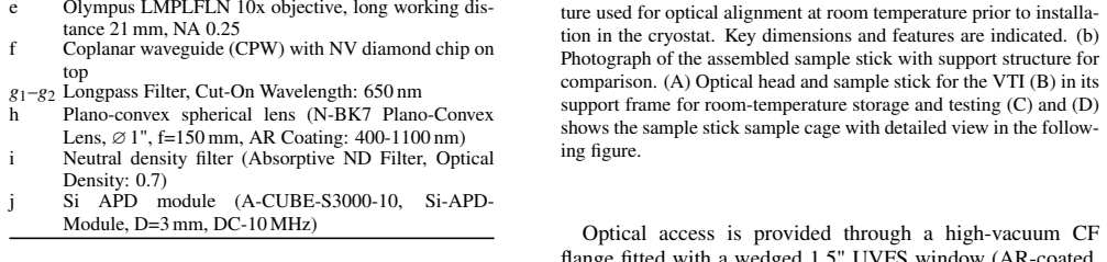
Optical Head All optical components located above the cryostat flange are mounted on a two-level breadboard construction, sepa- rating the detection (A) and excitation (B) paths vertically as shown in Fig. 1. The breadboards are enclosed in opaque cov- ers to prevent stray light contamination and ensure signal in- tegrity. This arrangement is referred to ...
-
[2]
Sample Stick The sample stick, shown in Fig. 2 together with its support frame, was custom-designed to meet the mechanical and spa- tial constraints imposed by the cryostat’s VTI, which limits the maximum outer diameter to 30 mm. It consists of three 160 cm long, stainless steel rods arranged in a triangular con- figuration and interference-fitted (press-...
-
[3]
Support Table and Frame for Pre-Alignment and Handling To enable reproducible optical alignment and safe han- dling of the setup components, we constructed two modu- lar aluminum support frames built from standard extrusion profiles (e.g. ITEM): a frame for pre-aligning and testing at room temperature, and a cryostat-mounted platform sys- tem. The pre-ali...
- [4]
-
[5]
L. Rondin, J.-P. Tetienne, T. Hingant, J.-F. Roch, P. Maletinsky, and V . Jacques, Rep. Prog. Phys.77, 056503 (2014)
work page 2014
-
[6]
Y . Xu, W. Zhang, and C. Tian, Photon. Res.11, 393 (2023)
work page 2023
- [7]
-
[8]
C. Du, T. van der Sar, T. X. Zhou, P. Upadhyaya, F. Casola, H. Zhang, M. C. Onbasli, C. A. Ross, R. L. Walsworth, and D. Wang, Science357, 195 (2017)
work page 2017
- [9]
-
[10]
Y . Dovzhenko, F. Casola, S. Schlotter, T. X. Zhou, F. Buttner, R. L. Walsworth, G. S. D. Beach, and A. Yacoby, Nature Com- munications9, 2712 (2018)
work page 2018
- [11]
- [12]
- [13]
-
[14]
K. Y . Yip, K. O. Ho, K. Y . Yu, Y . Chen, W. Zhang, S. Kasa- hara, Y . Mizukami, T. Shibauchi, Y . Matsuda, S. K. Goh, and S. Yang, Science366, 1355 (2019)
work page 2019
-
[15]
A. Batalov, V . Jacques, F. Kaiser, P. Siyushev, P. Neumann, L. J. Rogers, R. L. McMurtrie, N. B. Manson, F. Jelezko, and J. Wrachtrup, Phys. Rev. Lett.102, 195506 (2009)
work page 2009
-
[16]
M. W. Doherty, V . M. Acosta, A. Jarmola, M. S. J. Barson, N. B. Manson, D. Budker, and L. C. L. Hollenberg, Phys. Rev. B90, 041201 (2014)
work page 2014
- [17]
- [18]
- [19]
- [20]
- [21]
-
[22]
S. Kunkemöller, D. Brüning, A. Stunault, A. A. Nugroho, T. Lorenz, and M. Braden, Phys. Rev. B96, 220406 (2017)
work page 2017
discussion (0)
Sign in with ORCID, Apple, or X to comment. Anyone can read and Pith papers without signing in.