Inclusion of Inter-crystal Scattering in PET: Analytical Models and Dedicated Reconstruction
Pith reviewed 2026-05-16 16:10 UTC · model grok-4.3
The pith
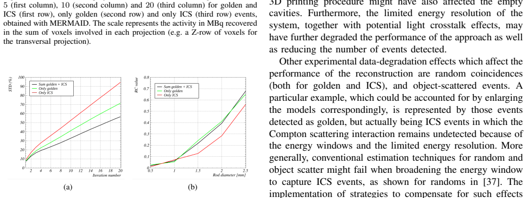
Inter-crystal scattering can be modeled analytically to improve PET image uniformity and reduce noise.
A machine-rendered reading of the paper's core claim, the machinery that carries it, and where it could break.
Core claim
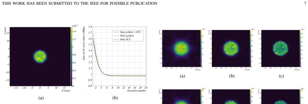
Analytical models derived from Compton scattering physics provide expressions for the sensitivity image and system matrix that incorporate inter-crystal scattering without requiring identification of the first interaction point. When used in a joint reconstruction algorithm with conventional PET events, these models yield images with lower noise and better uniformity on both simulated and real data from the MERMAID scanner.
What carries the argument
Analytical expressions for the ICS contribution to the system matrix and sensitivity image, based on Compton-scattering physics.
If this is right
- Reconstruction algorithms can now account for ICS events to increase effective sensitivity.
- Image uniformity improves and noise decreases in small-animal PET.
- No need for data-driven or machine-learning methods to locate scattering sites.
- Applicable to list-mode MLEM for joint processing of ICS and standard events.
Where Pith is reading between the lines
- The method might generalize to other PET scanner geometries with similar modifications to the models.
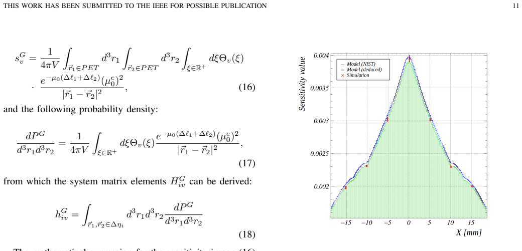
- It could enable lower dose imaging protocols by leveraging more events.
- Similar analytical approaches might apply to other imaging modalities affected by scattering.
Load-bearing premise
The derived analytical expressions accurately capture the ICS events for the specific geometry and energy window of the MERMAID scanner without introducing bias.
What would settle it
Reconstructing the same phantom data with and without the ICS model and measuring if the noise reduction and uniformity improvement persist or if new artifacts emerge.
Figures
read the original abstract
Inter-crystal scattering (ICS) in Positron Emission Tomography (PET) is commonly regarded as a degradation effect that might compromise the image spatial resolution. In parallel, the inclusion of ICS events has also been recognized as a potential approach to increase PET sensitivity, which could be especially beneficial in scenarios where the latter is a limiting factor, such as very small animal imaging. Several methods for the recovery of ICS events have been proposed, many of which aim to locate the first interaction, i.e., the Compton scattering site, usually limited by their success rate, computational burden or data and training dependency. Conversely, this work proposes a physics-based model for ICS events, leading to analytical expressions of the sensitivity image and the system matrix (required by statistical reconstruction algorithms), without the need to identify the original line of response. After validating the model, the work shows how ICS events can be integrated into a joint image reconstruction algorithm (based on list-mode MLEM) together with conventional PET events, for which dedicated analytical models are also developed. To assess the performance of the proposed approach, Monte-Carlo simulated and experimental data of an image quality phantom were obtained with the MERMAID small-fish PET scanner prototype. Both simulation and experimental results indicate that, while slightly decreasing the recovery coefficient values, the inclusion of ICS clearly reduces statistical noise and improves uniformity.
Editorial analysis
A structured set of objections, weighed in public.
Referee Report
Summary. The manuscript develops physics-based analytical models for inter-crystal scattering (ICS) events in PET, deriving closed-form expressions for the sensitivity image and system matrix from Compton kinematics and scanner geometry. These models enable joint list-mode MLEM reconstruction of ICS events together with conventional coincidences without explicit first-interaction localization. Validation uses Monte Carlo simulations and experimental data from the MERMAID small-animal PET scanner on an image-quality phantom, reporting reduced statistical noise and improved uniformity with a modest decrease in recovery coefficients.
Significance. If the analytical ICS contributions are unbiased for the MERMAID geometry and energy window, the work supplies a parameter-free route to recover sensitivity lost to inter-crystal scattering. This is especially relevant for small-animal PET where sensitivity is limiting, and the reported noise and uniformity gains are obtained within a standard statistical reconstruction framework.
minor comments (2)
- The derivation steps leading from Compton differential cross-section to the explicit system-matrix element for ICS events are only summarized; expanding one representative calculation (e.g., the integral over scattering angle for a given crystal pair) would improve reproducibility.
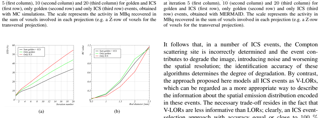
- Quantitative noise and uniformity metrics (e.g., coefficient of variation or standard deviation within ROIs) are mentioned qualitatively; reporting numerical values alongside the recovery-coefficient tables would strengthen the experimental claims.
Simulated Author's Rebuttal
We thank the referee for the positive summary of our work on physics-based analytical models for inter-crystal scattering in PET and for recommending minor revision. The assessment correctly captures the derivation of closed-form expressions for the sensitivity image and system matrix, the joint list-mode MLEM approach, and the observed gains in noise and uniformity on MERMAID data. No specific major comments appear in the report, so we provide no point-by-point rebuttals.
Circularity Check
No significant circularity; derivation is self-contained
full rationale
The paper derives analytical expressions for the ICS contribution to the system matrix and sensitivity image directly from Compton-scattering kinematics and the known MERMAID scanner geometry. These expressions are not fitted to the target image-quality metrics (recovery coefficients, noise, uniformity) but are instead validated against independent Monte-Carlo simulations and experimental phantom data. No load-bearing self-citation, self-definitional step, or renaming of a fitted result appears in the reported workflow; the central claim therefore rests on external physics and separate validation datasets rather than on its own outputs.
Axiom & Free-Parameter Ledger
axioms (2)
- standard math Compton scattering kinematics govern the probability and energy deposition of inter-crystal scattering events in PET detectors
- domain assumption The system response for both ICS and non-ICS events can be expressed analytically for the given scanner geometry and energy window
Reference graph
Works this paper leans on
-
[1]
E. Enlow and S. Abbaszadeh, “State-of-the-art challenges and emerging technologies in radiation detection for nuclear medicine imaging: A review,”Frontiers in Physics, vol. V olume 11 - 2023, 2023
work page 2023
-
[2]
Dose Reduction in Pediatric Oncology Patients with Delayed Total-Body [18F]FDG PET/CT,
C. Mingelset al., “Dose Reduction in Pediatric Oncology Patients with Delayed Total-Body [18F]FDG PET/CT,”Journal of Nuclear Medicine, vol. 65, no. 7, pp. 1101–1106, 2024
work page 2024
-
[3]
Fetal Dose from PET and CT in Pregnant Patients,
C. S. Burtonet al., “Fetal Dose from PET and CT in Pregnant Patients,” Journal of Nuclear Medicine, vol. 64, no. 2, pp. 312–319, 2023
work page 2023
-
[4]
T. Carlier, K. P. Willowson, E. Fourkal, D. L. Bailey, M. Doss, and M. Conti, “90Y -PET imaging: Exploring limitations and accuracy under conditions of low counts and high random fraction,”Medical Physics, vol. 42, no. 7, pp. 4295–4309, 2015
work page 2015
-
[5]
Real-Time PET Imaging for Range Verification of Helium Radiotherapy,
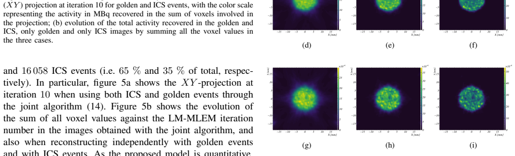
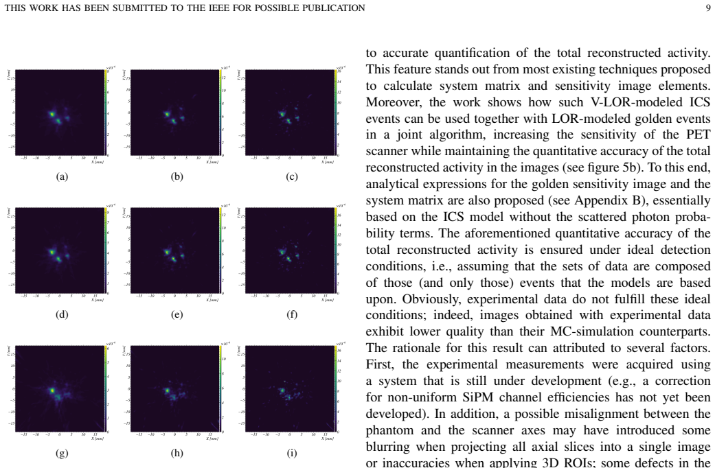
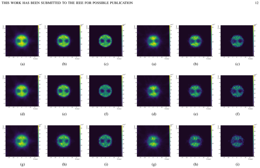
I. Ozoemelamet al., “Real-Time PET Imaging for Range Verification of Helium Radiotherapy,”Frontiers in Physics, vol. V olume 8 - 2020, 2020. THIS WORK HAS BEEN SUBMITTED TO THE IEEE FOR POSSIBLE PUBLICATION 12 (a) (b) (c) (d) (e) (f) (g) (h) (i) Fig. 13. Transversal (XY) projections over the NEMA inserts region at iteration5(first column),10(second column...
work page 2020
-
[6]
Development of a digital zebrafish phantom and its application to dedicated small-fish PET,
M. Zvolsk ´y, M. Schaar, S. Seeger, S. Rakers, and M. Rafecas, “Development of a digital zebrafish phantom and its application to dedicated small-fish PET,”Physics in Medicine and Biology, vol. 67, no. 17, p. 175005, aug 2022. [Online]. Available: https://doi.org/10.1088/1361-6560/ac71ee
-
[7]
Total Body PET/CT: Future Aspects,
F. Godinez, C. Mingels, R. Bayerlein, B. Mehadji, and L. Nardo, “Total Body PET/CT: Future Aspects,”Seminars in Nuclear Medicine, vol. 55, no. 1, pp. 107–115, 2025, total body PET/CT
work page 2025
-
[8]
The quest for multifunc- tional and dedicated PET instrumentation with irregular geometries,
A. Sanaat, M. Amini, H. Arabi, and H. Zaidi, “The quest for multifunc- tional and dedicated PET instrumentation with irregular geometries,” Annals of Nuclear Medicine, vol. 38, no. 1, pp. 31–70, 2024
work page 2024
-
[9]
Dedicated prostate DOI-TOF-PET based on the ProVision detection concept,
H. Phuc V o, T. Williams, K. Doroud, C. Williams, and M. Rafecas, “Dedicated prostate DOI-TOF-PET based on the ProVision detection concept,”Physics in Medicine and Biology, vol. 70, no. 18, p. 185001, sep 2025
work page 2025
-
[10]
A. Sanaat, H. Arabi, M. Reza Ay, and H. Zaidi, “Novel preclinical PET geometrical concept using a monolithic scintillator crystal offering concurrent enhancement in spatial resolution and detection sensitivity: a simulation study,”Physics in Medicine and Biology, vol. 65, no. 4, p. 045013, 2020
work page 2020
-
[11]
Physical and imaging performance of SIAT aPET under different energy windows and timing windows,
Z. Kuanget al., “Physical and imaging performance of SIAT aPET under different energy windows and timing windows,”Medical Physics, vol. 49, no. 3, pp. 1432–1444, 2022
work page 2022
-
[12]
Resolving inter-crystal scatter in a light-sharing depth-encoding PET detector,
E. Petersen, A. LaBella, Y . Li, Z. Wang, and A. H. Goldan, “Resolving inter-crystal scatter in a light-sharing depth-encoding PET detector,” Physics in Medicine and Biology, vol. 69, no. 3, p. 035024, feb 2024
work page 2024
-
[13]
H. Zouet al., “Impact of inter-crystal scattering on image quality of a dedicated brain PET scanner with DOI capability,”Physics in Medicine and Biology, vol. 70, no. 23, p. 235003, 2025
work page 2025
-
[14]
Inter-crystal scatter in a dual layer, high resolution LSO- APD positron emission tomograph,
M. Rafecas, G. B ¨oning, B. J. Pichler, E. Lorenz, M. Schwaiger, and S. I. Ziegler, “Inter-crystal scatter in a dual layer, high resolution LSO- APD positron emission tomograph,”Physics in Medicine and Biology, vol. 48, no. 7, p. 821, mar 2003
work page 2003
-
[15]
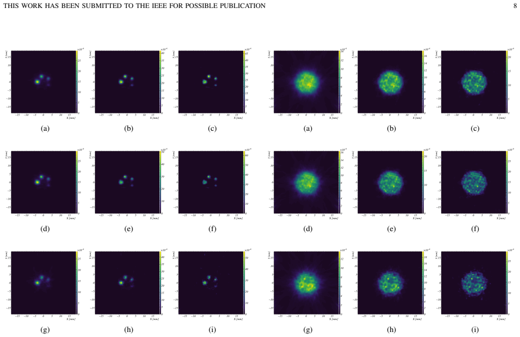
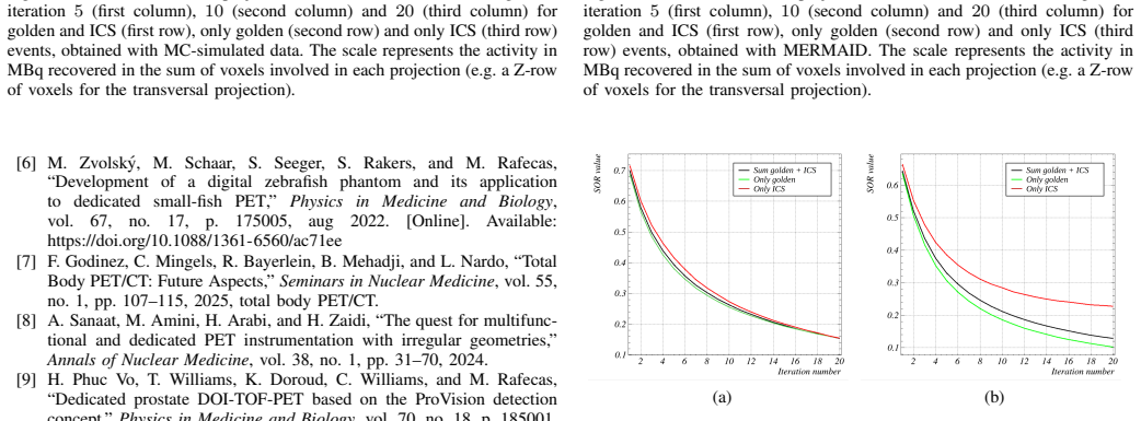
M. S. Lee, H. S. Shim, and J. S. Lee, “Strategies for mitigating inter- (a) (b) (c) (d) (e) (f) (g) (h) (i) Fig. 14. Transversal (XY) projections over the NEMA inserts region at iteration5(first column),10(second column) and20(third column) for golden and ICS (first row), only golden (second row) and only ICS (third row) events, obtained with MERMAID. The...
work page 2024
-
[16]
Algorithms to identify detector Compton scatter in PET modules,
K. Comanor, P. Virador, and W. Moses, “Algorithms to identify detector Compton scatter in PET modules,”IEEE Transactions on Nuclear Science, vol. 43, no. 4, pp. 2213–2218, 1996
work page 1996
-
[17]
Bayesian reconstruction of photon interaction sequences for high-resolution pet detectors,
G. Pratx and C. S. Levin, “Bayesian reconstruction of photon interaction sequences for high-resolution pet detectors,”Physics in Medicine and Biology, vol. 54, no. 17, p. 5073, 2009
work page 2009
-
[18]
Recovery and normalization of triple coincidences in PET,
E. Lageet al., “Recovery and normalization of triple coincidences in PET,”Medical Physics, vol. 42, no. 3, pp. 1398–1410, 2015
work page 2015
-
[19]
Novel inter-crystal scattering event identification method for PET detectors,
M. S. Lee, S. K. Kang, and J. S. Lee, “Novel inter-crystal scattering event identification method for PET detectors,”Physics in Medicine and Biology, vol. 63, no. 11, p. 115015, 2018
work page 2018
-
[20]
J.-B. Michaudet al., “Sensitivity Increase Through a Neural Network Method for LOR Recovery of ICS Triple Coincidences in High- THIS WORK HAS BEEN SUBMITTED TO THE IEEE FOR POSSIBLE PUBLICATION 13 Resolution Pixelated- Detectors PET Scanners,”IEEE Transactions on Nuclear Science, vol. 62, no. 1, pp. 82–94, 2015
work page 2015
-
[21]
C. Wu, M. S. Lee, and C. S. Levin, “Neural Network-based Inter-crystal Scatter Event Positioning in a PET System Design Based on 3D Position Sensitive Detectors,” in2020 IEEE Nuclear Science Symposium and Medical Imaging Conference (NSS/MIC), 2020, pp. 1–3
work page 2020
-
[22]
S. Lee and J. S. Lee, “Inter-crystal scattering recovery of light-sharing PET detectors using convolutional neural networks,”Physics in Medicine and Biology, vol. 66, no. 18, p. 185004, sep 2021
work page 2021
-
[23]
Sensitivity recovery for the AX-PET prototype using inter-crystal scattering events,
J. E. Gillamet al., “Sensitivity recovery for the AX-PET prototype using inter-crystal scattering events,”Physics in Medicine and Biology, vol. 59, no. 15, p. 4065, 2014
work page 2014
-
[24]
Evaluation of a high resolution silicon PET insert module,
M. Grkovskiet al., “Evaluation of a high resolution silicon PET insert module,”Nuclear Instruments and Methods in Physics Research Section A: Accelerators, Spectrometers, Detectors and Associated Equipment, vol. 788, pp. 86–94, 2015
work page 2015
-
[25]
Joint image reconstruction algorithm in compton cameras,
J. Roseret al., “Joint image reconstruction algorithm in compton cameras,”Physics in Medicine and Biology, vol. 67, no. 15, p. 155009, jul 2022
work page 2022
-
[26]
——, “Image reconstruction for a multi-layer Compton telescope: an analytical model for three interaction events,”Physics in Medicine and Biology, vol. 65, no. 14, p. 145005, jul 2020
work page 2020
-
[27]
Prism representation: a 3D ray-tracing algorithm for radiotherapy applications,
R. L. Siddon, “Prism representation: a 3D ray-tracing algorithm for radiotherapy applications,”Physics in Medicine and Biology, vol. 30, no. 8, p. 817, 1985
work page 1985
-
[28]
A Quantum Theory of the Scattering of X-rays by Light Elements,
A. H. Compton, “A Quantum Theory of the Scattering of X-rays by Light Elements,”Phys. Rev., vol. 21, pp. 483–502, May 1923
work page 1923
-
[29]
Characterisation of the Upgraded MERMAID Prototype, a PET/CT Device for Small Aquatic Animals,
S. Seeger, H. P. V o, A. Bolke, and M. Rafecas, “Characterisation of the Upgraded MERMAID Prototype, a PET/CT Device for Small Aquatic Animals,” in2022 IEEE Nuclear Science Symposium and Medical Imaging Conference (NSS/MIC), 2022, pp. 1–2
work page 2022
-
[30]
Performance measurements of small animal positron emission tomographs,
N. E. M. Associationet al., “Performance measurements of small animal positron emission tomographs,”NEMA Standards Publication, NU4-2008, pp. 1–23, 2008. [Online]. Available: https://www.nema.org/ docs/default-source/standards-document-library/nu-4-2008-website.pdf
work page 2008
-
[31]
Dedicated 3D-Printed Radioactive Phantoms With 18F-FDG for Ultrahigh-Resolution PET,
E. Elmoujarkachet al., “Dedicated 3D-Printed Radioactive Phantoms With 18F-FDG for Ultrahigh-Resolution PET,”IEEE Transactions on Radiation and Plasma Medical Sciences, vol. 9, no. 3, pp. 362–371, 2025
work page 2025
-
[32]
P. Hallen, D. Schug, and V . Schulz, “Comments on the NEMA NU 4- 2008 standard on performance measurement of small animal positron emission tomographs,”EJNMMI physics, vol. 7, no. 1, p. 12, 2020
work page 2008
-
[33]
GATE: a simulation toolkit for PET and SPECT,
S. Janet al., “GATE: a simulation toolkit for PET and SPECT,”Physics in Medicine and Biology, vol. 49, no. 19, p. 4543, 2004
work page 2004
-
[34]
XCOM: Photon Cross Section Database (version 1.5),
M. Bergeret al., “XCOM: Photon Cross Section Database (version 1.5),” 2010, NIST, PML, Radiation Physics Division (available online)
work page 2010
-
[35]
R. Ota, T. Omura, R. Yamada, T. Miwa, and M. Watanabe, “Evaluation of a Sub-Millimeter Resolution PET Detector With a 1.2 mm Pitch TSV- MPPC Array One-to-One Coupled to LFS Scintillator Crystals and Inter- Crystal Scatter Studies With Individual Signal Readout,”IEEE Trans. Rad. Plasma. Med. Sci., vol. 1, no. 1, pp. 15–22, 2017
work page 2017
-
[36]
Single-Ended Readout PET Detector Based on Pixelated Crystals With TOF and DOI Capabilities,
N. Cucarella, J. Barrio, D. Sanchez, J. M. Benlloch, and A. J. Gonzalez, “Single-Ended Readout PET Detector Based on Pixelated Crystals With TOF and DOI Capabilities,”TRPMS, vol. 9, no. 7, pp. 866–871, 2025
work page 2025
-
[37]
I. Torres-Espallardo, M. Rafecas, V . Spanoudaki, D. McElroy, and S. Ziegler, “Effect of inter-crystal scatter on estimation methods for random coincidences and subsequent correction,”Physics in Medicine & Biology, vol. 53, no. 9, p. 2391, 2008
work page 2008
-
[38]
A. Lougovski, F. Hofheinz, J. Maus, G. Schramm, and J. van den Hoff, “On the relation between Kaiser–Bessel blob and tube of response based modelling of the system matrix in iterative PET image reconstruction,” Physics in Medicine and Biology, vol. 60, no. 10, p. 4209, 2015
work page 2015
-
[39]
3d image reconstruction for a compton spect camera model,
A. Sauve, A. Hero, W. Rogers, S. Wilderman, and N. Clinthorne, “3d image reconstruction for a compton spect camera model,”IEEE Transactions on Nuclear Science, vol. 46, no. 6, pp. 2075–2084, 1999
work page 2075
-
[40]
Evaluation of a Compton scattering camera using 3-D position sensitive CdZnTe de- tectors,
Y . Du, Z. He, G. Knoll, D. Wehe, and W. Li, “Evaluation of a Compton scattering camera using 3-D position sensitive CdZnTe de- tectors,”Nuclear Instruments and Methods in Physics Research Section A: Accelerators, Spectrometers, Detectors and Associated Equipment, vol. 457, no. 1, pp. 203–211, 2001
work page 2001
discussion (0)
Sign in with ORCID, Apple, or X to comment. Anyone can read and Pith papers without signing in.