Inexpensive Optical Projection Tomography on a Mobile Phone Platform
Pith reviewed 2026-05-10 13:52 UTC · model grok-4.3
The pith
A mobile phone with $50 in add-ons performs optical projection tomography at 3.91 micrometer resolution.
A machine-rendered reading of the paper's core claim, the machinery that carries it, and where it could break.
Core claim
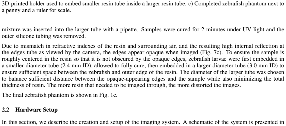
The completed system achieved a resolution of 3.91 μm and produced volumetric reconstructions in which anatomical features of the zebrafish phantom, including the spine, were clearly visible, using only an iPhone camera, low-cost lens attachment, stepper motor, LED illumination, and custom 3D-printed components with total added cost around 50 dollars.
What carries the argument
The low-cost OPT setup that rotates the sample under phone-camera view, converts projections to attenuation images, applies field nonuniformity correction, and reconstructs each slice by filtered backprojection before stacking.
If this is right
- Volumetric 3D images of small biological samples become obtainable without dedicated laboratory tomography instruments.
- Filtered backprojection on corrected phone-camera projections can resolve anatomical structures at the scale of a few micrometers.
- Portable, battery-powered 3D microscopy becomes feasible for education and field applications.
- Low-cost phantom fabrication by embedding fixed larvae in UV-cured resin provides a repeatable test object for system validation.
Where Pith is reading between the lines
- The same hardware approach might adapt to other small transparent specimens such as embryos or plant sections.
- On-device reconstruction software could turn the phone into a self-contained 3D imager without external computers.
- Calibration routines developed here could transfer to other phone-based optical systems that need consistent projection geometry.
Load-bearing premise
The low-cost phone camera, lens attachment, and custom corrections produce projection data of sufficient quality and consistency for filtered backprojection to yield accurate 3D volumes without major artifacts or distortions.
What would settle it
Reconstruction of the zebrafish phantom that fails to resolve the spine or shows large geometric distortions or missing features compared to the known phantom structure.
Figures


read the original abstract
This work presents an inexpensive optical projection tomography (OPT) system built on a mobile phone platform for three-dimensional optical microscopy. The system uses an iPhone camera together with a low-cost commercial microscope lens attachment, a stepper motor for sample rotation, LED illumination, and custom 3D-printed components, with a total component cost of approximately 50 US dollars excluding the phone. To support system evaluation, we also developed a low-cost method for fabricating a zebrafish phantom by embedding fixed larvae in UV-cured resin. Camera calibration was performed using a checkerboard target, and effective magnification was estimated with images of a 1951 Air Force resolution target. Projection images acquired during sample rotation were converted to attenuation images and corrected for field nonuniformity. Each slice was reconstructed with filtered backprojection and the resulting slices were stacked into a 3D volume. The completed system achieved a resolution of 3.91 $\mu m$ and produced volumetric reconstructions in which anatomical features of the zebrafish phantom, including the spine, were clearly visible. These results demonstrate that mobile-phone-based OPT can provide accessible, portable, and low-cost 3D microscopy, with potential utility for education, field work, and resource-limited settings.
Editorial analysis
A structured set of objections, weighed in public.
Referee Report
Summary. The manuscript describes construction of a low-cost optical projection tomography (OPT) system using an iPhone camera, commercial microscope lens attachment, stepper motor, LED illumination, and 3D-printed parts (total component cost ~$50 excluding phone). It reports a low-cost zebrafish phantom fabrication method, checkerboard camera calibration, Air Force target magnification estimation, projection-to-attenuation conversion with field nonuniformity correction, slice-by-slice filtered backprojection, and stacking into 3D volumes. The central claims are a measured resolution of 3.91 μm and clear visibility of anatomical features including the spine in the reconstructed zebrafish phantom volumes.
Significance. If the reported resolution and feature visibility are confirmed to be free of reconstruction artifacts, the work would establish that portable, sub-5 μm 3D optical microscopy is achievable with consumer-grade hardware and minimal cost, directly supporting applications in education, field biology, and resource-limited laboratories.
major comments (1)
- [Abstract and reconstruction procedure] Abstract and reconstruction procedure: no description is given of how the stepper-motor rotation axis (center of rotation) was located, calibrated, or corrected prior to filtered backprojection. Sub-pixel misalignment in FBP is known to produce streaking or blurring artifacts that can either obscure or fabricate linear structures such as a zebrafish spine; the claim that anatomical features are 'clearly visible' therefore rests on an unverified assumption of perfect alignment.
minor comments (2)
- [Abstract] The abstract states that effective magnification was estimated with a 1951 Air Force resolution target, but does not report the specific group/element used or the line-pair frequency at which contrast fell to a defined threshold (e.g., 10 % or Rayleigh criterion).
- [Results] No quantitative metrics (e.g., contrast-to-noise ratio, edge sharpness, or comparison against a commercial OPT system) are mentioned to support the visual assessment that features are 'clearly visible'.
Simulated Author's Rebuttal
We thank the referee for their detailed and constructive review. The major comment highlights an important omission in the description of our reconstruction procedure. We address this point below and commit to revisions that strengthen the manuscript.
read point-by-point responses
-
Referee: [Abstract and reconstruction procedure] Abstract and reconstruction procedure: no description is given of how the stepper-motor rotation axis (center of rotation) was located, calibrated, or corrected prior to filtered backprojection. Sub-pixel misalignment in FBP is known to produce streaking or blurring artifacts that can either obscure or fabricate linear structures such as a zebrafish spine; the claim that anatomical features are 'clearly visible' therefore rests on an unverified assumption of perfect alignment.
Authors: We agree that the manuscript lacks a description of how the center of rotation was located, calibrated, or corrected, and that this is a substantive gap. Sub-pixel misalignment in filtered backprojection can indeed introduce streaking or blurring that might affect interpretation of linear features such as the spine. In the revised manuscript we will add a dedicated subsection under the reconstruction procedure that details the alignment protocol, including the method used to determine the rotation axis, any verification steps performed with test projections, and the approach taken to minimize residual misalignment before applying filtered backprojection. This addition will enable readers to evaluate the likelihood of reconstruction artifacts and will support the reported visibility of anatomical structures. revision: yes
Circularity Check
No circularity: purely experimental hardware demonstration with no derivation chain
full rationale
The paper reports construction of a low-cost OPT system (iPhone + lens + stepper motor + 3D-printed parts), standard camera calibration on a checkerboard, magnification estimation on an Air Force target, projection acquisition with field nonuniformity correction, slice-by-slice filtered backprojection, and stacking into volumes. All results are direct physical measurements and standard reconstruction; no equations, fitted parameters renamed as predictions, self-citations, or ansatzes are invoked to derive the claimed 3.91 μm resolution or feature visibility. The work is self-contained against external benchmarks (resolution target, phantom imaging) with no reduction of outputs to inputs by construction.
Axiom & Free-Parameter Ledger
axioms (1)
- domain assumption Filtered backprojection can accurately reconstruct 3D volumes from 2D attenuation projections when field nonuniformity is corrected and the sample is rotated through sufficient angles.
Reference graph
Works this paper leans on
-
[1]
Fatima A Merchant and Alberto Diaspro. Three-dimensional imaging. InMicroscope image processing, pages 247–317. Elsevier, 2023
work page 2023
-
[2]
Introduction to confocal microscopy.Journal of Investigative Dermatology, 132(12):1–5, 2012
Adaobi Nwaneshiudu, Christiane Kuschal, Fernanda H Sakamoto, R Rox Anderson, Kathryn Schwarzenberger, and Roger C Young. Introduction to confocal microscopy.Journal of Investigative Dermatology, 132(12):1–5, 2012
work page 2012
-
[3]
Optical coherence tomography.science, 254(5035):1178–1181, 1991
David Huang, Eric A Swanson, Charles P Lin, Joel S Schuman, William G Stinson, Warren Chang, Michael R Hee, Thomas Flotte, Kenton Gregory, Carmen A Puliafito, et al. Optical coherence tomography.science, 254(5035):1178–1181, 1991
work page 1991
-
[4]
Optical projection tomography.Annu
James Sharpe. Optical projection tomography.Annu. Rev. Biomed. Eng., 6(1):209–228, 2004
work page 2004
-
[5]
Abbas Cheddad, Christoffer Svensson, James Sharpe, Fredrik Georgsson, and Ulf Ahlgren. Image processing assisted algorithms for optical projection tomography.IEEE Transactions on Medical Imaging, 31(1):1–15, 2011
work page 2011
-
[6]
Johnathon R Walls, John G Sled, James Sharpe, and R Mark Henkelman. Correction of artefacts in optical projection tomography.Physics in Medicine & Biology, 50(19):4645–4665, 2005
work page 2005
-
[7]
Pedro P Vallejo Ramirez, Joseph Zammit, Oliver Vanderpoorten, Fergus Riche, Francois-Xavier Bl´e, Xiao-Hong Zhou, Bogdan Spiridon, Christopher Valentine, Simeon E Spasov, Pelumi W Oluwasanya, et al. OptiJ: Open- source optical projection tomography of large organ samples.Scientific reports, 9(1):15693, 2019
work page 2019
-
[8]
Brian W Pogue and Michael S Patterson. Review of tissue simulating phantoms for optical spectroscopy, imaging and dosimetry.Journal of biomedical optics, 11(4):041102–041102, 2006
work page 2006
-
[9]
Gary CF Lee, Gennifer T Smith, Monica Agrawal, Theodore Leng, and Audrey K Ellerbee. Fabrication of healthy and disease-mimicking retinal phantoms with tapered foveal pits for optical coherence tomography.Journal of Biomedical Optics, 20(8):085004, 2015
work page 2015
-
[10]
Gennifer T Smith, Nicholas Dwork, Daniel O’Connor, Uzair Sikora, Kristen L Lurie, John M Pauly, and Au- drey K Ellerbee. Automated, depth-resolved estimation of the attenuation coefficient from optical coherence tomography data.IEEE transactions on medical imaging, 34(12):2592–2602, 2015
work page 2015
-
[11]
Multi- modal 3D cancer-mimicking optical phantom.Biomedical optics express, 7(2):648–662, 2016
Gennifer T Smith, Kristen L Lurie, Dimitar V Zlatev, Joseph C Liao, and Audrey K Ellerbee Bowden. Multi- modal 3D cancer-mimicking optical phantom.Biomedical optics express, 7(2):648–662, 2016. 10 APREPRINT- APRIL17, 2026
work page 2016
-
[12]
Alex Y Lin, Yifu Ding, Daniel J Vanselow, Spencer R Katz, Maksim A Yakovlev, Darin P Clark, David Mandrell, Jean E Copper, Damian B van Rossum, and Keith C Cheng. Rigid embedding of fixed and stained, whole, millimeter-scale specimens for section-free 3d histology by micro-computed tomography.Journal of visualized experiments: JoVE, (140):58293, 2018
work page 2018
-
[13]
Cambridge university press, 2003
Richard Hartley and Andrew Zisserman.Multiple view geometry in computer vision. Cambridge university press, 2003
work page 2003
-
[14]
Gerard Medioni and Sing Bing Kang.Emerging topics in computer vision. Prentice Hall PTR, 2004
work page 2004
-
[15]
Avinash C Kak and Malcolm Slaney.Principles of computerized tomographic imaging. SIAM, 2001. 11
work page 2001
discussion (0)
Sign in with ORCID, Apple, or X to comment. Anyone can read and Pith papers without signing in.