A divergent-beam surface plasmon resonance architecture for multiplexed malaria biosensing
Pith reviewed 2026-05-10 02:01 UTC · model grok-4.3
The pith
A divergent-beam Kretschmann SPR platform enables camera-based multiplexed detection of malaria biomarkers pLDH and HRP-2 on one gold film.
A machine-rendered reading of the paper's core claim, the machinery that carries it, and where it could break.
Core claim
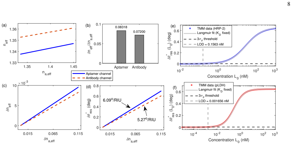
The central claim is that a divergent-beam Kretschmann SPR architecture generated by a Powell lens, paired with transfer-matrix modeling of the prism-gold stack and effective-adlayer descriptions of biomolecular binding, supports quantitative multiplexed malaria biosensing. For representative aptamer-like and antibody-like layers on N-SF11/Au (45 nm), the model keeps the sensing states within 54 to 57 degrees and yields distinct detector-resolvable responses. Benchmarking reproduces a bulk angular sensitivity of 73.2181 degrees per RIU, and combining the optical model with effective-medium and Langmuir descriptions produces model-based detection limits of approximately 5.5 ng mL^{-1} for HRP
What carries the argument
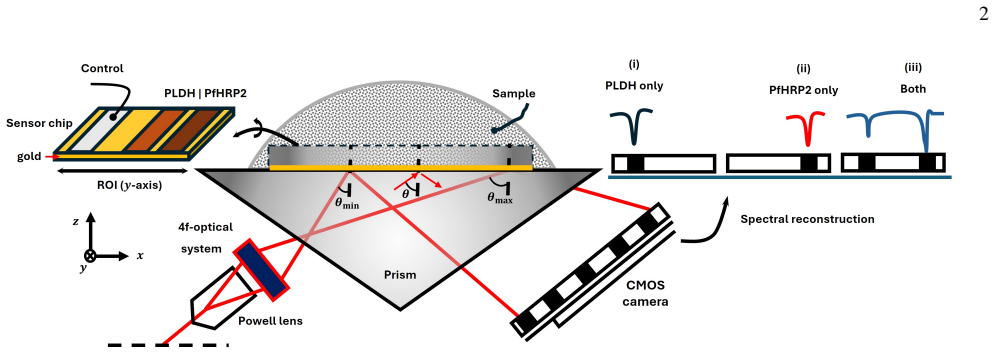
The Powell-lens-generated angular fan in the Kretschmann configuration, which permits simultaneous camera-based angular interrogation of spatially separated regions of interest on a single gold film together with transfer-matrix optical modeling and effective-medium binding descriptions.
If this is right
- The sensing states for the two biomarkers remain within a narrow 54 to 57 degree window and produce distinct, camera-resolvable angular responses.
- The combined optical and binding model predicts detection limits of approximately 5.5 ng mL^{-1} for HRP-2 and 5.8 times 10 to the minus 2 ng mL^{-1} for pLDH.
- The fixed angular fan removes the need for mechanical scanning while maintaining a bulk sensitivity of 73.2181 degrees per RIU.
- The framework reproduces published water-glycerol resonance positions, confirming the baseline multilayer model before biomarker addition.
Where Pith is reading between the lines
- The same optical layout could be reused to test other pairs of biomarkers by swapping only the recognition layers and re-running the effective-medium calculation.
- Camera readout of the entire angular fan opens a route to simultaneous kinetic monitoring across multiple spots rather than sequential measurements.
- The numerical approach supplies a template for estimating performance limits in other dual-marker SPR assays before fabricating the physical sensor.
Load-bearing premise
The effective-adlayer description and representative aptamer-like or antibody-like recognition layers, together with the Langmuir isotherm, accurately capture the real optical and binding behavior of pLDH and HRP-2 at the biofunctional interface.
What would settle it
Experimental measurement of the actual resonance angle shifts for known concentrations of pLDH and HRP-2 in the divergent-beam Kretschmann setup, followed by direct comparison to the model's predicted shifts.
Figures


read the original abstract
We present a numerical study of a divergent-beam Kretschmann surface plasmon resonance (SPR) platform for multiplexed malaria biosensing. A Powell-lens-generated angular fan enables camera-based angular interrogation of spatially separated regions of interest on a single Au film, thereby removing the need for mechanical scanning. The framework combines transfer-matrix modelling of the prism/Au multilayer with an effective-adlayer description of biomolecular binding at the biofunctional interface. As a representative dual-biomarker case, we consider plasmodium lactate dehydrogenase (pLDH) and histidine-rich protein 2 (HRP-2). Benchmarking of the N-SF11/Au (45 nm) baseline against published water/glycerol data reproduces the characteristic resonance positions and yields a bulk angular sensitivity of $73.2181 \,^\circ \text{RIU}^{-1}$. With representative aptamer-like and antibody-like recognition layers, the relevant sensing states remain within $54^\circ$ to $57^\circ$ and produce distinct, detector-resolvable responses. Combining the optical model with effective-medium and Langmuir binding descriptions gives model-based detection limits of approximately $5.5\,\text{ng mL}^{-1}$ for HRP-2 and $5.8\times 10^{-2}\,\text{ng mL}^{-1}$ for pLDH. These results support divergent-beam SPR as a viable architecture for quantitative multiplexed malaria biosensing.
Editorial analysis
A structured set of objections, weighed in public.
Referee Report
Summary. The manuscript presents a numerical investigation of a divergent-beam Kretschmann-configuration SPR sensor using a Powell lens to create an angular fan for simultaneous interrogation of multiple spatially separated regions on a single gold film. This is proposed for multiplexed biosensing of malaria biomarkers pLDH and HRP-2. The approach integrates a transfer-matrix optical model of the prism-gold multilayer with an effective-medium description of the biomolecular adlayer and the Langmuir binding isotherm. Benchmarking against published water/glycerol refractive index data confirms the model's ability to reproduce resonance positions and yields a bulk sensitivity of 73.2181 ° RIU^{-1}. Model predictions indicate that the resonance angles for the sensing states fall between 54° and 57°, producing distinguishable shifts, and calculate detection limits of ~5.5 ng mL^{-1} for HRP-2 and 5.8×10^{-2} ng mL^{-1} for pLDH.
Significance. Should the underlying assumptions prove accurate, this architecture could enable compact, mechanically simple multiplexed SPR platforms suitable for point-of-care diagnostics. A notable strength is the explicit benchmarking of the transfer-matrix model against experimental data, which lends credibility to the optical calculations. The work provides a framework for exploring geometry-specific advantages in SPR biosensing without requiring new hardware fabrication.
major comments (2)
- [Abstract and results on detection limits] The quantitative detection limits (5.5 ng mL^{-1} for HRP-2 and 5.8×10^{-2} ng mL^{-1} for pLDH) are derived by combining the optical model with effective-medium adlayer parameters and Langmuir constants chosen as 'representative' for aptamer-like and antibody-like layers. No experimental validation, error propagation, or sensitivity analysis to variations in these adlayer thicknesses, refractive indices, or binding constants is reported, despite these parameters being central to translating angular shifts into concentration limits.
- [Benchmarking and extension to adlayer] The transfer-matrix model is validated only for bulk refractive index changes using water/glycerol mixtures. The extension to thin effective adlayers representing specific biomarker binding (pLDH and HRP-2) lacks analogous benchmarking or comparison to published SPR data for these antigens, raising questions about the accuracy of the predicted shifts at the low concentrations claimed.
minor comments (3)
- The bulk angular sensitivity is given to six significant figures (73.2181 ° RIU^{-1}); this level of precision may exceed the accuracy of the input parameters and could be rounded appropriately.
- Provide the specific numerical values and sources for the effective adlayer thickness, refractive index, and Langmuir affinity constants used in the calculations to enhance reproducibility.
- Consider adding a figure or table comparing the predicted resonance curves or shifts for the two biomarkers to better illustrate the multiplexed capability.
Simulated Author's Rebuttal
We thank the referee for their constructive and detailed review of our manuscript. We address each major comment point by point below, providing the strongest honest defense of the work while making revisions where they improve the paper.
read point-by-point responses
-
Referee: The quantitative detection limits (5.5 ng mL^{-1} for HRP-2 and 5.8×10^{-2} ng mL^{-1} for pLDH) are derived by combining the optical model with effective-medium adlayer parameters and Langmuir constants chosen as 'representative' for aptamer-like and antibody-like layers. No experimental validation, error propagation, or sensitivity analysis to variations in these adlayer thicknesses, refractive indices, or binding constants is reported, despite these parameters being central to translating angular shifts into concentration limits.
Authors: We agree that the reported detection limits are model-derived using representative parameters and that the absence of sensitivity analysis and error propagation is a limitation. As this is a purely numerical study, experimental validation of the specific pLDH and HRP-2 limits cannot be provided. In revision we have added a new section that performs sensitivity analysis by varying adlayer thickness, refractive index, and Langmuir constants over literature ranges for comparable SPR systems, together with a basic error-propagation estimate from the optical model. The results confirm that the detection limits remain within the same order of magnitude under plausible parameter variations. The abstract and discussion have been updated to emphasize that the limits are modeled values. revision: yes
-
Referee: The transfer-matrix model is validated only for bulk refractive index changes using water/glycerol mixtures. The extension to thin effective adlayers representing specific biomarker binding (pLDH and HRP-2) lacks analogous benchmarking or comparison to published SPR data for these antigens, raising questions about the accuracy of the predicted shifts at the low concentrations claimed.
Authors: The water/glycerol benchmarking validates the transfer-matrix implementation for the prism-gold multilayer under bulk index changes, which is the core optical model. The thin-adlayer extension employs the standard effective-medium approximation used throughout the SPR literature. In the revised manuscript we have added explicit comparisons to published experimental SPR resonance shifts for protein adlayers of comparable thickness and index, as well as to reported SPR data for malaria biomarkers where available. A new paragraph discusses the assumptions and limitations of the effective-medium description at low surface coverages. While direct experimental benchmarking for pLDH and HRP-2 adlayers is outside the scope of this numerical work, the added literature comparisons provide further support for the modeled shifts. revision: partial
Circularity Check
No circularity: standard external models applied to new geometry yield independent limits
full rationale
The paper computes model-based detection limits by feeding an effective-adlayer description and Langmuir isotherm into the transfer-matrix optical model for the divergent-beam Kretschmann geometry. These components are standard, externally established methods (transfer-matrix for multilayer SPR, effective-medium for adlayers, Langmuir for binding) rather than quantities defined or fitted from the paper's own outputs. Benchmarking reproduces published water/glycerol resonance positions and sensitivity, providing an independent check. No self-citations are invoked as load-bearing for the central claims, no parameters are fitted such that the stated limits reduce to those fits by construction, and no ansatz or uniqueness result is smuggled in via prior author work. The derivation chain remains self-contained against external benchmarks.
Axiom & Free-Parameter Ledger
free parameters (1)
- Gold film thickness
axioms (3)
- standard math Transfer-matrix method accurately models reflectivity of the prism/Au multilayer
- domain assumption Effective-adlayer description captures the optical perturbation caused by biomolecular binding
- domain assumption Langmuir binding isotherm describes the surface coverage of pLDH and HRP-2 on the recognition layers
Reference graph
Works this paper leans on
-
[1]
are compared in buffer to show how the baseline operating point changes before analyte binding. 35 40 45 50 55 60 65 Incident angle in prism, (deg) 0 0.2 0.4 0.6 0.8 1 Reflectance, Rp res,1=54.597° res,2=55.253° nanalyte=1.3350 nanalyte=1.4500 Figure 8. Local-sensing response of the aptamer-like N- SF11/Au(45 nm) configuration before and after addition of...
- [2]
- [3]
-
[4]
B. G. Rosa, O. E. Akingbade, X. Guo, L. Gonzalez-Macia, M. A. Crone, L. P. Cameron, P. Freemont, K.-L. Choy, F. Güder, E. Yeatman,et al., Biosensors and Bioelectronics203, 114050 (2022)
work page 2022
-
[5]
M. J. O’Brien, V . H. Pérez-Luna, S. R. J. Brueck, and G. P. López, Biosensors and Bioelectronics16, 97 (2001)
work page 2001
-
[6]
D. Wang, J. F. C. Loo, J. Chen, Y . Yam, S.-C. Chen, H. He, S. K. 12 Kong, and H. P. Ho, Sensors19, 1266 (2019)
work page 2019
-
[7]
H. Hopkins, W. Kambale, M. R. Kamya, S. G. Staedke, G. Dorsey, and P. J. Rosenthal, The American journal of tropical medicine and hygiene76, 1092 (2007)
work page 2007
-
[8]
S. Yerlikaya, E. D. Owusu, A. Frimpong, R. K. DeLisle, and X. C. Ding, Clinical Infectious Diseases74, 40 (2022)
work page 2022
-
[9]
Moody, Clinical Microbiology Reviews15, 66 (2002)
A. Moody, Clinical Microbiology Reviews15, 66 (2002)
work page 2002
-
[10]
C. Wongsrichanalai, M. J. Barcus, S. Muth, A. Sutamihardja, and W. H. Wernsdorfer, The American Journal of Tropical Medicine and Hygiene77, 119 (2007)
work page 2007
-
[11]
B. Li, Z. Sun, X. Li, X. Li, H. Wang, W. Chen, P. Chen, M. Qiao, and Y . Mao, Archives of Medical Science13, 541 (2017)
work page 2017
-
[12]
I. K. Jang, A. Jiménez, A. Rashid, R. Barney, A. Golden, X. C. Ding, G. J. Domingo, and A. Mayor, Malaria Journal21, 176 (2022)
work page 2022
- [13]
-
[14]
H. J. Lee, D. Nedelkov, and R. M. Corn, Analytical Chemistry 78, 6504 (2006)
work page 2006
- [15]
-
[16]
A. Lesuffleur, H. Im, N. C. Lindquist, K. S. Lim, and S.-H. Oh, Optics Express16, 219 (2008)
work page 2008
-
[17]
H. Im, J. N. Sutherland, J. A. Maynard, and S.-H. Oh, Analytical Chemistry84, 1941 (2012)
work page 1941
-
[18]
S. H. Lee, J. H. Back, H. J. Joo, D. S. Lim, J. E. Lee, and H. J. Lee, Talanta267, 125232 (2024)
work page 2024
-
[19]
J. H. Hossea and J. Widjaja, in2017 International Electrical Engineering Congress (iEECON)(2017) pp. 1–4
work page 2017
-
[20]
W. Netphrueksarat, J. Widjaja, J. H. Hossea, and P. Meemon, Optik270, 169936 (2022)
work page 2022
-
[21]
H. Koresawa, K. Seki, E. Hase, Y . Tokizane, T.-A. Yano, T. Kajisa, T. Minamikawa, and T. Yasui, Optics Continuum 1, 565 (2022)
work page 2022
-
[22]
H. Koresawa, K. Seki, K. Nishimoto, E. Hase, Y . Tokizane, T.-A. Yano, T. Kajisa, T. Minamikawa, and T. Yasui, Scientific Reports 13, 15655 (2023)
work page 2023
-
[23]
A. S. Kiyumbi and M. S. Tame, Advanced Metamaterials1, 3 (2025)
work page 2025
-
[24]
Raether, inSurface plasmons on smooth and rough surfaces and on gratings(Springer, 2006) pp
H. Raether, inSurface plasmons on smooth and rough surfaces and on gratings(Springer, 2006) pp. 4–39
work page 2006
-
[25]
S. A. Maier,Plasmonics: fundamentals and applications, V ol. 1 (Springer, 2007)
work page 2007
-
[26]
H. H. Nguyen, J. Park, S. Kang, and M. Kim, Sensors15, 10481 (2015)
work page 2015
-
[27]
J. H. Hossea, East African Journal of Information Technology7, 412 (2024)
work page 2024
-
[28]
J. H. Hossea and G. Rugumira, East African Journal of Engin- eering7, 148 (2024)
work page 2024
-
[29]
C. H. Park, J. W. Choi, and Y .-H. Cho, Applied Optics49, 2470 (2010)
work page 2010
-
[30]
L. Liu, Y . He, Y . Zhang, S. Ma, H. Ma, and J. Guo, Applied Optics47, 5616 (2008)
work page 2008
- [31]
-
[32]
A. Karabchevsky, S. Karabchevsky, and I. Abdulhalim, Journal of Nanophotonics5, 051813 (2011)
work page 2011
- [33]
-
[34]
N. Vashistha, M. J. Abuleil, A. M. Shrivastav, A. Bajaj, and I. Abdulhalim, Biosensors13, 173 (2023)
work page 2023
- [35]
-
[36]
T. G. Mackay and A. Lakhtakia,The Transfer-Matrix Method in Electromagnetics and Optics(Springer, Cham, 2022)
work page 2022
-
[37]
H. Wang, Y . Liu, Y . Yang, T. Deng, G. Shen, and R. Yu, Analyt- ical Biochemistry324, 219 (2004)
work page 2004
-
[38]
S. Gao, J. M. Guisán, and J. Rocha-Martin, Analytica Chimica Acta1189, 338907 (2022)
work page 2022
-
[39]
W. Choe, T. A. Durgannavar, and S. J. Chung, Materials9, 994 (2016)
work page 2016
-
[40]
B. Ghafoor, G. Hamasalih,et al., Engineering and Technology Journal40, 1334 (2022)
work page 2022
- [41]
-
[42]
S. Lee, K. M. Song, W. Jeon, H. Jo, Y .-B. Shim, and C. Ban, Biosensors and Bioelectronics35, 291 (2012)
work page 2012
- [43]
-
[44]
P. Jain, B. Chakma, N. K. Singh, S. Patra, and P. Goswami, Molecular Biotechnology58, 497 (2016)
work page 2016
-
[45]
P. Jain, S. Das, B. Chakma, and P. Goswami, Analytical Bio- chemistry514, 32 (2016)
work page 2016
-
[46]
K. A. Frith, R. Fogel, J. P. D. Goldring, R. G. E. Krause, M. Khati, H. Hoppe, M. E. Cromhout, M. Jiwaji, and J. L. Limson, Malaria Journal17, 191 (2018)
work page 2018
-
[47]
A. S. Kiyumbi and M. S. Tame, Nanoscale Advances8, 1871 (2026)
work page 2026
- [48]
-
[49]
N. T. P. Linh, H. I. Park, J. Lee, D.-X. Liu, H.-J. Sohn, H.-N. Kim, J. W. Jang, B.-K. Na, E.-T. Han, and S.-J. Yeo, The Korean Journal of Parasitology55, 623 (2017)
work page 2017
- [50]
-
[51]
J. Mu, L. L. Yu, and T. E. Wellems, Frontiers in Cellular and Infection Microbiology10, 620419 (2021)
work page 2021
- [52]
-
[53]
A. Minopoli, B. Della Ventura, B. Lenyk, F. Gentile, J. A. Tanner, A. Offenhäusser, D. Mayer, and R. Velotta, Nature Communica- tions11, 6134 (2020)
work page 2020
-
[54]
A. Minopoli, B. Della Ventura, R. Campanile, J. A. Tanner, A. Offenhäusser, D. Mayer, and R. Velotta, Microchimica Acta 188, 88 (2021)
work page 2021
-
[55]
A. Minopoli, E. Scardapane, B. Della Ventura, J. A. Tanner, A. Offenhäusser, D. Mayer, and R. Velotta, ACS Applied Mater- ials & Interfaces14, 6417 (2022)
work page 2022
- [56]
- [57]
-
[58]
M. K. Sharma, V . K. Rao, G. S. Agarwal, G. P. Rai, N. Gopalan, S. Prakash, S. K. Sharma, and R. Vijayaraghavan, Journal of Clinical Microbiology46, 3759 (2008)
work page 2008
-
[59]
E. Ravaoarisoa, H. Zamanka, T. Fusai, J. Bellalou, H. Bedouelle, O. Mercereau-Puijalon, and T. Fandeur, mAbs2, 416 (2010)
work page 2010
-
[60]
C. H. Leow, M. Jones, Q. Cheng, S. Mahler, and J. McCarthy, Malaria Journal13, 277 (2014)
work page 2014
-
[61]
K. Kang, E. E. Dzakah, W. Li, M. Xie, X. Luo, and H. Liu, BMC Microbiology15, 98 (2015)
work page 2015
- [62]
-
[63]
C. M. Kifude, H. G. Rajasekariah, J. Sullivan, David J., V . A. 13 Stewart, E. Angov, S. K. Martin, C. L. Diggs, and J. N. Waitumbi, Clinical and Vaccine Immunology15, 1012 (2008)
work page 2008
-
[64]
B. Sikarwar, P. K. Sharma, A. Srivastava, M. Boopathi, B. Singh, and Y . K. Jaiswal, Biosensors and Bioelectronics60, 201 (2014)
work page 2014
- [65]
-
[66]
M. E. Parra, C. B. Evans, and D. W. Taylor, Journal of Clinical Microbiology29, 1629 (1991)
work page 1991
-
[67]
S. Arshavsky-Graham, K. Urmann, R. Salama, N. Massad-Ivanir, J.-G. Walter, T. Scheper, and E. Segal, Analyst145, 4991 (2020)
work page 2020
- [68]
-
[69]
N. A. Kaushal and D. C. Kaushal, Immunological Investigations 43, 556 (2014)
work page 2014
-
[70]
W. Y . Royero-Bermeo, M. M. Sánchez-Jiménez, and J. D. Ospina-Villa, Biology Methods and Protocols10, bpaf025 (2025)
work page 2025
-
[71]
L. S. Jung, C. T. Campbell, T. M. Chinowsky, M. N. Mar, and S. S. Yee, Langmuir14, 5636 (1998)
work page 1998
-
[72]
J. C. Maxwell Garnett, Philosophical Transactions of the Royal Society of London A203, 385 (1904)
work page 1904
-
[73]
Langmuir, Journal of the American Chemical Society40, 1361 (1918)
I. Langmuir, Journal of the American Chemical Society40, 1361 (1918)
work page 1918
- [74]
-
[75]
W. Park, K. Nam, and S. Choi, Journal of the Korean Physical Society76, 1010 (2020)
work page 2020
-
[76]
Z. Liu, J. Wu, C. Cai, B. Yang, and Z.-M. Qi, Nature Commu- nications13, 6475 (2022)
work page 2022
-
[77]
Y . Park, M. Diez-Silva, G. Popescu, G. Lykotrafitis, W. Choi, M. S. Feld, and S. Suresh, Proceedings of the National Academy of Sciences of the United States of America105, 13730 (2008)
work page 2008
-
[78]
M. A. Agnero, K. Konan, Z. G. C. S. Tokou, Y . T. A. Kossonou, B. S. Dion, K. A. Kaduki, and J. T. Zoueu, Sensors19, 3045 (2019)
work page 2019
-
[79]
M. J. Linman, A. Abbas, and Q. Cheng, Analyst135, 2759 (2010)
work page 2010
-
[80]
S. H. Lee, N. C. Lindquist, N. J. Wittenberg, L. R. Jordan, and S.-H. Oh, Lab on a Chip12, 3882 (2012)
work page 2012
discussion (0)
Sign in with ORCID, Apple, or X to comment. Anyone can read and Pith papers without signing in.