Designing interferometers within a single optical beam
Pith reviewed 2026-05-09 20:49 UTC · model grok-4.3
The pith
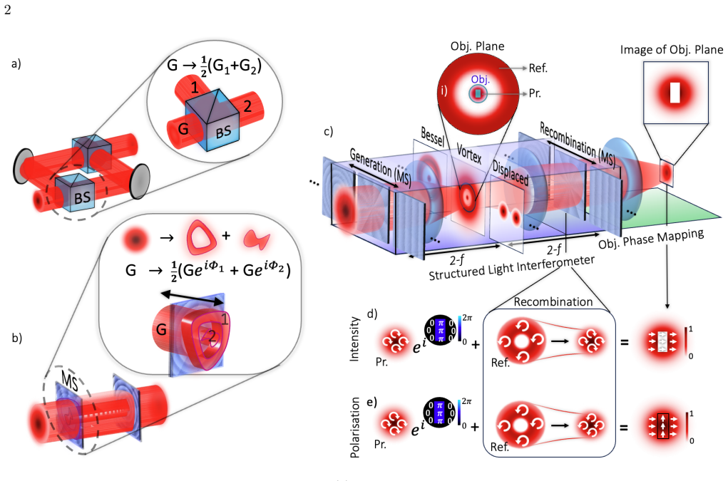
Structured light allows custom interferometers to be designed within a single optical beam.
A machine-rendered reading of the paper's core claim, the machinery that carries it, and where it could break.
Core claim
The central claim is that structured light enables the design of custom interferometers within a single beam via modal conversion, providing robust common-path configurations adaptable to different needs and applicable to accurate phase imaging.
What carries the argument
Structured light modes and modal conversion optics that allow phase information to interfere within one beam.
If this is right
- Produces compact, robust common-path interferometers integrable into existing optical setups.
- Bypasses the need for complex post-processing in phase measurements.
- Supports a range of interferometer types tailored by the structured mode.
- Enables mapping phase to amplitude or polarization for flexible detection.
Where Pith is reading between the lines
- This single-beam approach may simplify integration of phase-sensitive metrology into compact devices or portable instruments.
- By avoiding multiple paths, it could reduce sensitivity to environmental vibrations in field applications.
Load-bearing premise
The modal conversion optics must be realized precisely enough to prevent uncontrolled phase distortions or losses that degrade the phase signal.
What would settle it
A set of phase imaging experiments where the structured light interferometer results differ substantially from atomic force microscopy measurements on identical samples.
Figures


read the original abstract
Interferometry provides highly sensitive access to optical phase and is central to much of modern metrology and phase imaging methods. Conventional implementations, however, often face trade-offs between mechanical stability and experimental or computational complexity. Here, we present a general framework for designing custom interferometers within a single optical beam by exploiting structured light. This approach yields compact, robust common-path configurations that bypass the need for complex post-processing and can easily be integrated into existing setups. We demonstrate the versatility of this concept by designing a range of interferometers, each tailored by the structured mode, and implement them through active and passive modal conversion optics, proving its adaptability to different experimental requirements. To showcase the practical utility of our framework, we apply it to quantitative phase imaging over a variety of physical samples, showing excellent agreement with atomic force microscopy benchmarks. Furthermore, we emphasise the flexibility of our structured light interferometers by mapping phase objects to a choice of either amplitude or polarisation, the latter providing a direct route toward real-time phase-retrieval. This cost-effective approach offers a practical, high-throughput solution for phase-sensitive metrology across fields such as fundamental physics, biology, and material science.
Editorial analysis
A structured set of objections, weighed in public.
Referee Report
Summary. The manuscript presents a general framework for designing custom interferometers inside a single optical beam by converting between structured light modes via active or passive optics. This produces compact common-path configurations for phase imaging that map sample-induced phase to measurable intensity or polarization contrast. The authors demonstrate multiple tailored interferometer designs, apply the approach to quantitative phase imaging on physical samples with reported agreement to AFM benchmarks, and highlight flexibility in choosing amplitude or polarization readouts for real-time retrieval.
Significance. If the central claims hold, the work offers a practical route to robust, integrable phase metrology that reduces mechanical complexity and post-processing demands compared with conventional interferometers. The experimental demonstrations on samples provide concrete validation of end-to-end performance for the tested cases, and the emphasis on structured-light mode conversion supplies a design principle that could be adapted across metrology, biology, and materials applications. The absence of general error characterization, however, confines the assessed significance to specific implementations rather than a fully general method.
major comments (2)
- [Abstract and demonstration sections] Abstract and demonstration sections: the claim of quantitative phase imaging with 'excellent agreement' to AFM is load-bearing for the central assertion that the method bypasses complex post-processing, yet no error budgets, sample exclusion criteria, or uncertainty quantification on the extracted phase are supplied; this leaves the quantitative fidelity only partially verifiable from the presented evidence.
- [Framework and modal-conversion sections] Framework and modal-conversion sections: the mapping from sample phase to observable (intensity or polarization) assumes that active/passive conversion optics introduce no uncontrolled phase distortions or losses that would degrade the extracted signal; the manuscript provides no general bound, characterization, or sensitivity analysis on conversion fidelity, which is required to support the claim of a broadly applicable design method beyond the specific samples shown.
minor comments (1)
- [Abstract] The abstract would be strengthened by briefly naming the specific structured modes employed in the demonstrations and the range of sample types tested.
Simulated Author's Rebuttal
We thank the referee for their careful reading of the manuscript and for the constructive comments, which have helped us improve the clarity and rigor of our presentation. We address each major comment below.
read point-by-point responses
-
Referee: [Abstract and demonstration sections] Abstract and demonstration sections: the claim of quantitative phase imaging with 'excellent agreement' to AFM is load-bearing for the central assertion that the method bypasses complex post-processing, yet no error budgets, sample exclusion criteria, or uncertainty quantification on the extracted phase are supplied; this leaves the quantitative fidelity only partially verifiable from the presented evidence.
Authors: We agree that a more explicit quantification of uncertainties would strengthen the quantitative claims. In the revised manuscript we have added an error budget subsection to the results, reporting standard deviations from repeated acquisitions on the same samples and point-wise differences with the AFM reference data. Sample selection criteria (surface flatness, lateral size compatibility with the field of view, and absence of strong scattering) are now stated in the methods. These additions allow readers to assess the fidelity directly while preserving the central demonstration that the structured-light approach avoids the usual post-processing overhead of conventional interferometry. revision: yes
-
Referee: [Framework and modal-conversion sections] Framework and modal-conversion sections: the mapping from sample phase to observable (intensity or polarization) assumes that active/passive conversion optics introduce no uncontrolled phase distortions or losses that would degrade the extracted signal; the manuscript provides no general bound, characterization, or sensitivity analysis on conversion fidelity, which is required to support the claim of a broadly applicable design method beyond the specific samples shown.
Authors: The framework is formulated as a design principle in which the conversion optics are selected to realize a prescribed mode transformation; the manuscript therefore focuses on the mapping itself rather than on a universal error bound that would require assumptions about arbitrary optics. For the specific implementations shown, we have now included experimental characterization of the SLM and wave-plate fidelity together with a sensitivity analysis demonstrating that phase errors below approximately 0.1 rad in the conversion step produce less than 5 % deviation in the retrieved sample phase. A fully general analytic bound independent of component quality is not provided, as it would be component-specific; we have added a brief discussion of this practical limitation in the revised text. revision: partial
Circularity Check
Framework grounded in standard modal optics; minor self-citation not load-bearing
full rationale
The derivation chain relies on established principles of structured light modes, modal orthogonality, and common-path interferometry to map phase objects to intensity or polarization observables. Experimental validation against AFM benchmarks provides independent falsifiability rather than self-referential fitting. No equations reduce the extracted phase to a quantity defined by construction from input parameters or prior self-citations. Any self-citation on modal conversion is peripheral and does not carry the central claim.
Axiom & Free-Parameter Ledger
axioms (1)
- domain assumption Structured light modes can be generated, propagated, and converted using linear optical elements without introducing uncontrolled aberrations
Reference graph
Works this paper leans on
-
[1]
with slopek= 6.67rad/mm) or a horizontal lin- ear phase ramp,Φ(x, y) = 2πx Λ (g-plate [57] with spa- tial periodΛ= 75µm). We label these interferometers Bessel and displaced, respectively, and show the trans- verse spatial structures (simulated) for each interferome- ter as they appear in the plane of the object (upper row of Fig. 4) together with each MS...
2022
-
[2]
Structured light,
A. Forbes, M. De Oliveira, and M. R. Dennis, “Structured light,”Nature Photonics, vol.15, no.4, pp.253–262, 2021
2021
-
[3]
Towards higher- dimensional structured light,
C. He, Y. Shen, and A. Forbes, “Towards higher- dimensional structured light,”Light: Science & Appli- cations, vol. 11, no. 1, p. 205, 2022
2022
-
[4]
Optical trapping with structured light: a re- view,
Y. Yang, Y.-X. Ren, M. Chen, Y. Arita, and C. Rosales- Guzmán, “Optical trapping with structured light: a re- view,”Advanced Photonics, vol. 3, no. 3, pp. 034001– 034001, 2021
2021
-
[5]
Orbitalangularmomentumoflightforcommunications,
A. E. Willner, K. Pang, H. Song, K. Zou, and H. Zhou, “Orbitalangularmomentumoflightforcommunications,” Applied Physics Reviews, vol. 8, no. 4, 2021
2021
-
[6]
Direct phase detecting sys- tem,
Y. Ichioka and M. Inuiya, “Direct phase detecting sys- tem,”Applied optics, vol. 11, no. 7, pp. 1507–1514, 1972
1972
-
[7]
Op- tical phase measurement in real time,
L. M. Frantz, A. A. Sawchuk, and W. von der Ohe, “Op- tical phase measurement in real time,”Applied Optics, vol. 18, no. 19, pp. 3301–3306, 1979
1979
-
[8]
Optical field characterization using off-axis dig- ital holography,
S. van der Heide, B. van Esch, M. van den Hout, T. Bradley, A. M. Velazquez-Benitez, N. K. Fontaine, R. Ryf, H. Chen, M. Mazur, J. E. Antonio-López, et al., “Optical field characterization using off-axis dig- ital holography,” inOptical Fiber Communication Con- ference, pp. M3Z–6, Optica Publishing Group, 2022
2022
-
[9]
Quan- titative phase imaging based on polarization encoding,
S. Cui, S. Gao, C. Li, W. Zhang, and X. S. Yao, “Quan- titative phase imaging based on polarization encoding,” Optics Express, vol. 30, no. 24, pp. 43622–43632, 2022
2022
-
[10]
Spatially re- solvedphaseobjectsusingmach–zehnderinterferometry,
V. Besse, C. Cassagne, and G. Boudebs, “Spatially re- solvedphaseobjectsusingmach–zehnderinterferometry,” Journal of Modern Optics, vol. 59, no. 10, pp. 887–892, 2012
2012
-
[11]
A review of optical metrology techniques for advanced manufacturing appli- cations,
F. Zhao, H. Tang, X. Zou, and X. Li, “A review of optical metrology techniques for advanced manufacturing appli- cations,”Micromachines, vol. 16, no. 11, p. 1224, 2025
2025
-
[12]
A review of optical interferometry for high- precision length measurement,
G. Huang, C. Cui, X. Lei, Q. Li, S. Yan, X. Li, and G. Wang, “A review of optical interferometry for high- precision length measurement,”Micromachines, vol. 16, no. 1, p. 6, 2024
2024
-
[13]
Optical coherence tomogra- phy,
D. Huang, E. A. Swanson, C. P. Lin, J. S. Schuman, W. G. Stinson, W. Chang, M. R. Hee, T. Flotte, K. Gre- gory, C. A. Puliafito,et al., “Optical coherence tomogra- phy,”science, vol. 254, no. 5035, pp. 1178–1181, 1991
1991
-
[14]
Optical coherence tomog- raphy,
B. E. Bouma, J. F. de Boer, D. Huang, I.-K. Jang, T. Yonetsu, C. L. Leggett, R. Leitgeb, D. D. Sampson, M. Suter, B. J. Vakoc,et al., “Optical coherence tomog- raphy,”Nature Reviews Methods Primers, vol. 2, no. 1, p. 79, 2022
2022
-
[15]
Principlesofinterferencemicroscopyforthe measurement of surface topography,
P.DeGroot, “Principlesofinterferencemicroscopyforthe measurement of surface topography,”Advances in Optics and Photonics, vol. 7, no. 1, pp. 1–65, 2015
2015
-
[16]
Quantitative phase imaging in biomedicine,
Y. Park, C. Depeursinge, and G. Popescu, “Quantitative phase imaging in biomedicine,”Nature photonics, vol. 12, no. 10, pp. 578–589, 2018
2018
-
[17]
Nanomanufacturing—perspective and applica- tions,
F. Fang, X. Zhang, W. Gao, Y. Guo, G. Byrne, and H. N. Hansen, “Nanomanufacturing—perspective and applica- tions,”CIRP Annals, vol. 66, no. 2, pp. 683–705, 2017
2017
-
[18]
Calibration of step height standards for nanometrology using interference mi- croscopy and stylus profilometry,
U. Brand and W. Hillmann, “Calibration of step height standards for nanometrology using interference mi- croscopy and stylus profilometry,”Precision engineering, vol. 17, no. 1, pp. 22–33, 1995
1995
-
[19]
Accurate and traceable calibration of one- dimensional gratings,
G. Dai, L. Koenders, F. Pohlenz, T. Dziomba, and H.-U. Danzebrink, “Accurate and traceable calibration of one- dimensional gratings,”Measurement Science and Tech- nology, vol. 16, no. 6, p. 1241, 2005
2005
-
[20]
Quantitative phase imaging: Recent advances and expanding potential in biomedicine,
T. L. Nguyen, S. Pradeep, R. L. Judson-Torres, J. Reed, M. A. Teitell, and T. A. Zangle, “Quantitative phase imaging: Recent advances and expanding potential in biomedicine,”ACS nano, vol. 16, no. 8, pp. 11516–11544, 2022
2022
-
[21]
Quantitative phase microscopies: accuracy comparison,
P. C. Chaumet, P. Bon, G. Maire, A. Sentenac, and G. Baffou, “Quantitative phase microscopies: accuracy comparison,”Light: Science & Applications, vol. 13, no. 1, p. 288, 2024
2024
-
[22]
Quantitative phase imaging based on holography: trends and new perspectives,
Z. Huang and L. Cao, “Quantitative phase imaging based on holography: trends and new perspectives,”Light: Sci- ence & Applications, vol. 13, no. 1, p. 145, 2024
2024
-
[23]
Quantitative phase microscopy of biolog- ical samples using a portable interferometer,
N. T. Shaked, “Quantitative phase microscopy of biolog- ical samples using a portable interferometer,”Optics let- ters, vol. 37, no. 11, pp. 2016–2018, 2012
2016
-
[24]
Com- mon path interferometer based on the modified michel- son configuration using a reflective grating,
H. Bai, M. Shan, Z. Zhong, L. Guo, and Y. Zhang, “Com- mon path interferometer based on the modified michel- son configuration using a reflective grating,”Optics and Lasers in Engineering, vol. 75, pp. 1–4, 2015. 9
2015
-
[25]
On-axis complex-amplitude modulation for the generation of super-stable vector modes,
V. Rodriguez-Fajardo, F. Arvizu, D. Daza-Salgado, B. Perez-Garcia, and C. Rosales-Guzmán, “On-axis complex-amplitude modulation for the generation of super-stable vector modes,”Journal of Optics, 2024
2024
-
[26]
An active interferometer-stabilization schemewithlinearphasecontrol,
V. V. Krishnamachari, E. R. Andresen, S. R. Keiding, and E. O. Potma, “An active interferometer-stabilization schemewithlinearphasecontrol,”Optics Express, vol.14, no. 12, pp. 5210–5215, 2006
2006
-
[27]
Phase-locking an interferometer with single-photon de- tections,
B. Hacker, K. Günthner, C. Rößler, and C. Marquardt, “Phase-locking an interferometer with single-photon de- tections,”New Journal of Physics, vol. 25, no. 11, p. 113007, 2023
2023
-
[28]
Quantitative phase imaging,
M. Mir, B. Bhaduri, R. Wang, R. Zhu, and G. Popescu, “Quantitative phase imaging,” inProgress in optics, vol. 57, pp. 133–217, Elsevier, 2012
2012
-
[29]
Phase imaging by the transport equation of intensity,
N. Streibl, “Phase imaging by the transport equation of intensity,”Optics communications, vol. 49, no. 1, pp. 6– 10, 1984
1984
-
[30]
Noninterferometric phase imaging with partially coherent light,
D. Paganin and K. A. Nugent, “Noninterferometric phase imaging with partially coherent light,”Physical review letters, vol. 80, no. 12, p. 2586, 1998
1998
-
[31]
Quantita- tive phase-amplitude microscopy i: optical microscopy,
E. Barone-Nugent, A. Barty, and K. Nugent, “Quantita- tive phase-amplitude microscopy i: optical microscopy,” Journal of microscopy, vol. 206, no. 3, pp. 194–203, 2002
2002
-
[32]
3d intensity and phase imaging fromlightfieldmeasurementsinanledarraymicroscope,
L. Tian and L. Waller, “3d intensity and phase imaging fromlightfieldmeasurementsinanledarraymicroscope,” optica, vol. 2, no. 2, pp. 104–111, 2015
2015
-
[33]
Computational super-resolution phase re- trieval from multiple phase-coded diffraction patterns: simulation study and experiments,
V. Katkovnik, I. Shevkunov, N. V. Petrov, and K. Egiazarian, “Computational super-resolution phase re- trieval from multiple phase-coded diffraction patterns: simulation study and experiments,”Optica, vol. 4, no. 7, pp. 786–794, 2017
2017
-
[34]
Wavelength-scanning lensfree on-chip mi- croscopy for wide-field pixel-super-resolved quantitative phase imaging,
X. Wu, J. Sun, J. Zhang, L. Lu, R. Chen, Q. Chen, and C. Zuo, “Wavelength-scanning lensfree on-chip mi- croscopy for wide-field pixel-super-resolved quantitative phase imaging,”Optics letters, vol. 46, no. 9, pp. 2023– 2026, 2021
2023
-
[35]
Digital in-line holo- graphic microscopy,
J. Garcia-Sucerquia, W. Xu, S. K. Jericho, P. Klages, M. H. Jericho, and H. J. Kreuzer, “Digital in-line holo- graphic microscopy,”Applied optics, vol. 45, no. 5, pp. 836–850, 2006
2006
-
[36]
Iterative projection meets sparsity regularization: towards practical single-shot quantitative phase imaging with in-line holography,
Y. Gao and L. Cao, “Iterative projection meets sparsity regularization: towards practical single-shot quantitative phase imaging with in-line holography,”Light: Advanced Manufacturing, vol. 4, no. 1, pp. 37–53, 2023
2023
-
[37]
Compressive holographicsens- ing simplifies quantitative phase imaging,
J. Sun and J.W. Czarske, “Compressive holographicsens- ing simplifies quantitative phase imaging,”Light: Science & Applications, vol. 12, no. 1, p. 121, 2023
2023
-
[38]
Optical path difference microscopy withashack–hartmannwavefrontsensor,
H. Gong, T. E. Agbana, P. Pozzi, O. Soloviev, M. Verhae- gen, and G. Vdovin, “Optical path difference microscopy withashack–hartmannwavefrontsensor,”Optics Letters, vol. 42, no. 11, pp. 2122–2125, 2017
2017
-
[39]
Common-path interferometer for testing pur- poses,
J. Dyson, “Common-path interferometer for testing pur- poses,”JOSA, vol. 47, no. 5, pp. 386–390, 1957
1957
-
[40]
Very stable common-path interferometers and applications,
J. Dyson, “Very stable common-path interferometers and applications,”JOSA, vol. 53, no. 6, pp. 690–694, 1963
1963
-
[41]
Quantitative phase imaging in common-path cross-referenced holographic microscopy using double- exposure method,
J. Běhal, “Quantitative phase imaging in common-path cross-referenced holographic microscopy using double- exposure method,”Scientific Reports, vol. 9, no. 1, p. 9801, 2019
2019
-
[42]
W-shaped common- path interferometer,
R. Wei, L. Di, N. Qiao, and S. Chen, “W-shaped common- path interferometer,”Applied Optics, vol. 59, no. 34, pp. 10973–10979, 2020
2020
-
[43]
Lateral shearing interfer- ometer based on two ronchi phase gratings in series,
H. Schreiber and J. Schwider, “Lateral shearing interfer- ometer based on two ronchi phase gratings in series,”Ap- plied optics, vol. 36, no. 22, pp. 5321–5324, 1997
1997
-
[44]
A wavefront shearing interferometer,
W. Bates, “A wavefront shearing interferometer,”Pro- ceedings of the Physical Society, vol. 59, no. 6, p. 940, 1947
1947
-
[45]
A compact lateral shearing interferometer based on the michelson interferometer,
M. Murty, “A compact lateral shearing interferometer based on the michelson interferometer,”Applied Optics, vol. 9, no. 5, pp. 1146–1148, 1970
1970
-
[46]
Shearing interferometry via geometric phase,
L. A. Alemán-Castaneda, B. Piccirillo, E. Santamato, L. Marrucci, and M. A. Alonso, “Shearing interferometry via geometric phase,”Optica, vol. 6, no. 4, pp. 396–399, 2019
2019
-
[47]
Lat- eral shearing digital holographic imaging of small biologi- cal specimens,
A. S. Singh, A. Anand, R.A. Leitgeb, and B. Javidi, “Lat- eral shearing digital holographic imaging of small biologi- cal specimens,”Optics express, vol. 20, no. 21, pp. 23617– 23622, 2012
2012
-
[48]
Quantitative phase imaging unit,
K. Lee and Y. Park, “Quantitative phase imaging unit,” Optics letters, vol. 39, no. 12, pp. 3630–3633, 2014
2014
-
[49]
Dielectric metasurface enabled compact, single-shot digital holography for quantitative phase imaging,
J. Sardana, S. Devinder, W. Zhu, A. Agrawal, and J. Joseph, “Dielectric metasurface enabled compact, single-shot digital holography for quantitative phase imaging,”Nano Letters, vol. 23, no. 23, pp. 11112–11119, 2023
2023
-
[50]
Spin-to-orbital angular momentum conver- sion in dielectric metasurfaces,
R. C. Devlin, A. Ambrosio, D. Wintz, S. L. Oscurato, A. Y. Zhu, M. Khorasaninejad, J. Oh, P. Maddalena, and F. Capasso, “Spin-to-orbital angular momentum conver- sion in dielectric metasurfaces,”Optics express, vol. 25, no. 1, pp. 377–393, 2017
2017
-
[51]
Arbitrary spin-to–orbital angular mo- mentum conversion of light,
R. C. Devlin, A. Ambrosio, N. A. Rubin, J. B. Mueller, and F. Capasso, “Arbitrary spin-to–orbital angular mo- mentum conversion of light,”Science, vol. 358, no. 6365, pp. 896–901, 2017
2017
-
[52]
Creation and detection of optical modes with spatial light modulators,
A. Forbes, A. Dudley, and M. McLaren, “Creation and detection of optical modes with spatial light modulators,” Advances in optics and photonics, vol. 8, no. 2, pp. 200– 227, 2016
2016
-
[53]
Photon spin-to-orbital angular mo- mentum conversion via an electrically tunable q-plate,
B. Piccirillo, V. D’Ambrosio, S. Slussarenko, L. Marrucci, and E. Santamato, “Photon spin-to-orbital angular mo- mentum conversion via an electrically tunable q-plate,” Applied Physics Letters, vol. 97, no. 24, 2010
2010
-
[54]
Tunable liquid crystal q- plates with arbitrary topological charge,
S. Slussarenko, A. Murauski, T. Du, V. Chigrinov, L. Marrucci, and E. Santamato, “Tunable liquid crystal q- plates with arbitrary topological charge,”Optics express, vol. 19, no. 5, pp. 4085–4090, 2011
2011
-
[55]
Design of optimal polarimeters: maximization of signal-to-noise ratio and minimization of systematic error,
J. S. Tyo, “Design of optimal polarimeters: maximization of signal-to-noise ratio and minimization of systematic error,”Applied optics, vol. 41, no. 4, pp. 619–630, 2002
2002
-
[56]
Optimal configu- rations for imaging polarimeters: impact of image noise and systematic errors,
J. Zallat, S. Aïnouz, and M. P. Stoll, “Optimal configu- rations for imaging polarimeters: impact of image noise and systematic errors,”Journal of Optics A: Pure and Applied Optics, vol. 8, no. 9, p. 807, 2006
2006
-
[57]
Micro-patterned liquid crystal pancharatnam– berry axilens,
J. Ren, W. Wang, W. Yang, C. Yuan, K. Zhou, X. Li, A. Mingwai Tam, C. Meng, J. Sun, V. G. Chigrinov, et al., “Micro-patterned liquid crystal pancharatnam– berry axilens,”Chinese Optics Letters, vol. 16, no. 6, p. 062301, 2018
2018
-
[58]
Two-dimensional topo- logical quantum walks in the momentum space of struc- tured light,
A. D’Errico, F. Cardano, M. Maffei, A. Dauphin, R. Barboza, C. Esposito, B. Piccirillo, M. Lewenstein, 10 P. Massignan, and L. Marrucci, “Two-dimensional topo- logical quantum walks in the momentum space of struc- tured light,”Optica, vol. 7, no. 2, pp. 108–114, 2020
2020
-
[59]
Ince–gaussian beams,
M. A. Bandres and J. C. Gutiérrez-Vega, “Ince–gaussian beams,”Optics letters, vol. 29, no. 2, pp. 144–146, 2004
2004
-
[60]
Airy beams and accelerating waves: an overview of recent advances,
N. K. Efremidis, Z. Chen, M. Segev, and D. N. Christodoulides, “Airy beams and accelerating waves: an overview of recent advances,”Optica, vol. 6, no. 5, pp. 686–701, 2019
2019
-
[61]
Az- imuthal decomposition with digital holograms,
I. A. Litvin, A. Dudley, F. S. Roux, and A. Forbes, “Az- imuthal decomposition with digital holograms,”Optics Express, vol. 20, no. 10, pp. 10996–11004, 2012
2012
-
[62]
Self- healing of structured light: a review,
Y. Shen, S. Pidishety, I. Nape, and A. Dudley, “Self- healing of structured light: a review,”Journal of Optics, vol. 24, no. 10, p. 103001, 2022
2022
-
[63]
Generation of long- distance stably propagating bessel beams,
N. Zhang, J.-S. Ye, S.-F. Feng, X.-K. Wang, P. Han, W.- F. Sun, Y. Zhang, and X.-C. Zhang, “Generation of long- distance stably propagating bessel beams,”OSA Contin- uum, vol. 4, no. 4, pp. 1223–1233, 2021
2021
-
[64]
Structured light: tailored for purpose,
A. Forbes, “Structured light: tailored for purpose,”Opt. Photon. News, vol. 31, no. 6, pp. 24–31, 2020
2020
-
[65]
Ultrabroadband, achro- matic, and non-diffracting perfect optical vortex genera- tion via radial momentum control in dielectric metasur- faces,
T. Xia, Z. Xie, Q. Zhang, W. Xiao, H. Yang, Y. Hu, H. Duan, Y. Cai, and X. Yuan, “Ultrabroadband, achro- matic, and non-diffracting perfect optical vortex genera- tion via radial momentum control in dielectric metasur- faces,”Nature Communications, 2025
2025
-
[66]
Edge- enhanced microscopy of complex objects using scalar and vectorial vortex filtering,
J. Zangpo, T. Kawabe, and H. Kobayashi, “Edge- enhanced microscopy of complex objects using scalar and vectorial vortex filtering,”Optics Express, vol. 31, no. 23, pp. 38388–38399, 2023
2023
-
[67]
Compact oam microscope for edge enhancement of biomedical and object samples,
R. Gozali, T.-A. Nguyen, E. Bendau, and R. R. Al- fano, “Compact oam microscope for edge enhancement of biomedical and object samples,”Review of Scientific Instruments, vol. 88, no. 9, 2017
2017
-
[68]
Spiral interferometry,
S. Fürhapter, A. Jesacher, S. Bernet, and M. Ritsch- Marte, “Spiral interferometry,”Optics letters, vol. 30, no. 15, pp. 1953–1955, 2005
1953
-
[69]
Measurement of nanometric heights by modal decomposition,
V. Rodríguez-Fajardo and A. Forbes, “Measurement of nanometric heights by modal decomposition,”Physical Review Applied, vol. 18, no. 6, p. 064068, 2022
2022
-
[70]
Quantitative imaging of complex sam- ples by spiral phase contrast microscopy,
S. Bernet, A. Jesacher, S. Fürhapter, C. Maurer, and M. Ritsch-Marte, “Quantitative imaging of complex sam- ples by spiral phase contrast microscopy,”Optics Express, vol. 14, no. 9, pp. 3792–3805, 2006
2006
-
[71]
Op- tical imaging using orbital angular momentum: interfer- ometry, holography and microscopy,
J. Zeng, Y. Dong, Y. Wang, J. Zhang, and J. Wang, “Op- tical imaging using orbital angular momentum: interfer- ometry, holography and microscopy,”Journal of Light- wave Technology, vol. 41, no. 7, pp. 2025–2040, 2022
2025
-
[72]
Path-separated electron interferometry in a scanning transmission electron microscope,
F. S. Yasin, T. R. Harvey, J. J. Chess, J. S. Pierce, and B. J. McMorran, “Path-separated electron interferometry in a scanning transmission electron microscope,”Journal of Physics D: Applied Physics, vol. 51, no. 20, p. 205104, 2018
2018
-
[73]
From nanoscopic to macroscopic photo-driven motion in azobenzene-containing materials,
S. L. Oscurato, M. Salvatore, P. Maddalena, and A. Am- brosio, “From nanoscopic to macroscopic photo-driven motion in azobenzene-containing materials,”Nanopho- tonics, vol. 7, no. 8, pp. 1387–1422, 2018
2018
-
[74]
Shapeshifting diffrac- tive optical devices,
S. L. Oscurato, F. Reda, M. Salvatore, F. Borbone, P. Maddalena, and A. Ambrosio, “Shapeshifting diffrac- tive optical devices,”Laser & Photonics Reviews, vol. 16, no. 4, p. 2100514, 2022
2022
-
[75]
Reprogrammable holograms from mask- less surface photomorphing,
F. Reda, M. Salvatore, M. Astarita, F. Borbone, and S. L. Oscurato, “Reprogrammable holograms from mask- less surface photomorphing,”Advanced Optical Materials, vol. 11, no. 21, p. 2300823, 2023. 1 Supplementary Information: Designing interferometers within a single optical beam Bereneice Sephton,1 Rakhi Thomas,1 Carlo Schiano,1 Francesco Reda,1 I Komang ...
2023
discussion (0)
Sign in with ORCID, Apple, or X to comment. Anyone can read and Pith papers without signing in.