A Wireless Reconfigurable Metasurface for Enhanced Parallel Magnetic Resonance Imaging
Pith reviewed 2026-06-29 23:23 UTC · model grok-4.3
The pith
A coaxial loop metasurface creates phase-coherent currents for up to 14.8-fold MRI SNR gains and works with existing parallel imaging arrays.
A machine-rendered reading of the paper's core claim, the machinery that carries it, and where it could break.
Core claim
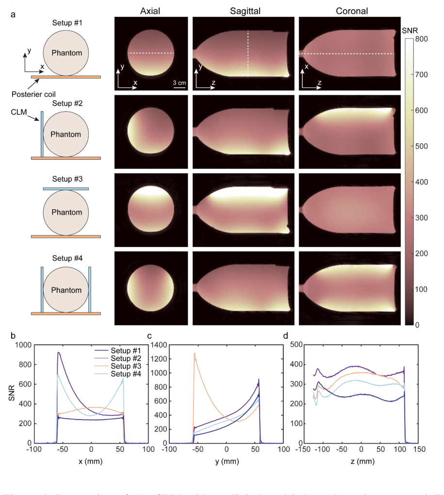
Through its coaxial architecture and shared current pathways, the CLM establishes a collective in-phase resonant mode that enforces phase-coherent current distributions across all loops, resulting in consistently constructive interference and SNR enhancements of up to 14.8-fold relative to the birdcage coil, with additional 2.9-fold gains and parallel-imaging compatibility when used as an add-on to clinical arrays.
What carries the argument
The coaxial loop metasurface (CLM), whose coaxial architecture and shared current pathways enforce a collective in-phase resonant mode for phase-coherent currents across loops.
Load-bearing premise
The coaxial architecture and shared current pathways establish a collective in-phase resonant mode that produces consistently constructive interference.
What would settle it
Measure SNR before and after severing the shared current pathways while keeping the coaxial geometry fixed; loss of the reported enhancement would falsify the mechanism.
Figures
read the original abstract
Modern magnetic resonance imaging (MRI) relies on application-specific multi-channel receive coils to achieve high performance, but these coils are typically costly, rigid, and difficult to generalize across anatomies. Recent wireless, low-cost metamaterials offer improved signal-to-noise ratio (SNR) but remain anatomy-dependent, are prone to destructive inter-element interference, and lack demonstrated compatibility with parallel imaging. Herein, a wireless, reconfigurable coaxial loop metasurface (CLM) is introduced as a platform for localized SNR enhancement that can operate either as a standalone element or as an insertable add-on alongside existing clinical receive systems. Through its coaxial architecture and shared current pathways, the CLM establishes a collective in-phase resonant mode that enforces phase-coherent current distributions across all loops, resulting in consistently constructive interference. Benchmarking on a 3.0 T MR system using an 8-loop CLM shows SNR enhancements of up to 14.8-fold and 14.02-fold in the sagittal and axial planes, relative to the birdcage coil (BC). As an add-on to a clinical posterior receive array, it further demonstrates up to 2.9-fold SNR enhancement and compatibility with parallel imaging across ex vivo and in vivo settings. The proposed CLM paves the way toward a new class of reconfigurable and insertable MRI hardware for flexible and system-compatible signal enhancement.
Editorial analysis
A structured set of objections, weighed in public.
Referee Report
Summary. The manuscript introduces a wireless reconfigurable coaxial loop metasurface (CLM) for localized SNR enhancement in MRI. The design uses a coaxial architecture and shared current pathways to establish a collective in-phase resonant mode that enforces phase-coherent currents and constructive interference across loops. An 8-loop CLM is benchmarked on a 3.0 T system, reporting SNR gains of up to 14.8-fold (sagittal) and 14.02-fold (axial) relative to a birdcage coil; as an add-on to a clinical posterior array it yields up to 2.9-fold enhancement while remaining compatible with parallel imaging in ex vivo and in vivo settings. Supporting EM simulations and bench measurements are provided.
Significance. If the reported SNR values and parallel-imaging compatibility are reproducible, the CLM offers a low-cost, wireless, and insertable platform that addresses rigidity and anatomy-dependence limitations of conventional receive coils. The reconfigurability and demonstrated add-on compatibility with existing clinical arrays are practically relevant strengths. Explicit credit is due for the inclusion of EM simulations and bench measurements that corroborate the headline performance numbers.
minor comments (2)
- [Abstract] Abstract: the maximum SNR values (14.8-fold, 14.02-fold) are stated without reference to the precise imaging plane locations, voxel sizes, or acceleration factors used; adding these qualifiers would improve reproducibility claims.
- The description of the collective in-phase mode would benefit from a brief circuit-equivalent diagram or explicit current-phase plot in the methods to make the mechanism more transparent to readers unfamiliar with metamaterial coil design.
Simulated Author's Rebuttal
We thank the referee for the thorough and positive assessment of our work on the coaxial loop metasurface for MRI. The recommendation for minor revision is noted. No major comments were provided in the report, so we have no specific points requiring point-by-point rebuttal or revision at this stage. We will incorporate any minor suggestions during the revision process.
Circularity Check
No significant circularity identified
full rationale
The manuscript introduces an experimental wireless metasurface device for MRI SNR enhancement and reports measured performance gains (up to 14.8-fold vs. birdcage coil) from bench and in vivo testing on a 3.0 T system. No derivation chain, predictive equations, fitted parameters presented as predictions, or self-citation load-bearing uniqueness theorems appear in the abstract or described content. The in-phase resonant mode is offered as a physical mechanism supported by EM simulations and measurements rather than a self-referential definition or ansatz smuggled via citation. The central claims rest on standard experimental reporting and are externally falsifiable via replication on clinical scanners.
Axiom & Free-Parameter Ledger
Reference graph
Works this paper leans on
-
[1]
P. C. Lauterbur, Image Formation by Induced Local Interactions: Examples Employing Nuclear Magnetic Resonance, Nature 242 (1973): 190–191, https://doi.org/10.1038/242190a0
-
[2]
N. K. Logothetis, What we can do and what we cannot do with fMRI , Nature 453, 869–878 (2008). https://doi.org/10.1038/nature06976
-
[3]
Young , Electron, Nuclear magnetic resonance imaging , Electron
I. Young , Electron, Nuclear magnetic resonance imaging , Electron. Power 1984, 30, 205. https://doi.org/10.1049/ep.1984.0112
-
[4]
M. H. Levitt, Spin Dynamics: Basics of Nuclear Magnetic Resonance, 2nd ed., John Wiley & Sons, Hoboken, NJ 2008
2008
-
[5]
D. I. Hoult, R. E. Richards, The signal -to-noise ratio of the nuclear magnetic resonance experiment, J. Magn. Reson. 1976, 24, 71. https://doi.org/10.1016/j.jmr.2011.09.018
-
[6]
Macovski, (1996), Noise in MRI
A. Macovski, (1996), Noise in MRI. Magn. Reson. Med., 36: 494 -497. https://doi.org/10.1002/mrm.1910360327
-
[7]
T. M. Link, S. Majumdar, C. Peterfy, H. E. Daldrup, M. Uffmann, C. Dowling, L. Steinbach, High Resolution MRI of Small Joints: Impact of Spatial Resolution on Diagnostic Performance and SNR,” H. K. Genant, Magn. Reson. Imaging 1998, 16, 147. https://doi.org/10.1016/S0730-725X(97)00244-0
-
[8]
T. W. Redpath, Signal -to-noise ratio in MRI, Brit. J. Radiol. 1998, 71, 704. https://doi.org/10.1259/bjr.71.847.9771379
-
[9]
P. B. Roemer , W. A. Edelstein , C. E. Hayes , S. P. Souza , O. M. Mueller , The NMR phased array, Magn. Resonance Med. 1990, 16, 192. https://doi.org/10.1002/mrm.1910160203
-
[10]
W. E. Kwok, Basic principles of and practical guide to clinical MRI radiofrequency coils, Radiographics 2022, 42, 898. https://doi.org/10.1148/rg.210180
-
[11]
J. Hennig, K. Zhong, O. Speck, NeuroImage (2007), MR-Encephalography: Fast multi - channel monitoring of brain physiology with magnetic resonance, NeuroImage, 34, 212–219. https://doi.org/10.1016/j.neuroimage.2006.08.036
-
[12]
B. Keil, J.N. Blau, S. Biber, P. Hoecht, V. Tountcheva, K. Setsompop, C. Triantafyllou and L.L. Wald (2013), A 64-channel 3T array coil for accelerated brain MRI. Magn Reson Med, 70: 248-258. https://doi.org/10.1002/mrm.24427
-
[13]
G.C. Wiggins, C. Triantafyllou, A. Potthast, A. Reykowski, M. Nittka and L.L. Wald (2006), 32-channel 3 Tesla receive -only phased-array head coil with soccer -ball element geometry. Magn. Reson. Med., 56: 216-223. https://doi.org/10.1002/mrm.20925
-
[14]
J. J. Hess, C. J. Moran, P. Shah, et al., Relative SNR Measurements in Supine vs. Prone Breast MRI, Magnetic Resonance in Medicine95, no. 5 (2026): 2718 –2725, https://doi.org/10.1002/mrm.70217
-
[15]
A.N. Nnewihe, T. Grafendorfer, B.L. Daniel, P. Calderon, M.T. Alley, F. Robb and B.A. Hargreaves (2011), Custom -fitted 16 -channel bilateral breast coil for bidirectional parallel imaging. Magn. Reson. Med., 66: 281-289. https://doi.org/10.1002/mrm.22771
-
[16]
R. Brown, K. Lakshmanan, G. Madelin, L. Alon, G. Chang, D.K. Sodickson, R.R. Regatte and G.C. Wiggins (2016), A flexible nested sodium and proton coil array with wideband matching for knee cartilage MRI at 3T. Magn. Reson. Med., 76: 1325 -1334. https://doi.org/10.1002/mrm.26017
-
[17]
C.J. Hardy, R.O. Giaquinto, J.E. Piel, K.W. Rohling AAS, L. Marinelli, D.J. Blezek, E.W. Fiveland, R.D. Darrow and T.K.F. Foo (2008), 128-channel body MRI with a flexible high - density receiver -coil array. J. Magn. Reson. Imaging, 28: 1219 -1225. https://doi.org/10.1002/jmri.21463
-
[18]
J. T. Vaughan and J. R. Griffiths, RF Coils for MRI (John Wiley & Sons, 2012)
2012
-
[19]
B. Gruber, M. Froeling , T. Leiner, & D. W. Klomp (2018). RF coils: A practical guide for nonphysicists. Journal of magnetic resonance imaging, 48(3), 590 -604. https://doi.org/10.1002/jmri.26187
-
[20]
E. Motovilova, E. T. Tan, V. Taracila, J. M. Vincent, T. Grafendorfer, J. Shin, ... & S. A. Winkler (2021). Stretchable self -tuning MRI receive coils based on liquid metal technology (LiquiTune). Scientific reports, 11(1), 16228. https://doi.org/10.1038/s41598-021-95335-6
-
[21]
J. Corea, A. Flynn, B. Lechê ne et al (2016), Screen-printed flexible MRI receive coils , Nat Commun 7, 10839. https://doi.org/10.1038/ncomms10839
-
[22]
P.A. Narayana, W.W. Brey, M.V. Kulkarni, C.L. Sievenpiper . (1988) Compensation for surface coil sensitivity variation in magnetic resonance imaging, Magnetic Resonance Imaging, 6(3):271-4. https://doi.org/10.1016/0730-725X(88)90401-8
-
[23]
A. Deshmane, V. Gulani, M.A. Griswold and N. Seiberlich (2012), Parallel MR imaging. J. Magn. Reson. Imaging, 36: 55-72. https://doi.org/10.1002/jmri.23639
-
[24]
K.P. Pruessmann, M. Weiger, M.B. Scheidegger and P. Boesiger (1999), SENSE: Sensitivity encoding for fast MRI. Magn. Reson. Med., 42: 952-962. https://doi.org/10.1002/(SICI)1522- 2594(199911)42:5<952::AID-MRM16>3.0.CO;2-S
-
[25]
D. J. Larkman, R. G. Nunes (2007). Parallel magnetic resonance imaging. Physics in Medicine & Biology, 52(7), R15-R55. https://doi.org/10.1088/0031-9155/52/7/R01
-
[26]
Microstructured Magnetic Materials for RF Flux Guides in Magnetic Resonance Imaging,
M. C. K. Wiltshire, J. B. Pendry, I. R. Young, D. J. Larkman, D. J. Gilderdale, and J. V. Hajnal, “Microstructured Magnetic Materials for RF Flux Guides in Magnetic Resonance Imaging,” Science 291 (2001): 849–851, https://doi.org/10.1126/science.291.5505.849
-
[27]
M. J. Freire, R. Marques, and L. Jelinek, Experimental Demonstration of a μ = −1 Metamaterial Lens for Magnetic Resonance Imaging, Applied Physics Letters 93, no. 23 (2008): 231108, https://doi.org/10.1063/1.3043725
-
[28]
M. J. Freire, L. Jelinek, R. Marques, and M. Lapine, On the Applications of Metamaterial Lenses for Magnetic Resonance Imaging, Journal of Magnetic Resonance 203 (2010): 81–90, https://doi.org/10.1016/j.jmr.2009.12.005
-
[29]
Duan, G., Zhao, X., Anderson, S.W. et al. (2019), Boosting magnetic resonance imaging signal-to-noise ratio using magnetic metamaterials. Commun Phys 2, 35. https://doi.org/10.1038/s42005-019-0135-7
-
[30]
Li, B., Xie, R., Sun, Z. et al. Nonlinear metamaterials enhanced surface coil array for parallel magnetic resonance imaging. Nat Commun 15, 7949 (2024). https://doi.org/10.1038/s41467- 024-52423-1
-
[31]
A. P. Slobozhanyuk, A. N. Poddubny, A. J. Raaijmakers, et al., Enhancement of Magnetic Resonance Imaging with Metasurfaces, Advanced Materials 28 (2016): 1832 –1838, https://doi.org/10.1002/adma.201504270
-
[32]
A. V. Shchelokova, A. P. Slobozhanyuk, I. V. Melchakova, et al., Locally Enhanced Image Quality with Tunable Hybrid Metasurfaces, Physical Review Applied 9 (2018): 014020, https://doi.org/10.1103/PhysRevApplied.9.014020
-
[33]
A. V. Shchelokova, C. A. van den Berg, D. A. Dobrykh, et al., Volumetric Wireless Coil Based on Periodically Coupled Split -Loop Resonators for Clinical Wrist Imaging, Magnetic Resonance in Medicine 80 (2018): 1726–1737, https://doi.org/10.1002/mrm.27140
-
[34]
H. Fujita, T. Zheng, X. Yang, M. J. Finnerty, and S. Handa, RF Surface Receive Array Coils: the Art of an LC Circuit, Journal of Magnetic Resonance Imaging 38 (2013): 12 –25, https://doi.org/10.1002/jmri.24159
-
[35]
X. Zhu, K. Wu, S. W. Anderson, X. Zhang (2023), Helmholtz Coil -Inspired Volumetric Wireless Resonator for Magnetic Resonance Imaging. Adv. Mater. Technol., 8, 2301053. https://doi.org/10.1002/admt.202301053
-
[36]
X. Zhao, G. Duan, K. Wu, S. W. Anderson, X. Zhang, Intelligent Metamaterials Based on Nonlinearity for Magnetic Resonance Imaging. Adv. Mater. 2019, 31, 1905461. https://doi.org/10.1002/adma.201905461
-
[37]
Z. Chi, Y. Yi, Y. Wang, M. Wu, L. Wang, X. Zhao, ... & J. Zhou (2021). Adaptive cylindrical wireless metasurfaces in clinical magnetic resonance imaging. Advanced Materials, 33(40), 2102469. https://doi.org/10.1002/adma.202102469
-
[38]
Y. Liu, X. Zhu, K. Wu, S. W. Anderson, and X. Zhang, Circularly Polarized Metamaterial Cage for Homogeneous Signal-to-Noise Ratio Enhancement in Magnetic Resonance Imaging. Advanced Materials (2026): e16569. https://doi.org/10.1002/adma.202516569
-
[39]
K. Wu, X. Zhu, X. Zhao, S. W. Anderson, X. Zhang. Conformal Metamaterials with Active Tunability and Self -Adaptivity for Magnetic Resonance Imaging. Research. 2024;7:0560. https://doi.org/10.34133/research.0560
-
[40]
X. Zhu, K. Wu, S. W. Anderson, X. Zhang, Wearable Coaxially-Shielded Metamaterial for Magnetic Resonance Imaging. Adv. Mater.2024, 36, 2313692. https://doi.org/10.1002/adma.202313692
-
[41]
K. Wu, X. Zhu, S. W. Anderson, X. Zhang, Wireless, customizable coaxially shielded coils for magnetic resonance imaging , Sci. Adv.10,eadn5195(2024). https://doi.org/10.1126/sciadv.adn5195
-
[42]
X. Zhu, K. Wu, S. W. Anderson, X. Zhang, Metamaterial-Enabled Hybrid Receive Coil for Enhanced Magnetic Resonance Imaging Capabilities. Adv. Sci.2025, 12, 2410907. https://doi.org/10.1002/advs.202410907
-
[43]
A. Shchelokova, V. Ivanov, A. Mikhailovskaya et al. (2020), Ceramic resonators for targeted clinical magnetic resonance imaging of the breast. Nat Commun 11, 3840. https://doi.org/10.1038/s41467-020-17598-3
-
[44]
K. Wu, X. Zhu, T. G. Bifano, S. W. Anderson, X. Zhang, Computational-Design Enabled Wearable and Tunable Metamaterials via Freeform Auxetics for Magnetic Resonance Imaging. Adv. Sci.2024, 11, 2400261. https://doi.org/10.1002/advs.202400261
-
[45]
Jia, Y., Pang, Y., Jin, R., Liu, Y., Kong, X., Shao, K., ... & Wang, Z. (2025). A unified circular-polarization metamaterial -inspired resonator for increasing SNR in breast MRI. Magnetic Resonance Imaging, 121, 110403. https://doi.org/10.1016/j.mri.2025.110403
discussion (0)
Sign in with ORCID, Apple, or X to comment. Anyone can read and Pith papers without signing in.