GRASP MRI: A Decade of Innovation from Bench to Bedside
Pith reviewed 2026-06-29 23:17 UTC · model grok-4.3
The pith
GRASP MRI enables free-breathing dynamic imaging through continuous golden-angle radial sampling combined with compressed sensing and parallel imaging.
A machine-rendered reading of the paper's core claim, the machinery that carries it, and where it could break.
Core claim
GRASP (Golden-angle RAdial Sparse Parallel) MRI has emerged as one of the most influential motion-robust dynamic MRI frameworks over the past decade. By combining continuous golden-angle radial sampling with compressed sensing and parallel imaging, GRASP enables free-breathing data acquisition with flexible retrospective image reconstruction. Since its original introduction, the framework has evolved substantially and has inspired a broad range of technical developments, including motion-resolved reconstruction, real-time imaging, quantitative MRI, deep learning-enabled reconstruction, and multidimensional cardiovascular imaging. These advances have further expanded the role of GRASP MRI in
What carries the argument
The GRASP framework, which integrates continuous golden-angle radial sampling with compressed sensing and parallel imaging to support motion-robust free-breathing dynamic MRI and flexible retrospective reconstruction.
If this is right
- Continuous data collection allows reconstruction at arbitrary temporal resolutions after acquisition.
- Motion-resolved methods can separate respiratory and cardiac phases without additional hardware.
- Quantitative parameter mapping becomes possible during free breathing for organs that move.
- Deep learning can accelerate reconstruction while preserving the radial sampling advantages.
- Multidimensional cardiovascular protocols extend dynamic coverage to multiple spatial dimensions.
Where Pith is reading between the lines
- The same radial-plus-compressed-sensing pattern could transfer to other motion-sensitive modalities such as CT or ultrasound.
- Standardization of golden-angle ordering across vendors would simplify multi-center trials.
- Real-time GRASP variants might reduce the need for sedation in pediatric or uncooperative patients.
- Quantitative free-breathing extensions could enable longitudinal studies without repeated breath-hold training.
Load-bearing premise
The listed technical developments in motion-resolved reconstruction, real-time imaging, quantitative MRI, deep learning, and multidimensional cardiovascular imaging have substantially expanded the clinical role of GRASP where conventional breath-hold imaging is challenging.
What would settle it
A controlled clinical comparison demonstrating that GRASP-based free-breathing scans produce no measurable gain in diagnostic quality, patient throughput, or feasibility over standard breath-hold protocols in the same patient population would falsify the claim of expanded clinical utility.
Figures
read the original abstract
GRASP (Golden-angle RAdial Sparse Parallel) MRI has emerged as one of the most influential motion-robust dynamic MRI frameworks over the past decade. By combining continuous golden-angle radial sampling with compressed sensing and parallel imaging, GRASP enables free-breathing data acquisition with flexible retrospective image reconstruction. Since its original introduction, the framework has evolved substantially and has inspired a broad range of technical developments, including motion-resolved reconstruction, real-time imaging, quantitative MRI, deep learning-enabled reconstruction, and multidimensional cardiovascular imaging. These advances have further expanded the role of GRASP MRI in a range of clinical applications where conventional breath-hold imaging is challenging. This review summarizes the technical evolution and clinical translation of GRASP MRI over the past decade, with a particular focus on the conceptual advantages of continuous radial acquisition, flexible retrospective reconstruction, and motion-robust imaging. Emerging developments in deep learning reconstruction, real-time volumetric imaging, and quantitative free-breathing MRI are also discussed together with future directions of motion-robust MRI acquisition and reconstruction.
Editorial analysis
A structured set of objections, weighed in public.
Referee Report
Summary. The manuscript is a review paper summarizing the technical evolution and clinical translation of GRASP (Golden-angle RAdial Sparse Parallel) MRI over the past decade. It claims that GRASP has emerged as one of the most influential motion-robust dynamic MRI frameworks by combining continuous golden-angle radial sampling with compressed sensing and parallel imaging, enabling free-breathing acquisition and flexible retrospective reconstruction. The review discusses subsequent developments including motion-resolved reconstruction, real-time imaging, quantitative MRI, deep learning-enabled reconstruction, and multidimensional cardiovascular imaging, which are presented as having expanded GRASP's clinical role in applications where breath-hold imaging is challenging. It covers conceptual advantages, emerging developments in deep learning and quantitative free-breathing MRI, and future directions.
Significance. If the cited literature and historical framing are accurate and balanced, the review provides a useful consolidation of advances in motion-robust MRI for the medical imaging community. The emphasis on the flexibility of continuous radial acquisition and retrospective reconstruction offers a coherent narrative thread across technical and clinical sections. As a descriptive synthesis rather than an empirical or theoretical contribution, its value lies in organizing a decade of work; no machine-checked proofs or new falsifiable predictions are present.
Simulated Author's Rebuttal
We thank the referee for their positive evaluation of our review manuscript and for recommending acceptance. The summary accurately reflects the scope and narrative of the paper as a descriptive synthesis of GRASP MRI developments over the past decade.
Circularity Check
No significant circularity; descriptive review with no derivations
full rationale
The paper is a review article summarizing the history and applications of the GRASP MRI framework. It contains no equations, no predictions, no fitted parameters, and no derivation chain. All statements are descriptive syntheses of prior literature. Self-citations exist (as expected for originators of the method) but are not load-bearing for any technical claim that reduces to itself; the influence and expansion statements are interpretive framing rather than self-referential results. This matches the default expectation of no circularity for a non-derivational paper.
Axiom & Free-Parameter Ledger
Reference graph
Works this paper leans on
-
[1]
2 Center for Advanced Imaging Innovation and Research (CAI2R), Department of Radiology, New York University Grossman School of Medicine, New York, NY, USA
1 GRASP MRI: A Decade of Innovation from Bench to Bedside Li Feng, Kai Tobias Block, Hersh Chandarana, Daniel K Sodickson 1 Bernard and Irene Schwartz Center for Biomedical Imaging, Department of Radiology, New York University Grossman School of Medicine, New York, NY, USA. 2 Center for Advanced Imaging Innovation and Research (CAI2R), Department of Radio...
2012
-
[2]
In the same year, NYU became the first academic institution to adopt Radial VIBE for clinical patient studies, and preliminary results were published the following year (32)
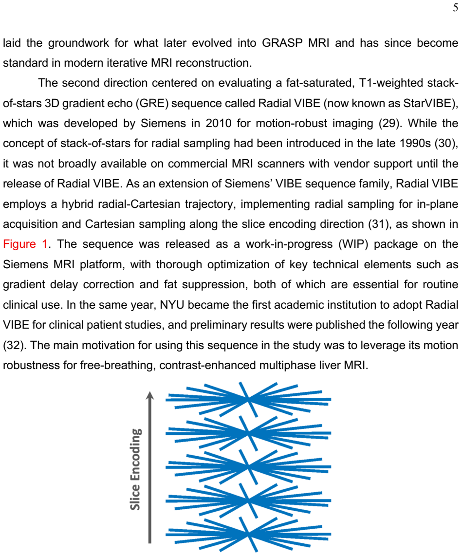
The sequence was released as a work-in-progress (WIP) package on the Siemens MRI platform, with thorough optimization of key technical elements such as gradient delay correction and fat suppression, both of which are essential for routine clinical use. In the same year, NYU became the first academic institution to adopt Radial VIBE for clinical patient st...
2011
-
[3]
gridding
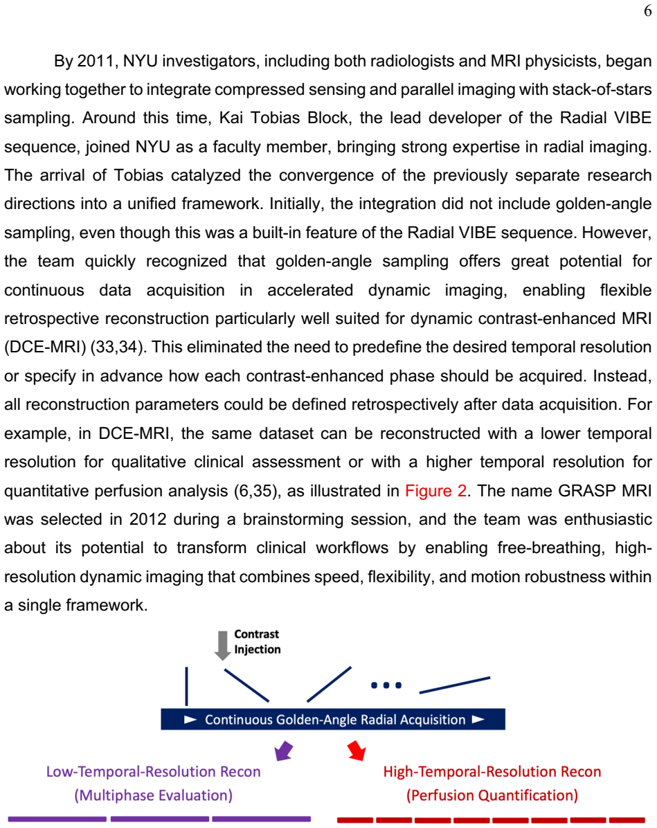
The name GRASP MRI was selected in 2012 during a brainstorming session, and the team was enthusiastic about its potential to transform clinical workflows by enabling free-breathing, high-resolution dynamic imaging that combines speed, flexibility, and motion robustness within a single framework. Figure 2: Flexibility of golden-angle radial sampling. The c...
2012
-
[4]
unstreaking
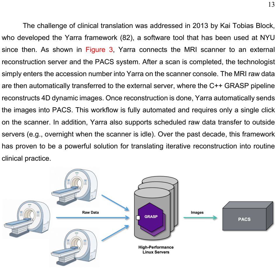
He re-implemented the entire GRASP reconstruction pipeline in C++, which substantially accelerated image reconstruction. With parallel computing, reconstruction time for an entire 3D volume was reduced to under 30 minutes, compared to 15-30 minutes per slice in MATLAB. This marked the first major step toward practical clinical translation. 13 The challeng...
2013
-
[5]
In addition to liver imaging, GRASP-Pro has been applied to DCE-MRI of the breast with sub-second temporal resolution to enable more accurate perfusion quantification (100)
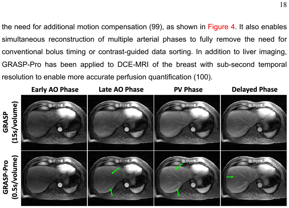
It also enables simultaneous reconstruction of multiple arterial phases to fully remove the need for conventional bolus timing or contrast-guided data sorting. In addition to liver imaging, GRASP-Pro has been applied to DCE-MRI of the breast with sub-second temporal resolution to enable more accurate perfusion quantification (100). Figure 4: Comparison be...
2024
-
[6]
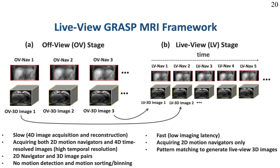
During the live-view stage, only 2D navigators are acquired in real time, which can be rapidly matched to the off-line database for retrieving the best-matching 3D image
During the off-line stage, free-breathing time-resolved 4D (3D + motion) images are acquired and reconstructed to form a motion-resolved image database, where each 3D image in the database is linked to a low-resolution 2D navigator that represents a specific respiratory state. During the live-view stage, only 2D navigators are acquired in real time, which...
2023
-
[7]
Golden-angle radial sparse parallel MRI: combination of compressed sensing, parallel imaging, and golden-angle radial sampling for fast and flexible dynamic volumetric MRI
Feng L, Grimm R, Block KT obias, Chandarana H, Kim S, Xu J, et al. Golden-angle radial sparse parallel MRI: combination of compressed sensing, parallel imaging, and golden-angle radial sampling for fast and flexible dynamic volumetric MRI. Magn Reson Med. 2014 Sep 1;72(3):707–17
2014
-
[8]
The rapid imaging renaissance: sparser samples, denser dimensions, and glimmerings of a grand unified tomography
Sodickson DK, Feng L, Knoll F, Cloos M, Ben-Eliezer N, Axel L, et al. The rapid imaging renaissance: sparser samples, denser dimensions, and glimmerings of a grand unified tomography. In: Gimi B, Molthen RC, editors. Medical Imaging 2015: Biomedical Applications in Molecular, Structural, and Functional Imaging. SPIE
2015
-
[9]
Compressed sensing for body MRI
Feng L, Benkert T, Block KT, Sodickson DK, Otazo R, Chandarana H. Compressed sensing for body MRI. Journal of Magnetic Resonance Imaging. 2017 Apr 1;45(4):966–87
2017
-
[10]
Golden-Angle Radial Sparse Parallel (GRASP) MRI differentiates head & neck paragangliomas from schwannomas
Demerath T, Blackham K, Anastasopoulos C, Block KT, Stieltjes B, Schubert T. Golden-Angle Radial Sparse Parallel (GRASP) MRI differentiates head & neck paragangliomas from schwannomas. Magn Reson Imaging [Internet]. 2020 Jul 1 [cited 2025 Sep 22];70:73. Available from: https://pmc.ncbi.nlm.nih.gov/articles/PMC8191413/
2020
-
[11]
Image Quality of High-Resolution 3-Dimensional Neck MRI Using CAIPIRINHA-VIBE and GRASP-VIBE: An Intraindividual Comparative Study
Seo M, Yoon J, Choi Y, Nickel D, Jang J, Shin NY, et al. Image Quality of High-Resolution 3-Dimensional Neck MRI Using CAIPIRINHA-VIBE and GRASP-VIBE: An Intraindividual Comparative Study. Invest Radiol [Internet]. 2022 Nov 1 [cited 2025 Sep 22];57(11):711–9. Available from: https://journals.lww.com/investigativeradiology/fulltext/2022/11000/image_quality...
2022
-
[12]
Kim SG, Feng L, Grimm R, Freed M, Block KT, Sodickson DK, et al. Influence of temporal regularization and radial undersampling factor on compressed sensing reconstruction in dynamic contrast enhanced MRI of the breast. Journal of Magnetic Resonance Imaging [Internet]. 2016 Jan 1 [cited 2021 Aug 15];43(1):261–9. Available from: https://onlinelibrary.wiley....
-
[13]
Free-breathing contrast-enhanced multiphase MRI of the liver using a combination of compressed sensing, parallel imaging, and golden-angle radial sampling
Chandarana H, Feng L, Block TK, Rosenkrantz AB, Lim RP, Babb JS, et al. Free-breathing contrast-enhanced multiphase MRI of the liver using a combination of compressed sensing, parallel imaging, and golden-angle radial sampling. Invest Radiol. 2013 Jan;48(1):10–6
2013
-
[14]
Comparison of image quality of subtracted and nonsubtracted breath hold VIBE and free breathing GRASP in the evaluation of renal masses
Huber S, Balcacer De la Cruz P, Mahan M, Spektor M, Lo R, Block KT, et al. Comparison of image quality of subtracted and nonsubtracted breath hold VIBE and free breathing GRASP in the evaluation of renal masses. Clin Imaging [Internet]. 25 2021 Jun 1 [cited 2025 Sep 22];74:15–8. Available from: https://www.sciencedirect.com/science/article/pii/S0899707120305313
2021
-
[15]
Ream JM, Doshi A, Lala S V., Kim S, Rusinek H, Chandarana H. High Spatiotemporal Resolution Dynamic Contrast-Enhanced MR Enterography in Crohn Disease Terminal Ileitis Using Continuous Golden-Angle Radial Sampling, Compressed Sensing, and Parallel Imaging. http://dx.doi.org/102214/AJR1413674 [Internet]. 2015 May 22 [cited 2021 Aug 16];204(6):W663–9. Avail...
2015
-
[16]
Rosenkrantz AB, Geppert C, Grimm R, Block TK, Glielmi C, Feng L, et al. Dynamic contrast-enhanced MRI of the prostate with high spatiotemporal resolution using compressed sensing, parallel imaging, and continuous golden-angle radial sampling: Preliminary experience. Journal of Magnetic Resonance Imaging [Internet]. 2015 May 1 [cited 2021 Aug 15];41(5):136...
-
[17]
Parikh N, Ream JM, Zhang HC, Block KT, Chandarana H, Rosenkrantz AB. Performance of simultaneous high temporal resolution quantitative perfusion imaging of bladder tumors and conventional multi-phase urography using a novel free-breathing continuously acquired radial compressed-sensing MRI sequence. Magn Reson Imaging [Internet]. 2016 Jun 1 [cited 2025 Se...
2016
-
[18]
Yoon JH, Nickel MD, Peeters JM, Lee JM. Rapid Imaging: Recent Advances in Abdominal MRI for Reducing Acquisition Time and Its Clinical Applications. Korean J Radiol [Internet]. 2019 Oct 4 [cited 2023 Jul 18];20(12):1597–615. Available from: https://synapse.koreamed.org/articles/1139311
-
[19]
Accelerated Abdominal MRI: A Review of Current Methods and Applications
Feng L, Chandarana H. Accelerated Abdominal MRI: A Review of Current Methods and Applications. Journal of Magnetic Resonance Imaging [Internet]. 2025 Sep 1 [cited 2025 Sep 22];62(3):654–72. Available from: /doi/pdf/10.1002/jmri.29750
-
[20]
XD-GRASP: Golden-angle radial MRI with reconstruction of extra motion-state dimensions using compressed sensing
Feng L, Axel L, Chandarana H, Block KT, Sodickson DK, Otazo R. XD-GRASP: Golden-angle radial MRI with reconstruction of extra motion-state dimensions using compressed sensing. Magn Reson Med. 2016 Feb 1;75(2):775–88
2016
-
[21]
4D GRASP MRI at Sub-Second Temporal Resolution
Feng L. 4D GRASP MRI at Sub-Second Temporal Resolution. NMR Biomed [Internet]. 2022 Oct 19 [cited 2022 Oct 30];e4844. Available from: https://onlinelibrary.wiley.com/doi/full/10.1002/nbm.4844
-
[22]
Feng L. Live-view 4D GRASP MRI: A framework for robust real-time respiratory motion tracking with a sub-second imaging latency. Magn Reson Med [Internet]. 2023 [cited 2023 Jul 2];90(3). Available from: https://pubmed.ncbi.nlm.nih.gov/37203314/
-
[23]
Magnetization-prepared GRASP MRI for rapid 3D T1 mapping and fat/water-separated T1 mapping
Feng L, Liu F, Soultanidis G, Liu C, Benkert T, Block KT, et al. Magnetization-prepared GRASP MRI for rapid 3D T1 mapping and fat/water-separated T1 mapping. Magn Reson Med [Internet]. 2021 Jul 1 [cited 2021 Jun 10];86(1):97–114. Available from: https://onlinelibrary.wiley.com/doi/full/10.1002/mrm.28679
-
[24]
DeepGrasp4D: A General Framework for Highly-Accelerated Real-Time 4D Golden-Angle Radial MRI Using Deep Learning
Pei H, Chandarana H, Sodickson D, Feng L. DeepGrasp4D: A General Framework for Highly-Accelerated Real-Time 4D Golden-Angle Radial MRI Using Deep Learning. In: Proc Intl Soc Mag Reson Med 31 (2024) p0040. 26
2024
-
[25]
Sparse MRI: The application of compressed sensing for rapid MR imaging
Lustig M, Donoho D, Pauly JM. Sparse MRI: The application of compressed sensing for rapid MR imaging. Magn Reson Med. 2007 Dec;58(6):1182–95
2007
-
[26]
Compressed sensing MRI: A look at how CS can improve on current imaging techniques
Lustig M, Donoho DL, Santos JM, Pauly JM. Compressed sensing MRI: A look at how CS can improve on current imaging techniques. IEEE Signal Process Mag. 2008;25(2):72–82
2008
-
[27]
Undersampled radial MRI with multiple coils
Block KT, Uecker M, Frahm J. Undersampled radial MRI with multiple coils. Iterative image reconstruction using a total variation constraint. Magn Reson Med. 2007;Jun;57(6):1086-98
2007
-
[28]
Combination of compressed sensing and parallel imaging for highly accelerated first-pass cardiac perfusion MRI
Otazo R, Kim D, Axel L, Sodickson DK. Combination of compressed sensing and parallel imaging for highly accelerated first-pass cardiac perfusion MRI. Magn Reson Med. 2010 Sep;64(3):767–76
2010
-
[29]
Combination of Compressed Sensing, Parallel Imaging & Partial Fourier for Highly-Accelerated 3D First-Pass Cardiac Perfusion MRI
Feng L; Xu J; Axel L; Sodickson DK; Otazo R. Combination of Compressed Sensing, Parallel Imaging & Partial Fourier for Highly-Accelerated 3D First-Pass Cardiac Perfusion MRI. In: Proc Intl Soc Mag Reson Med 19 (2011), p4368
2011
-
[30]
Highly accelerated real-time cardiac cine MRI using k–t SPARSE-SENSE
Feng L, Srichai MB, Lim RP, Harrison A, King W, Adluru G, et al. Highly accelerated real-time cardiac cine MRI using k–t SPARSE-SENSE. Magn Reson Med [Internet]. 2013 Jul 1 [cited 2022 Feb 22];70(1):64–74. Available from: https://onlinelibrary.wiley.com/doi/full/10.1002/mrm.24440
-
[31]
Liu J, Feng L, Shen HW, Zhu C, Wang Y, Mukai K, et al. Highly-accelerated self-gated free-breathing 3D cardiac cine MRI: validation in assessment of left ventricular function. Magnetic Resonance Materials in Physics, Biology and Medicine [Internet]. 2017 Aug 1 [cited 2022 Feb 21];30(4):337–46. Available from: https://link.springer.com/article/10.1007/s103...
-
[32]
Fast Real-Time Cardiac MRI: a Review of Current Techniques and Future Directions
Wang X, Uecker M, Feng L. Fast Real-Time Cardiac MRI: a Review of Current Techniques and Future Directions. Investig Magn Reson Imaging. 2021;25(4):252
2021
-
[33]
Accelerated phase-contrast cine MRI using k-t SPARSE-SENSE
Kim D, Dyvorne HA, Otazo R, Feng L, Sodickson DK, Lee VS. Accelerated phase-contrast cine MRI using k-t SPARSE-SENSE. Magn Reson Med [Internet]. 2012 Apr 1 [cited 2022 Feb 22];67(4):1054–64. Available from: https://onlinelibrary.wiley.com/doi/full/10.1002/mrm.23088
-
[34]
Feng L, Otazo R, Jung H, Jensen JH, Ye JC, Sodickson DK, et al. Accelerated cardiac T2 mapping using breath-hold multiecho fast spin-echo pulse sequence with k-t FOCUSS. Magn Reson Med [Internet]. 2011 Jun 1 [cited 2022 Feb 22];65(6):1661–9. Available from: https://onlinelibrary.wiley.com/doi/full/10.1002/mrm.22756
-
[35]
Undersampled Projection Reconstruction Applied to MR Angiography
Peters DC, Korosec FR, Grist TM, Block WF, Holden JE, Vigen KK, et al. Undersampled Projection Reconstruction Applied to MR Angiography. Magn Reson Med [Internet]. 2000 [cited 2025 Sep 22];43:91–101. Available from: /doi/pdf/10.1002/%28SICI%291522-2594%28200001%2943%3A1%3C91%3A%3AAID-MRM11%3E3.0.CO%3B2-4
2000
-
[36]
Golden-Angle Radial MRI: Basics, Advances, and Applications
Feng L. Golden-Angle Radial MRI: Basics, Advances, and Applications. Journal of Magnetic Resonance Imaging [Internet]. 2022 Apr 9 [cited 2022 May 30]; Available from: https://onlinelibrary.wiley.com/doi/full/10.1002/jmri.28187 27
-
[37]
Free-breathing radial 3D fat-suppressed T1-weighted gradient echo sequence: A viable alternative for contrast-enhanced liver imaging in patients unable to suspend respiration
Chandarana H, Block TK, Rosenkrantz AB, Lim RP, Kim D, Mossa DJ, et al. Free-breathing radial 3D fat-suppressed T1-weighted gradient echo sequence: A viable alternative for contrast-enhanced liver imaging in patients unable to suspend respiration. Invest Radiol [Internet]. 2011 Oct [cited 2021 Aug 15];46(10):648–53. Available from: https://journals.lww.co...
2011
-
[38]
An optimal radial profile order based on the golden ratio for time-resolved MRI
Winkelmann S, Schaeffter T, Koehler T, Eggers H, Doessel O. An optimal radial profile order based on the golden ratio for time-resolved MRI. IEEE Trans Med Imaging. 2007 Jan;26(1):68–76
2007
-
[39]
A 3D golden-angle projection reconstruction technique for dynamic contrast-enhanced MRI
Song H, Lin W, Dougherty L, Schnall M. A 3D golden-angle projection reconstruction technique for dynamic contrast-enhanced MRI. In: Proc Intl Soc Mag Reson Med, 2006, p3364
2006
-
[40]
Chandarana H, Block TK, Ream J, Mikheev A, Sigal SH, Otazo R, et al. Estimating Liver Perfusion From Free–Breathing Continuously Acquired Dynamic Gadolinium-Ethoxybenzyl-Diethylenetriamine Pentaacetic Acid–Enhanced Acquisition With Compressed Sensing Reconstruction. Invest Radiol. 2015 Feb;50(2):88–94
2015
-
[41]
Image Formation by Induced Local Interactions: Examples Employing Nuclear Magnetic Resonance
Lauterbur PC. Image Formation by Induced Local Interactions: Examples Employing Nuclear Magnetic Resonance. Nature 1973 242:5394 [Internet]. 1973 [cited 2022 Feb 20];242(5394):190–1. Available from: https://www.nature.com/articles/242190a0
1973
-
[42]
Projection Reconstruction Techniques for Reduction of Motion Effects in MRI
Glover GH, Pauly JM. Projection Reconstruction Techniques for Reduction of Motion Effects in MRI. Magn Reson Med [Internet]. 1992 Dec 1 [cited 2022 Feb 21];28(2):275–89. Available from: https://onlinelibrary.wiley.com/doi/full/10.1002/mrm.1910280209
-
[43]
Use of a projection reconstruction method to decrease motion sensitivity in diffusion-weighted MRI
Gmitro AF, Alexander AL. Use of a projection reconstruction method to decrease motion sensitivity in diffusion-weighted MRI. Magn Reson Med [Internet]. 1993 [cited 2025 Sep 22];29(6):835–8. Available from: https://pubmed.ncbi.nlm.nih.gov/8350730/
-
[44]
Continuous radial data acquisition for dynamic MRI
Rasche V, De Boer RW, Holz D, Proksa R. Continuous radial data acquisition for dynamic MRI. Magn Reson Med [Internet]. 1995 [cited 2025 Sep 22];34(5):754–61. Available from: https://pubmed.ncbi.nlm.nih.gov/8544697/
-
[45]
MR Fluoroscopy Using Projection Reconstruction Multi-Gradient-Echo (prMGE) MRI
Rasche V, Holz D, Proksa R. MR Fluoroscopy Using Projection Reconstruction Multi-Gradient-Echo (prMGE) MRI. Magn Reson Med [Internet]. 1999 [cited 2025 Sep 22];42:324–34. Available from: /doi/pdf/10.1002/%28SICI%291522-2594%28199908%2942%3A2%3C324%3A%3AAID-MRM15%3E3.0.CO%3B2-R
1999
-
[46]
Barger A V., Block WF, Toropov Y, Grist TM, Mistretta CA. Time-resolved contrast-enhanced imaging with isotropic resolution and broad coverage using an undersampled 3D projection trajectory. Magn Reson Med [Internet]. 2002 [cited 2025 Sep 22];48(2):297–305. Available from: https://pubmed.ncbi.nlm.nih.gov/12210938/
-
[47]
Radial fast spin-echo method for T2-weighted imaging and T2 mapping of the liver
Altbach MI, Outwater EK, Trouard TP, Krupinski EA, Theilmann RJ, Stopeck AT, et al. Radial fast spin-echo method for T2-weighted imaging and T2 mapping of the liver. J Magn Reson Imaging [Internet]. 2002 [cited 2025 Sep 22];16(2):179–89. Available from: https://pubmed.ncbi.nlm.nih.gov/12203766/ 28
-
[48]
Radial GRASE: implementation and applications
Gmitro AF, Kono M, Theilmann RJ, Altbach MI, Li Z, Trouard TP. Radial GRASE: implementation and applications. Magn Reson Med [Internet]. 2005 [cited 2025 Sep 22];53(6):1363–71. Available from: https://pubmed.ncbi.nlm.nih.gov/15906298/
-
[49]
Model-based iterative reconstruction for radial fast spin-echo MRI
Block KT, Uecker M, Frahm J. Model-based iterative reconstruction for radial fast spin-echo MRI. IEEE Trans Med Imaging. 2009 Nov;28(11):1759–69
2009
-
[50]
Rapid Water and Lipid Imaging with T2 Mapping Using a Radial IDEAL-GRASE Technique
Li Z, Graff C, Gmitro AF, Squire SW, Bilgin A, Outwater EK, et al. Rapid Water and Lipid Imaging with T2 Mapping Using a Radial IDEAL-GRASE Technique. Magnetic resonance in medicine : official journal of the Society of Magnetic Resonance in Medicine / Society of Magnetic Resonance in Medicine [Internet]. 2009 [cited 2025 Sep 22];61(6):1415. Available from...
2009
-
[51]
Boron-11 imaging with a three-dimensional reconstruction method
Glover GH, Pauly JM, Bradshaw KM. Boron-11 imaging with a three-dimensional reconstruction method. J Magn Reson Imaging [Internet]. 1992 [cited 2025 Sep 22];2(1):47–52. Available from: https://pubmed.ncbi.nlm.nih.gov/1623280/
-
[52]
Lung parenchyma: projection reconstruction MR imaging
Bergin CJ, Pauly JM, Macovski A. Lung parenchyma: projection reconstruction MR imaging. Radiology [Internet]. 1991 [cited 2025 Sep 22];179(3):777–81. Available from: https://pubmed.ncbi.nlm.nih.gov/2027991/
-
[53]
Lu A, Grist TM, Block WF. Fat/water separation in single acquisition steady-state free precession using multiple echo radial trajectories. Magn Reson Med [Internet]. 2005 [cited 2025 Sep 22];54(5):1051–7. Available from: https://pubmed.ncbi.nlm.nih.gov/16217786/
-
[54]
Larson AC, White RD, Laub G, McVeigh ER, Li D, Simonetti OP. Self-gated cardiac cine MRI. Magn Reson Med [Internet]. 2004 [cited 2025 Sep 22];51(1):93–102. Available from: https://pubmed.ncbi.nlm.nih.gov/14705049/
-
[55]
Highly constrained backprojection for time-resolved MRI
Mistretta CA, Wieben O, Velikina J, Block W, Perry J, Wu Y, et al. Highly constrained backprojection for time-resolved MRI. Magn Reson Med [Internet]. 2006 [cited 2025 Sep 22];55(1):30–40. Available from: https://pubmed.ncbi.nlm.nih.gov/16342275/
-
[56]
Single Shot T1-Mapping, using a Radial Look-Locker Sequence and an optimal Profile Order determined by the Golden Cut Rule
Winkelmann S, Schaeffter T, Eggers H, Nielsen T, Doessel O. Single Shot T1-Mapping, using a Radial Look-Locker Sequence and an optimal Profile Order determined by the Golden Cut Rule. In: Proc Intl Soc Mag Reson Med 13 (2005), p2196
2005
-
[57]
A small surrogate for the golden angle in time-resolved radial MRI based on generalized fibonacci sequences
Wundrak S, Paul J, Ulrici J, Hell E, Rasche V. A small surrogate for the golden angle in time-resolved radial MRI based on generalized fibonacci sequences. IEEE Trans Med Imaging. 2015 Jun 1;34(6):1262–9
2015
-
[58]
Golden ratio sparse MRI using tiny golden angles
Wundrak S, Paul J, Ulrici J, Hell E, Geibel MA, Bernhardt P, et al. Golden ratio sparse MRI using tiny golden angles. Magn Reson Med. 2016 Jun 1;75(6):2372–8
2016
-
[59]
Temporal stability of adaptive 3D radial MRI using multidimensional golden means
Chan RW, Ramsay EA, Cunningham CH, Plewes DB. Temporal stability of adaptive 3D radial MRI using multidimensional golden means. Magn Reson Med. 2009 Feb;61(2):354–63
2009
-
[60]
Spiral phyllotaxis: The natural way to construct a 3D radial trajectory in MRI
Piccini D, Littmann A, Nielles-Vallespin S, Zenge MO. Spiral phyllotaxis: The natural way to construct a 3D radial trajectory in MRI. Magn Reson Med. 2011 Oct;66(4):1049–56. 29
2011
-
[61]
ECG and navigator-free four-dimensional whole-heart coronary MRA for simultaneous visualization of cardiac anatomy and function
Pang J, Sharif B, Fan Z, Bi X, Arsanjani R, Berman DS, et al. ECG and navigator-free four-dimensional whole-heart coronary MRA for simultaneous visualization of cardiac anatomy and function. Magn Reson Med. 2014 Nov 1;72(5):1208–17
2014
-
[62]
Piccini D, Littmann A, Nielles-Vallespin S, Zenge MO. Respiratory self-navigation for whole-heart bright-blood coronary MRI: Methods for robust isolation and automatic segmentation of the blood pool. Magn Reson Med [Internet]. 2012 Aug 1 [cited 2022 Feb 21];68(2):571–9. Available from: https://onlinelibrary.wiley.com/doi/full/10.1002/mrm.23247
-
[63]
A double echo ultra short echo time (UTE) acquisition for respiratory motion-suppressed high resolution imaging of the lung
Delacoste J, Chaptinel J, Beigelman-Aubry C, Piccini D, Sauty A, Stuber M. A double echo ultra short echo time (UTE) acquisition for respiratory motion-suppressed high resolution imaging of the lung. Magn Reson Med. 2018 Apr 1;79(4):2297–305
2018
-
[64]
Motion robust high resolution 3D free-breathing pulmonary MRI using dynamic 3D image self-navigator
Jiang W, Ong F, Johnson KM, Nagle SK, Hope TA, Lustig M, et al. Motion robust high resolution 3D free-breathing pulmonary MRI using dynamic 3D image self-navigator. Magn Reson Med. 2018 Jun 1;79(6):2954–67
2018
-
[65]
k-Space Weighted Image Contrast (KWIC) for Contrast Manipulation in Projection Reconstruction MRI
Song HK, Dougherty L. k-Space Weighted Image Contrast (KWIC) for Contrast Manipulation in Projection Reconstruction MRI. 2000 [cited 2022 Oct 26]; Available from: https://onlinelibrary.wiley.com/doi/10.1002/1522-2594
-
[66]
Simultaneous acquisition of spatial harmonics (SMASH): Fast imaging with radiofrequency coil arrays
Sodickson DK, Manning WJ. Simultaneous acquisition of spatial harmonics (SMASH): Fast imaging with radiofrequency coil arrays. Magn Reson Med. 1997 Oct;38(4):591–603
1997
-
[67]
SENSE: Sensitivity encoding for fast MRI
Pruessmann KP, Weiger M, Scheidegger MB, Boesiger P. SENSE: Sensitivity encoding for fast MRI. Magn Reson Med. 1999 Nov;42(5):952–62
1999
-
[68]
Generalized Autocalibrating Partially Parallel Acquisitions (GRAPPA)
Griswold MA, Jakob PM, Heidemann RM, Nittka M, Jellus V, Wang J, et al. Generalized Autocalibrating Partially Parallel Acquisitions (GRAPPA). Magn Reson Med. 2002 Jun;47(6):1202–10
2002
-
[69]
Advances in sensitivity encoding with arbitrary k-space trajectories
Pruessmann KP, Weiger M, Börnert P, Boesiger P. Advances in sensitivity encoding with arbitrary k-space trajectories. Magn Reson Med [Internet]. 2001 Oct 1 [cited 2022 Oct 26];46(4):638–51. Available from: https://onlinelibrary.wiley.com/doi/full/10.1002/mrm.1241
-
[70]
Inherently self-calibrating non-Cartesian parallel imaging
Yeh EN, Stuber M, McKenzie CA, Botnar RM, Leiner T, Ohliger MA, et al. Inherently self-calibrating non-Cartesian parallel imaging. Magn Reson Med [Internet]. 2005 [cited 2025 Sep 22];54(1):1–8. Available from: https://pubmed.ncbi.nlm.nih.gov/15968671/
-
[71]
Direct parallel imaging reconstruction of radially sampled data using GRAPPA with relative shifts
Griswold M, Heidemann R, Jakob P. Direct parallel imaging reconstruction of radially sampled data using GRAPPA with relative shifts. In: Proceedings of the 11th Annual Meeting of the ISMRM, 2003 p2349
2003
-
[72]
Non-Cartesian parallel imaging reconstruction
Wright KL, Hamilton JI, Griswold MA, Gulani V, Seiberlich N. Non-Cartesian parallel imaging reconstruction. J Magn Reson Imaging. 2014 Nov 1;40(5):1022–40
2014
-
[73]
Hybrid Radial-Parallel 3D Imaging
Cashen TA, Carroll TJ. Hybrid Radial-Parallel 3D Imaging. In: Proc Intl Soc Mag Reson Med 13 (2005) p288
2005
-
[74]
Faster Imaging with Randomly Perturbed, Undersampled Spirals and |L|_1 Reconstruction
Lustig M, Lee JH, Donoho DL, Pauly JM. Faster Imaging with Randomly Perturbed, Undersampled Spirals and |L|_1 Reconstruction. In: Proc Intl Soc Mag Reson Med 13 (2005) p685
2005
-
[75]
VAMPIRE: Variation Minimizing Parallel Imaging Reconstruction
Velikina J V. VAMPIRE: Variation Minimizing Parallel Imaging Reconstruction. In: Proc Intl Soc Mag Reson Med 13 (2005) p2424. 30
2005
-
[76]
Compressed Sensing
Lustig M, Donoho DL, Pauly JM. Rapid MR Imaging with “Compressed Sensing” and Randomly Under-Sampled 3DFT Trajectories. In: Proc Intl Soc Mag Reson Med 14 (2006) p695
2006
-
[77]
k-t SPARSE: High frame rate dynamic MRI exploiting spatio-temporal sparsity
Lustig M, Santos JM, Donoho DL, Pauly JM. k-t SPARSE: High frame rate dynamic MRI exploiting spatio-temporal sparsity. In: ISMRM 2006 Annual Meeting Proceedings p2420
2006
-
[78]
MR Image Reconstruction from Sparse Radial Samples Using Bregman Iteration
Chang TC, He L, Fang T. MR Image Reconstruction from Sparse Radial Samples Using Bregman Iteration. In: Proc Intl Soc Mag Reson Med 14 (2006) p696 [Internet]. [cited 2025 Sep 22]. Available from: http://www.acm.caltech.edu/~emmanuel/papers/SparseRecovery.pdf
2006
-
[79]
Projection reconstruction MR imaging using FOCUSS
Jong CY, Tak S, Han Y, Hyun WP. Projection reconstruction MR imaging using FOCUSS. Magn Reson Med [Internet]. 2007 [cited 2025 Sep 22];57(4):764–75. Available from: https://pubmed.ncbi.nlm.nih.gov/17390360/
-
[80]
Accelerating SENSE using compressed sensing
Liang D, Liu B, Wang J, Ying L. Accelerating SENSE using compressed sensing. Magn Reson Med. 2009 Dec;62(6):1574–84
2009
discussion (0)
Sign in with ORCID, Apple, or X to comment. Anyone can read and Pith papers without signing in.