Mid-Infrared Single-Photon Edge Enhanced Imaging based on Nonlinear Vortex Filtering
Pith reviewed 2026-06-28 13:28 UTC · model grok-4.3
The pith
Nonlinear upconversion in a crystal with a spiral-phase pump achieves single-photon mid-IR edge-enhanced imaging.
A machine-rendered reading of the paper's core claim, the machinery that carries it, and where it could break.
Core claim
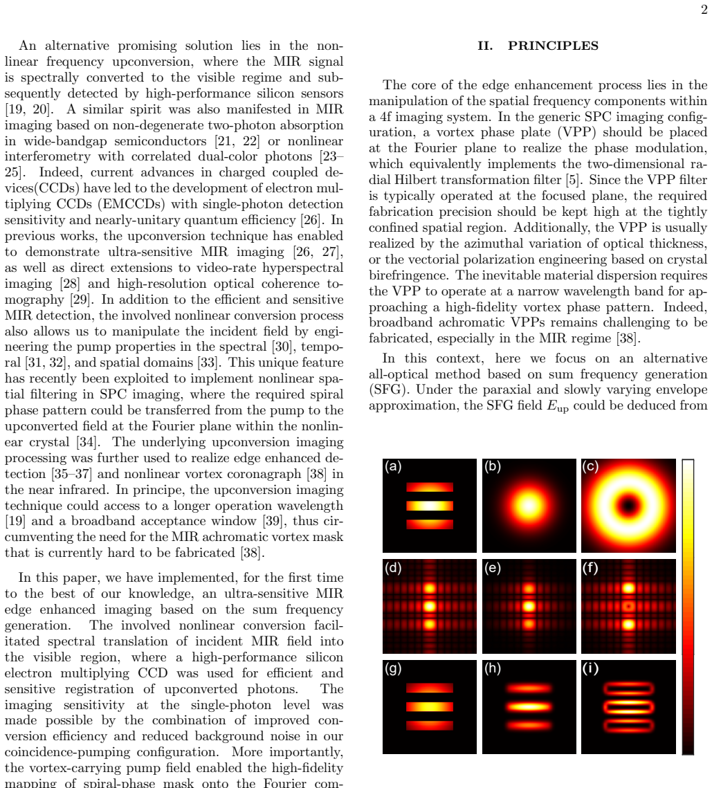
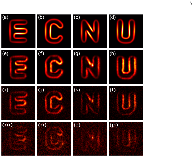
Ultra-sensitive MIR imaging at the single-photon level is achieved based on nonlinear frequency upconversion, where the spectrally converted replica of the 3070 nm MIR object image is captured by a silicon EMCCD; the edge enhancement is realized by imprinting the spiral phase pattern of the pump onto the upconverted field at the Fourier plane within the nonlinear crystal, providing both high-fidelity vortex screening and efficient low-noise visible rendering of the MIR object.
What carries the argument
Nonlinear vortex filtering performed by imprinting the pump's spiral phase at the Fourier plane inside the upconversion crystal
If this is right
- MIR object illumination is rendered into a visible image in an efficient and low-noise fashion.
- Imaging sensitivity is significantly improved by coincidence pulsed pumping with spectro-temporal optimization.
- The nonlinear spatial filter implements high-fidelity vortex screening for edge-enhanced detection.
- The approach facilitates label-free histopathological diagnosis and non-destructive defect inspection at low light levels.
Where Pith is reading between the lines
- The requirement for coincidence detection implies that synchronized pulsed sources are necessary for reaching the reported sensitivity.
- Placing the phase pattern inside the crystal removes the need for external vortex optics after conversion.
- The same crystal-based filter could be tested at other MIR wavelengths by matching crystal and pump parameters.
Load-bearing premise
The nonlinear frequency upconversion process inside the crystal faithfully transfers both amplitude and phase information from the 3070 nm MIR object to the visible domain while preserving single-photon statistics and allowing the pump's spiral phase to act as a high-fidelity spatial filter.
What would settle it
If upconverted images taken with a spiral-phase pump show no edge enhancement compared with a Gaussian pump, or if coincidence counts fall below the single-photon regime under the reported pulsed pumping conditions, the central claim would be falsified.
Figures


read the original abstract
Edge enhanced imaging via the spiral phase contrast enables to reveal the phase or amplitude gradients of a target, which has been proved useful in feature recognition, machine vision, and object identification. A long quest is to extend the operation wavelength into the mid-infrared (MIR) region, as highly demanded in various fields including infrared sensing, astronomic observation, and biomedical diagnosis. Here, we demonstrated ultra-sensitive MIR imaging at the single-photon level based on nonlinear frequency upconversion, where the spectrally converted replica of the MIR object image at 3070 nm was captured by a silicon electron multiplying charged coupled device. The imaging sensitivity was significantly improved by the coincidence pulsed pumping with a spectro-temporal optimization. Furthermore, the edge enhancement has been realized by imprinting the spiral phase pattern of the pump onto the upconverted field at the Fourier plane within the nonlinear crystal. Such a nonlinear spatial filter not only provided an effective way to implement the required high-fidelity vortex screening in the edge enhanced detection, but also rendered the MIR illumination into a visible image in an efficient and low-noise fashion. The presented system for MIR edge enhanced imaging might facilitate immediate applications in label-free histopathological diagnosis and non-destructive defect inspection.
Editorial analysis
A structured set of objections, weighed in public.
Referee Report
Summary. The manuscript presents an experimental demonstration of mid-infrared single-photon edge-enhanced imaging at 3070 nm using nonlinear frequency upconversion in a crystal. The MIR object image is converted to the visible domain and captured by a silicon EMCCD, with sensitivity improved via coincidence pulsed pumping and spectro-temporal optimization. Edge enhancement is realized by imprinting the pump's spiral phase pattern onto the upconverted field at the Fourier plane inside the nonlinear crystal, acting as a nonlinear vortex filter for high-fidelity spatial filtering while converting the illumination efficiently and with low noise.
Significance. If validated, the work integrates nonlinear upconversion with vortex filtering to enable single-photon-level MIR edge-enhanced imaging without direct MIR detectors. This could support applications in label-free histopathological diagnosis and non-destructive inspection by providing an efficient, low-noise method for phase-sensitive MIR imaging. The approach addresses a long-standing challenge in extending spiral phase contrast techniques to the MIR regime.
minor comments (3)
- [Abstract] Abstract: The phrasing 'enables to reveal' is grammatically awkward; consider revising to 'enables revealing' or 'allows the revelation of'.
- [Abstract] Abstract: The abstract would be strengthened by including at least one key quantitative metric (e.g., coincidence rate, SNR, or photon flux) to support the 'single-photon level' and 'ultra-sensitive' claims, even if detailed data appear later in the manuscript.
- [Experimental Methods] The manuscript should clarify the exact placement and alignment tolerances for the Fourier-plane filtering inside the crystal, as small misalignments could affect the fidelity of the spiral phase imprinting.
Simulated Author's Rebuttal
We thank the referee for their positive summary and recommendation of minor revision. No major comments were provided in the report.
Circularity Check
No significant circularity: experimental demonstration without load-bearing derivations
full rationale
The manuscript presents an experimental demonstration of MIR single-photon edge-enhanced imaging via nonlinear upconversion and vortex filtering in a crystal. No derivation chain, equations, or first-principles results are claimed that reduce to fitted parameters, self-definitions, or self-citation chains. The central claims rest on direct measurements (coincidence detection, image comparisons) rather than any mathematical reduction to inputs. Self-citations, if present, are not invoked to justify uniqueness theorems or ansatzes that bear the result. This is a standard experimental optics paper whose validity is assessed by reproducibility of the setup, not internal definitional closure.
Axiom & Free-Parameter Ledger
Reference graph
Works this paper leans on
-
[1]
E”, “C”, “N
and a broadband acceptance window [39], thus cir- cumventing the need for the MIR achromatic vortex mask that is currently hard to be fabricated [38]. In this paper, we have implemented, for the first time to the best of our knowledge, an ultra-sensitive MIR edge enhanced imaging based on the sum frequency generation. The involved nonlinear conversion fac...
1951
-
[2]
Phase contrast, a new method for the mi- croscopic observation of transparent objects,
F. Zernike, “Phase contrast, a new method for the mi- croscopic observation of transparent objects,” Physica9, 686-698 (1942)
1942
-
[3]
Spiral phase contrast imaging in microscopy,
S. F¨ urhapter, A. Jesacher, S. Bernet, and M. Ritsch- Marte, “Spiral phase contrast imaging in microscopy,” Opt. Express13, 689-694 (2005)
2005
-
[4]
Optical vortex coronagraph,
G. Foo, D. M. Palacios, and G. A. Swartzlander, “Optical vortex coronagraph,” Opt. Lett.30, 3308-3310 (2005)
2005
-
[5]
The phase rotor filter,
S. N. Khonina, V. V. Kotlyar, M. V. Shinkaryev, V. A. Soifer, and G. V. Uspleniev, “The phase rotor filter,” J. Mod. Opt.39, 1147-1154 (1992)
1992
-
[6]
Image processing with the radial Hilbert transform: theory and experiments,
Jeffrey A. Davis, Dylan E. McNamara, Don M. Cottrell, and Juan Campos, “Image processing with the radial Hilbert transform: theory and experiments,” Opt. Lett. 25, 99-101 (2000)
2000
-
[7]
Plasmonic computing of spatial differentiation,
T. Zhu, Y. Zhou, Y. Lou, H. Ye, M. Qiu, Z. Ruan, and S. Fan, “Plasmonic computing of spatial differentiation,” Nat. Commun.8, 15391 (2017)
2017
-
[8]
Ra- dial Hilbert transform with Laguerre-Gaussian spatial fil- ters,
C.-S. Guo, Y.-J. Han, J.-B. Xu, and J. Ding, “Ra- dial Hilbert transform with Laguerre-Gaussian spatial fil- ters,” Opt. Lett.31, 1394-1396 (2006)
2006
-
[9]
Shadow Effects in Spiral Phase Contrast Mi- croscopy,
A. Jesacher, S. Furhapter, S. Bernet, and M. Ritsch- Marte, “Shadow Effects in Spiral Phase Contrast Mi- croscopy,” Phys. Rev. Lett.94, 233902 (2005)
2005
-
[10]
Spiral phase filtering and orientation-selective edge detection/enhancement,
G. Situ, G. Pedrini, and W. Osten, “Spiral phase filtering and orientation-selective edge detection/enhancement,” J. Opt. Soc. Am. A26, 1788 (2009)
2009
-
[11]
Imple- menting selective edge enhancement in nonlinear optics,
D. Xu, T. Ma, X. Qiu, W. Zhang, and L. Chen, “Imple- menting selective edge enhancement in nonlinear optics,” Opt. Express28, 32377-32385 (2020)
2020
-
[12]
Mid-infrared photonics using 2D materials: status and challenges,
Y. Fang, Y. Ge, C. Wang, and H. Zhang, “Mid-infrared photonics using 2D materials: status and challenges,” Laser Photon. Rev.14, 1900098 (2020)
2020
-
[13]
Advances in mid- infrared detection and imaging: a key issues review,
M. Razeghi and B.-M. Nguyen, “Advances in mid- infrared detection and imaging: a key issues review,” Rep. Prog. Phys.77, 082401 (2014)
2014
-
[14]
Efficient Single Pho- ton Detection from 500 nm to 5µm Wavelength,
F. Marsili, F. Bellei, F. Najafi, A. E. Dane, E. A. Dauler, R. J. Molnar, and K. K. Berggren, “Efficient Single Pho- ton Detection from 500 nm to 5µm Wavelength,” Nano Lett.12, 4799-4804 (2012)
2012
-
[15]
Mid-infrared single photon detector with superconductor Mo 80Si20 nanowire,
Q. Chenet al., “Mid-infrared single photon detector with superconductor Mo 80Si20 nanowire,” arXiv:2011.06699 (2020)
-
[16]
M. Long, A. Gao, P. Wang, H. Xia, C. Ott, C. Pan, Y. Fu, E. Liu, X. Chen, W. Lu, T. Nilges, J. Xu, X. Wang, W. Hu, and F. Miao, ‘Room temperature high- detectivity mid-infrared photodetectors based on black arsenic phosphorus,” Sci. Adv.3, e1700589 (2017)
2017
-
[17]
Polarization-resolved black phos- phorus/molybdenum disulfide mid-wave infrared photo- diodes with high detectivity at room temperature,
J. Bullock, M. Amani, J. Cho, Y.-Z. Chen, G. H. Ahn, V. Adinolfi, V. R. Shrestha, Y. Gao, K. B. Crozier, Y.-L. Chueh, and A. Javey, “Polarization-resolved black phos- phorus/molybdenum disulfide mid-wave infrared photo- diodes with high detectivity at room temperature,” Na- ture Photon.12, 601-607 (2018)
2018
-
[18]
Efficient electrical detection of mid-infrared graphene plasmons at room temperature,
Q. Guo, R. Yu, C. Li, S. Yuan, B. Deng, F. Javier Garc´ ıa de Abajo, and F. Xia, “Efficient electrical detection of mid-infrared graphene plasmons at room temperature,” Nature Mater.17, 986-992 (2018)
2018
-
[19]
Stable mid-infrared polarization imag- ing based on quasi-2D tellurium at room temperature,
L. Tong et al., “Stable mid-infrared polarization imag- ing based on quasi-2D tellurium at room temperature,” Nature Comm.11, 2308 (2020)
2020
-
[20]
Room- Temperature, High-SNR Upconversion Spectrometer in the 6-12µm Region,
P. Rodrigo, L. Høgstedt, S. Friis, L. Lindvold, P. Tidemand-Lichtenberg, and C. Pedersen, “Room- Temperature, High-SNR Upconversion Spectrometer in the 6-12µm Region,” Laser Photon. Rev.15, 2000443 (2021). 9
2021
-
[21]
Mid-infrared photon counting and resolving via efficient frequency upconver- sion,
K. Huang, Y. Wang, J. Fang, W. Kang, Y. Sun, Y. Liang, Q. Hao, M. Yan, and H. Zeng, “Mid-infrared photon counting and resolving via efficient frequency upconver- sion,” Photonics Res.9, 259 (2021)
2021
-
[22]
Sensitive mid-infrared detection in wide-bandgap semi- conductors using extreme non-degenerate two-photon ab- sorption,
D. A. Fishman, C. M. Cirloganu, S. Webster, L. A. Padilha, M. Monroe, D. J. Hagan, and E. W. V. Stryland, “Sensitive mid-infrared detection in wide-bandgap semi- conductors using extreme non-degenerate two-photon ab- sorption,” Natrue Photon.5, 561-565 (2011)
2011
-
[23]
Infrared chemical imaging through non-degenerate two-photon absorption in silicon-based cameras,
D. Knez, A. M. Hanninen, R. C. Prince, E. O. Potma, and D. A. Fishman, “Infrared chemical imaging through non-degenerate two-photon absorption in silicon-based cameras,” Light Sci. Appl.9, 125 (2020)
2020
-
[24]
Hyperspectral infrared microscopy with visible light,
A. V. Paterova, S. M. Maniam, H. Yang, G. Grenci, and L. A. Krivitsky, “Hyperspectral infrared microscopy with visible light,” Sci. Adv.6, eabd0460 (2020)
2020
-
[25]
Microscopy with un- detected photons in the mid-infrared,
I. Kviatkovsky, H. M. Chrzanowski, E. G. Avery, H. Bartolomaeus, and S. Ramelow, “Microscopy with un- detected photons in the mid-infrared,” Sci. Adv.6, eabd0264, (2020)
2020
-
[26]
Video-Rate Imaging with Undetected Photons,
M. G. Basset, A. Hochrainer, S. T¨ opfer, F. Riexinger, P. Bickert, J. R. Le´ on-Torres, F. Steinlechner, and M. Gr¨ afe, “Video-Rate Imaging with Undetected Photons,” Laser Photon. Rev. 2000327 (2021)
2021
-
[27]
Ul- trasensitive mid-infrared up-conversion imaging at few- photon level,
Q. Zhou, K. Huang, H. Pan, E. Wu, and H. Zeng, “Ul- trasensitive mid-infrared up-conversion imaging at few- photon level,” Appl. Phys. Lett.102, 241110 (2013)
2013
-
[28]
Room-temperature mid-infrared single-photon spectral imaging,
J. S. Dam, P. Tidemand-Lichtenberg, and C. Pedersen, “Room-temperature mid-infrared single-photon spectral imaging,” Nature Photon.6, 788-793(2012)
2012
-
[29]
Video-rate, mid-infrared hyperspectral upconversion imaging,
S. Junaid, S. Chaitanya Kumar, M. Mathez, M. Her- mes, N. Stone, N. Shepherd, M. Ebrahim-Zadeh, P. Tidemand-Lichtenberg, and C. Pedersen, “Video-rate, mid-infrared hyperspectral upconversion imaging,” Op- tica6, 702-708 (2019)
2019
-
[30]
Real-time high- resolution mid-infrared optical coherence tomography,
N. M. Israelsen, C. R. Petersen, A. Barh, D. Jain, M. Jensen, G. Hannesschl¨ ager, P. Tidemand-Lichtenberg, C. Pedersen, A. Podoleanu, and O. Bang, “Real-time high- resolution mid-infrared optical coherence tomography,” Light. Sci. Appl.8, 11 (2019)
2019
-
[31]
Frequency and bandwidth conversion of single photons in a room- temperature diamond quantum memory,
K. A. G. Fisher, D. G. England, J.-P. W. MacLean, P. J. Bustard, K. J. Resch, and B. J. Sussman, “Frequency and bandwidth conversion of single photons in a room- temperature diamond quantum memory,” Nature Comm. 7, 1-6 (2016)
2016
-
[32]
Tailoring nonlinear processes for quantum optics with pulsed temporal-mode encodings,
V. Ansari, J. M. Donohue, B. Brecht, and C. Silberhorn, “Tailoring nonlinear processes for quantum optics with pulsed temporal-mode encodings,” Optica5, 534 (2018)
2018
-
[33]
Mul- tidimensional mode-separable frequency conversion for high-speed quantum communication,
P. Manurkar, N. Jain, M. Silver, Y.-P. Huang, C. Lan- grock, M. M. Fejer, P. Kumar, and G. S. Kanter, “Mul- tidimensional mode-separable frequency conversion for high-speed quantum communication,” Optica3, 1300- 1307 (2016)
2016
-
[34]
Orbital angular momentum photonic quantum interface,
Z. Zhou, Y. Li, D. Ding, W. Zhang, S. Shi, B. Shi, and G. Guo, “Orbital angular momentum photonic quantum interface,” Light. Sci. Appl.5, e16019-7 (2016)
2016
-
[35]
Spiral phase contrast imaging in nonlinear optics: seeing phase objects using invisible illumination,
X. Qiu, F. Li, W. Zhang, Z. Zhu, and L. Chen, “Spiral phase contrast imaging in nonlinear optics: seeing phase objects using invisible illumination,” Optica5, 208-212 (2018)
2018
-
[36]
In- frared upconverted image edge enhancement using spiral phase filter,
L. Liu, H. Y. Wang, Y. Ning, C. Guo, and G. Ren, “In- frared upconverted image edge enhancement using spiral phase filter,” Laser Phys.29, 015401 (2019)
2019
-
[37]
Up-conversion imaging processing with field-of-view and edge enhancement,
S. Liu, C. Yang, S. Liu, Z. Zhou, Y. Li, Y. H. Li, Z. H. Xu, G. Guo, and B. Shi, “Up-conversion imaging processing with field-of-view and edge enhancement,” Phys. Rev. Appl.11, 044013 (2019)
2019
-
[38]
Upconversion dark-field imaging with extended field of view at video frame rate,
S. Junaid, P. Tidemand-Lichtenberg, C. Pedersen, and P. John Rodrigo, “Upconversion dark-field imaging with extended field of view at video frame rate,” Appl. Opt. 59, 2157-2164 (2020)
2020
-
[39]
Nonlinear optical vortex coronagraph,
E. Engay and P. John Rodrigo, “Nonlinear optical vortex coronagraph,” Opt. Lett.45, 1579-1582 (2020)
2020
-
[40]
Multicolor Time-Resolved Upconversion Imaging by Adiabatic Sum Frequency Conversion,
M. Mrejen, Y. Erlich, A. Levanon, and H. Suchowski, “Multicolor Time-Resolved Upconversion Imaging by Adiabatic Sum Frequency Conversion,” Laser Photon. Rev.14, 2000040 (2020)
2020
-
[41]
Real-time quantum edge enhanced imag- ing,
S. Liu, Y. Li, S. Liu, Z. Zhou, Y. Li, C. Yang, G. Guo, and B. Shi, “Real-time quantum edge enhanced imag- ing,” Opt. Express28, 35415-35426 (2020)
2020
-
[42]
Metasurface enabled quantum edge detection,
J. Zhou, S. Liu, H. Qian, Y. Li, H. Luo, S. Wen, Z. Zhou, G. Guo, B. Shi, and Z. Liu, “Metasurface enabled quantum edge detection,” Sci. Adv.6, eabc4385 (2020)
2020
discussion (0)
Sign in with ORCID, Apple, or X to comment. Anyone can read and Pith papers without signing in.