AtlasGS: Brain MRI Spatial Resolution Harmonization With Shared Gaussian Geometry
Pith reviewed 2026-06-28 11:50 UTC · model grok-4.3
The pith
A shared Gaussian scaffold learned from one isotropic MRI scan enables high-fidelity reconstruction of sparse-slice images across multiple modalities.
A machine-rendered reading of the paper's core claim, the machinery that carries it, and where it could break.
Core claim
The paper establishes that an explicit, subject-specific Gaussian scaffold encoding anatomical geometry can be learned once from an isotropic structural scan and then reused to fit appearance parameters for target modalities acquired with sparse slices, producing state-of-the-art reconstructions, arbitrary-view images with structural consistency, and potential for self-supervised in-plane super-resolution.
What carries the argument
The subject-specific Gaussian scaffold, an explicit representation of anatomical geometry that is learned from one scan and held fixed while appearance is fitted for other modalities.
If this is right
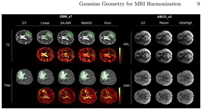
- State-of-the-art reconstruction fidelity is achieved on UK Biobank, GBM and ABCD datasets across degradation factors of ×3, ×5 and ×7 and in the presence of glioblastoma.
- Arbitrary-view generation for target modalities maintains strong structural consistency with the original geometry.
- Self-supervised in-plane super-resolution becomes feasible using the same fixed scaffold.
- Explicit geometry-guided representations offer a flexible pathway for retrospective multi-contrast MRI harmonization and clinical reference construction.
Where Pith is reading between the lines
- The separation of geometry from appearance could be tested on longitudinal scans to check whether a single scaffold remains valid when anatomy changes slowly over time.
- The same two-stage pattern might apply outside the brain if an initial isotropic reference scan can be obtained for any anatomy.
- Because the scaffold is explicit, it could be inspected or edited by clinicians to correct for known anatomical variants before appearance fitting.
Load-bearing premise
An anatomical geometry scaffold learned from one isotropic scan can be reused for other modalities without introducing structural distortions or loss of fidelity.
What would settle it
A side-by-side experiment on the same sparse-slice volumes that compares reconstructions produced by the shared scaffold against reconstructions produced by modality-specific scaffolds learned from scratch; if the shared-scaffold versions show measurably lower fidelity or visible anatomical mismatches, the central claim is falsified.
Figures
read the original abstract
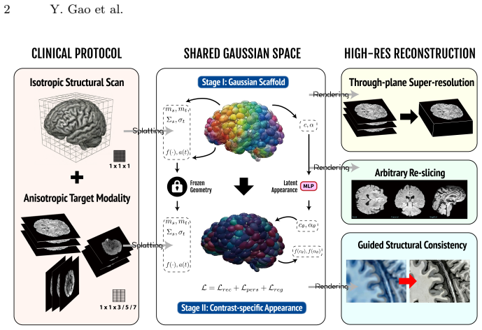
Splatting (GS)-based shared geometry framework adopts a two-stage training strategy, in which an explicit, subject-specific Gaussian scaffold encoding anatomical geometry is first learned from the isotropic structural scan and then reused to fit appearance for target modalities acquired with sparse slices. Experiments on the UK Biobank, GBM, and ABCD datasets for through-plane super-resolution across multiple modalities (T2-weighted, FLAIR, DWI, ASL), degradation factors ($\times 3$, $\times 5$, $\times 7$), and pathological abnormalities (glioblastoma) demonstrate state-of-the-art reconstruction fidelity. The shared Gaussian geometry enables arbitrary-view generation for target modalities with strong structural consistency and further shows potential for self-supervised in-plane super-resolution. This work establishes explicit geometry-guided representations as a novel, flexible, and interpretable pathway toward retrospective multi-contrast MRI harmonization and reliable clinical reference construction. Source code is available at: https://github.com/yfgao76/AtlasGS
Editorial analysis
A structured set of objections, weighed in public.
Referee Report
Summary. The paper introduces AtlasGS, a Gaussian Splatting (GS)-based framework for brain MRI spatial resolution harmonization. It employs a two-stage strategy: first learning an explicit, subject-specific Gaussian scaffold that encodes anatomical geometry from an isotropic structural (T1) scan, then freezing this scaffold and optimizing only appearance parameters (opacity, color) to fit target modalities acquired with sparse slices. Experiments on the UK Biobank, GBM, and ABCD datasets claim state-of-the-art through-plane super-resolution performance for T2-weighted, FLAIR, DWI, and ASL modalities across degradation factors of ×3, ×5, and ×7, including cases with glioblastoma pathology. The shared geometry is said to enable arbitrary-view generation with structural consistency and shows potential for self-supervised in-plane super-resolution. Source code is released at https://github.com/yfgao76/AtlasGS.
Significance. If the results hold, this work provides a novel explicit geometry-guided representation for retrospective multi-contrast MRI harmonization that is interpretable and flexible compared to implicit methods. The public code release supports reproducibility. The approach could aid clinical reference construction by enabling consistent structural scaffolds across modalities and views. Significance is tempered by the need to validate the core modality-invariance assumption for the fixed scaffold.
major comments (1)
- [Method (two-stage training) and Experiments (UK Biobank/GBM/ABCD results)] The two-stage training strategy (abstract and method description) fixes the Gaussian scaffold positions/scales learned from T1 while optimizing only appearance for other modalities. This assumption that anatomical geometry is modality-invariant at the scale of the primitives is load-bearing for the harmonization and SOTA fidelity claims, yet no ablation is reported that relaxes the fixed-geometry constraint (e.g., allowing small per-modality position/scale offsets or joint geometry-appearance optimization) to quantify any fidelity loss, especially on DWI/ASL or the GBM glioblastoma cases where tissue contrast and boundaries differ from T1.
minor comments (1)
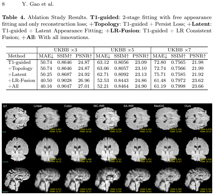
- [Abstract] The abstract asserts state-of-the-art reconstruction fidelity without summarizing any quantitative metrics (e.g., PSNR, SSIM), baseline comparisons, error bars, or train/validation split details; these should be included in the abstract for a self-contained claim.
Simulated Author's Rebuttal
We thank the referee for the constructive comment regarding validation of the fixed-geometry assumption in our two-stage training strategy.
read point-by-point responses
-
Referee: [Method (two-stage training) and Experiments (UK Biobank/GBM/ABCD results)] The two-stage training strategy (abstract and method description) fixes the Gaussian scaffold positions/scales learned from T1 while optimizing only appearance for other modalities. This assumption that anatomical geometry is modality-invariant at the scale of the primitives is load-bearing for the harmonization and SOTA fidelity claims, yet no ablation is reported that relaxes the fixed-geometry constraint (e.g., allowing small per-modality position/scale offsets or joint geometry-appearance optimization) to quantify any fidelity loss, especially on DWI/ASL or the GBM glioblastoma cases where tissue contrast and boundaries differ from T1.
Authors: The fixed-geometry constraint is a deliberate and core design choice of AtlasGS: the subject-specific Gaussian scaffold is learned once from the isotropic T1 scan to capture anatomical structure, then frozen so that only appearance parameters are optimized for each target modality. This enforces the structural consistency that enables arbitrary-view synthesis and multi-modal harmonization. The modality-invariance assumption at the primitive scale is empirically supported by the reported state-of-the-art results on T2-weighted, FLAIR, DWI, and ASL across UK Biobank, ABCD, and the GBM dataset (which includes glioblastoma cases with altered tissue boundaries). These outcomes demonstrate that appearance-only optimization suffices for high-fidelity reconstruction even when contrast differs markedly from T1. An ablation that relaxes the fixed scaffold (e.g., per-modality offsets or joint optimization) would test a different method and incur substantial additional compute; we therefore did not perform it, as the existing cross-modality and cross-pathology results already validate the shared-geometry premise. revision: no
Circularity Check
No circularity: two-stage geometry-appearance optimization is a standard fitting procedure with independent validation
full rationale
The paper's core procedure learns a Gaussian scaffold from an isotropic T1 scan then optimizes only appearance parameters on sparse-slice targets. This is a conventional two-stage optimization on external multi-modal datasets (UK Biobank, GBM, ABCD) rather than any self-definitional loop, fitted-input-renamed-as-prediction, or load-bearing self-citation chain. No equations are presented that reduce the reported fidelity metrics to quantities defined by the same fitted values; the claimed arbitrary-view consistency and SOTA results are evaluated against held-out data and therefore remain falsifiable outside the fitting process itself.
Axiom & Free-Parameter Ledger
Reference graph
Works this paper leans on
-
[1]
Abbasi, S., et al.: Deep learning for the harmonization of structural MRI scans: a survey. Biomed. Eng. Online23, 90 (2024).https://doi.org/10.1186/ s12938-024-01280-6
2024
-
[2]
Bakas, S., et al.: The university of pennsylvania glioblastoma (UPenn-GBM) co- hort: advanced MRI, clinical, genomics, & radiomics. Scientific Data9, 453 (2022). https://doi.org/10.1038/s41597-022-01560-7
-
[3]
Neural Comput.15(6), 1373–1396 (2003).https://doi.org/10
Belkin, M., Niyogi, P.: Laplacian eigenmaps for dimensionality reduction and data representation. Neural Comput.15(6), 1373–1396 (2003).https://doi.org/10. 1162/089976603321780317
2003
-
[4]
Casey, B., et al.: The adolescent brain cognitive development (ABCD) study: Imaging acquisition across 21 sites. Dev. Cogn. Neurosci.32, 43–54 (2018). https://doi.org/10.1016/j.dcn.2018.03.001
-
[5]
arXiv:2506.14432 (2026),https://arxiv.org/ abs/2506.14432
Cerri, S., et al.: A large-scale heterogeneous 3D magnetic resonance brain imaging dataset for self-supervised learning. arXiv:2506.14432 (2026),https://arxiv.org/ abs/2506.14432
Pith/arXiv arXiv 2026
-
[6]
In: MICCAI 2025
Choi, Y., et al.: TESLA: Test-time reference-free through-plane super- resolution for multi-contrast brain MRI. In: MICCAI 2025. LNCS, vol. 15972, pp. 584–593. Springer Nature Switzerland (2025).https://doi.org/10.1007/ 978-3-032-05169-1_56
2025
-
[7]
Clough, J., et al.: A topological loss function for deep-learning based image segmen- tation using persistent homology. IEEE Trans. Pattern Anal. Mach. Intell.44(12), 8766–8778 (2022).https://doi.org/10.1109/TPAMI.2020.3013679
-
[8]
Greenspan, H., Peled, S., Oz, G., Kiryati, N.: MRI inter-slice reconstruction using super-resolution. Magn. Reson. Imaging20, 437–446 (2002).https://doi.org/ 10.1016/S0730-725X(02)00511-8
-
[9]
Hu, F., et al.: Image harmonization: a review of statistical and deep learning meth- ods for removing batch effects and evaluation metrics for effective harmonization. 10 Y. Gao et al. NeuroImage274,120125(2023).https://doi.org/10.1016/j.neuroimage.2023. 120125
-
[10]
Isensee,F.,etal.:AutomatedbrainextractionofmultisequenceMRIusingartificial neural networks. Hum. Brain Mapp.40(17), 4952–4964 (2019).https://doi.org/ 10.1002/hbm.24750
-
[11]
Isensee, F., et al.: nnu-net: self-configuring biomedical image segmentation. Nat. Methods18(2), 203–211 (2021).https://doi.org/10.1038/s41592-020-01008-z
-
[12]
3D Gaussian splatting for real-time radiance field rendering,
Kerbl, B., et al.: 3D gaussian splatting for real-time radiance field rendering. ACM Trans. Graph. (SIGGRAPH) (2023).https://doi.org/10.1145/3592433
-
[13]
arXiv:2511.16854 (2025).https://doi.org/10.48550/arXiv.2511.16854
Khateri, M., et al.: MRI super-resolution with deep learning: A comprehensive survey. arXiv:2511.16854 (2025).https://doi.org/10.48550/arXiv.2511.16854
-
[14]
Adam: A Method for Stochastic Optimization
Kingma, D., Ba, J.: Adam: A method for stochastic optimization. arXiv:1412.6980 (2014).https://doi.org/10.48550/arXiv.1412.6980
work page internal anchor Pith review Pith/arXiv arXiv doi:10.48550/arxiv.1412.6980 2014
-
[15]
Lehmann,T.,Gönner,C.,Spitzer,K.:Addendum:B-splineinterpolationinmedical imageprocessing.IEEETrans.Med.Imaging20(7),660–665(2001).https://doi. org/10.1109/42.932749
-
[16]
Liu, X., et al.: Deep unregistered multi-contrast MRI reconstruction. Magn. Reson. Imaging81, 33–41 (2021).https://doi.org/10.1016/j.mri.2021.05.005
-
[17]
Mahmoudzadeh, A., Kashou, N.: Interpolation-based super-resolution reconstruc- tion: effects of slice thickness. J. Med. Imaging (Bellingham)1(3), 034007 (2014). https://doi.org/10.1117/1.JMI.1.3.034007
-
[18]
MedGS: Gaussian Splatting for Multi-Modal 3D Medical Imaging
Marzol, K., et al.: MedGS: Gaussian splatting for multi-modal 3D medical imaging. arXiv:2509.16806 (2025).https://doi.org/10.48550/arXiv.2509.16806
work page internal anchor Pith review Pith/arXiv arXiv doi:10.48550/arxiv.2509.16806 2025
-
[19]
In: Greenspan, H., et al
McGinnis, J., et al.: Single-subject multi-contrast MRI super-resolution via im- plicit neural representations. In: Greenspan, H., et al. (eds.) MICCAI 2023. LNCS, vol. 14229, pp. 173–183. Springer, Cham (2023).https://doi.org/10. 1007/978-3-031-43993-3_17
2023
- [20]
-
[21]
In: Wolterink, J., et al
Remedios, S., et al.: Self-supervised super-resolution for anisotropic MR im- ages with and without slice gap. In: Wolterink, J., et al. (eds.) SASHIMI 2023. LNCS, vol. 14288, pp. 118–128. Springer, Cham (2023).https://doi.org/10. 1007/978-3-031-44689-4_12
2023
-
[22]
arXiv:2412.16619 (2024).https://doi.org/10.48550/arXiv.2412.16619
Shen, T., Liu, S., Feng, J., Ma, Z., An, N.: Topology-aware 3D gaussian splatting: Leveraging persistent homology for optimized structural integrity. arXiv:2412.16619 (2024).https://doi.org/10.48550/arXiv.2412.16619
-
[23]
arXiv:2411.11024 (2024).https://doi.org/10.48550/arXiv.2411.11024
Smolak-Dyżewska, W., et al.: VeGaS: Video gaussian splatting. arXiv:2411.11024 (2024).https://doi.org/10.48550/arXiv.2411.11024
-
[24]
PLoS Med.12(3), e1001779 (2015).https://doi.org/10.1371/journal.pmed.1001779
Sudlow, C., et al.: UK biobank: an open access resource for identifying the causes of a wide range of complex diseases of middle and old age. PLoS Med.12(3), e1001779 (2015).https://doi.org/10.1371/journal.pmed.1001779
-
[25]
In: CVPR Workshop (2025),https://github.com/kushalvyas/alpine, last accessed 2026/02/25
Vyas, K., et al.: Alpine: distributed PyTorch library for implicit neural representa- tions. In: CVPR Workshop (2025),https://github.com/kushalvyas/alpine, last accessed 2026/02/25
2025
-
[26]
Wang, X., et al.: SA-INR for arbitrary MRI slice-spacing reduction. Med. Image Anal.94, 103158 (2024).https://doi.org/10.1016/j.media.2024.103158
-
[27]
arXiv:2507.16962 (2025).https: //doi.org/10.48550/arXiv.2507.16962
Yang, Q., Shomal-Zadeh, F., Gholipour, A.: MRI harmonization: a survey of ac- quisition, image-level, and feature-level methods. arXiv:2507.16962 (2025).https: //doi.org/10.48550/arXiv.2507.16962
discussion (0)
Sign in with ORCID, Apple, or X to comment. Anyone can read and Pith papers without signing in.