A Positron Range Correction with Texture Preservation Framework in PET Imaging
Pith reviewed 2026-06-26 05:59 UTC · model grok-4.3
The pith
A neural framework corrects positron range blurring in PET while re-injecting matching texture via an auxiliary model.
A machine-rendered reading of the paper's core claim, the machinery that carries it, and where it could break.
Core claim
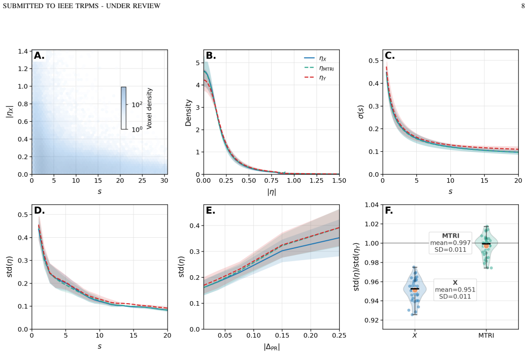
PRC-TP decouples deterministic resolution recovery performed by an nnFormer network trained on patient-derived Monte Carlo simulations from stochastic texture restoration performed by Model-consistent Texture Re-Injection derived from an auxiliary Noise2Noise estimate; the combined pipeline restores contrast recovery to 98.96-99.04 percent of ground truth, returns noise and CNR closer to reference values, and achieves near-unity global texture amplitude agreement of 0.997 plus or minus 0.011 while reducing input bias.
What carries the argument
Model-consistent Texture Re-Injection (MTRI), which isolates the smoothing induced by the main resolution-recovery network and transfers the original noise texture back to the corrected output to maintain acquisition-consistent statistics.
If this is right
- Radiomics features from texture-sensitive families show improved numerical agreement with ground-truth values.
- Clinical 82Rb evaluations exhibit contrast-ratio increases comparable to simulation results together with restored texture.
- The same decoupled correction-plus-re-injection structure can be applied to other high-energy positron emitters.
- Noise and CNR values are returned closer to the reference distribution than resolution recovery alone.
Where Pith is reading between the lines
- The same separation of deterministic recovery from stochastic texture could be tested on other resolution-degrading effects such as motion or partial-volume correction.
- If the MTRI step proves robust across scanners, it may reduce the need for scanner-specific texture post-processing in multi-center studies.
- Extending the Monte Carlo training set to include measured rather than simulated patient data would directly test whether the reported agreement holds outside the simulation domain.
Load-bearing premise
The auxiliary Noise2Noise model accurately isolates only the smoothing caused by the main network without adding new biases or mismatches to the underlying acquisition physics.
What would settle it
A head-to-head comparison on real clinical 82Rb patient scans that measures texture amplitude or radiomics features against an independent ground-truth reference and finds deviation beyond the reported 0.997 plus or minus 0.011 agreement.
Figures


read the original abstract
Positron range (PR) blurring is a fundamental resolution limitation in PET imaging with high-energy positron emitters such as 82Rb, causing contrast loss and spill-out effects across heterogeneous tissue interfaces. We propose PRC-TP, a positron range correction (PRC) framework with explicit texture preservation that decouples deterministic resolution recovery from stochastic texture restoration. A nnFormer-based neural network (NN) was trained on patient-derived Monte Carlo simulations to map PR-degraded 82Rb reconstructions to PR-free references using attenuation maps as anatomical context. However, this NN also significantly removed the noise in the images, which could impact some texture analysis methods or make the images look unrealistic. An auxiliary Noise2Noise model estimates that smoothing effect, enabling texture extraction and transfer to the PR-corrected prediction through Model-consistent Texture Re-Injection (MTRI). In simulated patients, PRC-TP preserved contrast recovery close to ground truth (GT) (98.96-99.04%) while restoring noise and CNR closer to the reference. The function-based MTRI formulation achieved near unity global texture amplitude agreement with GT (0.997 +/- 0.011), reducing the input texture amplitude bias (0.951 +/- 0.011). Radiomics analysis showed improved agreement with GT across texture-sensitive feature families. A clinical 82Rb evaluation showed trends consistent with simulations, including comparable contrast-ratio increase (10.18% vs. 10.99%) and restoration of texture suppressed by PRC. These results support PRC-TP as a practical framework for resolution recovery with acquisition-consistent texture preservation in PET imaging. Submitted to IEEE TRPMS.
Editorial analysis
A structured set of objections, weighed in public.
Referee Report
Summary. The manuscript proposes PRC-TP, a positron range correction framework for 82Rb PET that trains an nnFormer network on patient-derived Monte Carlo simulations to map PR-degraded images to PR-free references (using attenuation maps as context), then uses an auxiliary Noise2Noise model to estimate and remove the network's smoothing effect before applying Model-consistent Texture Re-Injection (MTRI) to restore acquisition-consistent texture. Quantitative results on simulated patients report contrast recovery of 98.96-99.04% with near-unity global texture amplitude (0.997 +/- 0.011) after MTRI, plus improved radiomics agreement; a single clinical case shows consistent trends in contrast ratio and texture restoration.
Significance. If the auxiliary model isolation and MTRI re-injection are shown to be physics-consistent, the framework offers a practical route to resolution recovery in high-energy positron PET while addressing the common side-effect of over-smoothing that affects texture analysis and visual realism; the use of patient-derived MC simulations for training and the function-based MTRI formulation are explicit strengths that support reproducibility of the texture amplitude result.
major comments (2)
- [Methods: auxiliary Noise2Noise and MTRI] Methods (auxiliary Noise2Noise and MTRI sections): the central claim that MTRI re-injects texture whose amplitude and statistics match the original acquisition physics rests on the unvalidated assumption that the Noise2Noise model isolates only the deterministic smoothing induced by nnFormer; no quantitative comparison of the estimated residual to Monte Carlo Poisson noise statistics, PR-free ground truth, or anatomy-dependent bias is reported, so the reported 0.997 +/- 0.011 texture amplitude agreement does not yet confirm consistency with the underlying physics.
- [Results: simulated patients] Results (simulated patients paragraph): the contrast recovery figures (98.96-99.04%) and CNR/noise restoration claims are presented without error bars, ablation studies on MTRI hyperparameters, or details on training/validation splits and Monte Carlo variance, which are load-bearing for assessing whether the preservation is robust rather than an artifact of the simulation setup.
minor comments (2)
- [Abstract and Results: clinical evaluation] Abstract and Results: the single clinical case is described only qualitatively ('trends consistent'); adding at least basic quantitative metrics with comparison to the simulation protocol would strengthen the generalizability statement.
- [Methods: MTRI] Notation: the distinction between the 'function-based MTRI formulation' and any alternative formulations is referenced but not defined with an equation; adding the explicit functional form would clarify how global texture amplitude is computed.
Simulated Author's Rebuttal
We thank the referee for their constructive comments on our manuscript. We address each major comment point by point below, agreeing where revisions are warranted to strengthen the presentation of physics consistency and robustness.
read point-by-point responses
-
Referee: Methods (auxiliary Noise2Noise and MTRI sections): the central claim that MTRI re-injects texture whose amplitude and statistics match the original acquisition physics rests on the unvalidated assumption that the Noise2Noise model isolates only the deterministic smoothing induced by nnFormer; no quantitative comparison of the estimated residual to Monte Carlo Poisson noise statistics, PR-free ground truth, or anatomy-dependent bias is reported, so the reported 0.997 +/- 0.011 texture amplitude agreement does not yet confirm consistency with the underlying physics.
Authors: We acknowledge the referee's point that the manuscript lacks an explicit quantitative validation of the Noise2Noise residual against Monte Carlo Poisson statistics or anatomy-dependent bias. The MTRI approach is formulated to extract and re-inject texture based on the auxiliary model's estimate of nnFormer-induced smoothing, with the reported amplitude agreement measured directly against PR-free ground truth. To address the concern, we will add a dedicated validation subsection (or supplementary figure) comparing the estimated residual noise power spectrum and amplitude to the known Poisson statistics from the patient-derived Monte Carlo simulations used in training. revision: yes
-
Referee: Results (simulated patients paragraph): the contrast recovery figures (98.96-99.04%) and CNR/noise restoration claims are presented without error bars, ablation studies on MTRI hyperparameters, or details on training/validation splits and Monte Carlo variance, which are load-bearing for assessing whether the preservation is robust rather than an artifact of the simulation setup.
Authors: We agree that error bars, ablation studies, and explicit details on data splits and simulation variance are necessary to demonstrate robustness. The reported contrast recovery range (98.96-99.04%) summarizes results across the simulated patient cohort; we will revise the text and figures to include mean values with standard deviations and error bars. Expanded Methods text will specify the patient-wise training/validation split and Monte Carlo variance (derived from the 10^8 decay simulations). Ablation results on MTRI hyperparameters (e.g., scaling factor) will be added to the supplementary material. revision: yes
Circularity Check
No significant circularity in derivation chain
full rationale
The paper presents an empirical ML framework (nnFormer + auxiliary Noise2Noise + MTRI) trained and evaluated on Monte Carlo simulations, with additional clinical data trends. No equations, definitions, or steps are shown that reduce outputs to inputs by construction, rename fitted parameters as predictions, or rely on load-bearing self-citations whose content is unverified. Reported metrics (contrast recovery 98.96-99.04%, texture amplitude 0.997) are measured results from the trained model rather than tautological identities. The derivation is therefore self-contained against the simulation benchmarks and external clinical checks.
Axiom & Free-Parameter Ledger
Reference graph
Works this paper leans on
-
[1]
State of the art in total body pet,
S. Vandenberghe, P. Moskal, and J. S. Karp, “State of the art in total body pet,”EJNMMI physics, vol. 7, no. 1, p. 35, 2020
2020
-
[2]
Pet molecular imaging: a holistic review of current practice and emerging perspectives for diagnosis, therapeutic evaluation and prognosis in clinical oncology,
V . Duclos, A. Iep, L. Gomez, L. Goldfarb, and F. L. Besson, “Pet molecular imaging: a holistic review of current practice and emerging perspectives for diagnosis, therapeutic evaluation and prognosis in clinical oncology,”International journal of molecular sciences, vol. 22, no. 8, p. 4159, 2021
2021
-
[3]
Clinical utility and future applications of pet/ct and pet/cmr in cardiology,
J. A. Pan and M. Salerno, “Clinical utility and future applications of pet/ct and pet/cmr in cardiology,”Diagnostics, vol. 6, no. 3, p. 32, 2016
2016
-
[4]
Pet/spect molecular imaging in clinical neuro- science: recent advances in the investigation of cns diseases,
F.-M. Lu and Z. Yuan, “Pet/spect molecular imaging in clinical neuro- science: recent advances in the investigation of cns diseases,”Quantita- tive imaging in medicine and surgery, vol. 5, no. 3, p. 433, 2015
2015
-
[5]
Calculation of positron range and its effect on the fundamental limit of positron emission tomography system spatial resolution,
C. S. Levin and E. J. Hoffman, “Calculation of positron range and its effect on the fundamental limit of positron emission tomography system spatial resolution,”Physics in Medicine & Biology, vol. 44, no. 3, p. 781, 1999
1999
-
[6]
Analytical positron range model for pet with cross-code monte carlo benchmark- ing,
R. J. Paneque-Yunta, N. Encina-Baranda, L. M. Carter, P. Galve, P. Ib´a˜nez, K. M. Abushab, J. M. Ud ´ıas, and J. L. Herraiz, “Analytical positron range model for pet with cross-code monte carlo benchmark- ing,”Physics in Medicine & Biology, vol. 70, no. 16, p. 165013, 2025
2025
-
[7]
Effect of the positron range of 18f, 68ga and 124i on pet/ct in lung-equivalent ma- terials,
G. J. Kemerink, M. G. Visser, R. Franssen, E. Beijer, M. Zamburlini, S. G. Halders, B. Brans, F. M. Mottaghy, and G. J. Teule, “Effect of the positron range of 18f, 68ga and 124i on pet/ct in lung-equivalent ma- terials,”European journal of nuclear medicine and molecular imaging, vol. 38, no. 5, pp. 940–948, 2011
2011
-
[8]
Positron range estimations with penelopet,
J. Cal-Gonz ´alez, J. Herraiz, S. Espa ˜na, P. G. Corzo, J. J. Vaquero, M. Desco, and J. M. Udias, “Positron range estimations with penelopet,” Physics in Medicine & Biology, vol. 58, no. 15, p. 5127, 2013
2013
-
[9]
Performance characteristics of the digital biograph vision pet/ct system,
J. Van Sluis, J. De Jong, J. Schaar, W. Noordzij, P. Van Snick, R. Dierckx, R. Borra, A. Willemsen, and R. Boellaard, “Performance characteristics of the digital biograph vision pet/ct system,”Journal of Nuclear Medicine, vol. 60, no. 7, pp. 1031–1036, 2019
2019
-
[10]
Mathematical removal of positron range blurring in high resolution tomography,
S. E. Derenzo, “Mathematical removal of positron range blurring in high resolution tomography,”IEEE Transactions on Nuclear Science, vol. 33, no. 1, pp. 565–569, 2007
2007
-
[11]
Study of ct-based positron range correction in high resolution 3d pet imaging,
J. Cal-Gonz ´alez, J. L. Herraiz, S. Espa ˜na, E. Vicente, E. Herranz, M. Desco, J. Vaquero, and J. M. Ud´ıas, “Study of ct-based positron range correction in high resolution 3d pet imaging,”Nuclear Instruments and Methods in Physics Research Section A: Accelerators, Spectrometers, Detectors and Associated Equipment, vol. 648, pp. S172–S175, 2011
2011
-
[12]
Simulation study of tissue-specific positron range correction for the new biograph mmr whole-body pet/mr system,
R. Kraus, G. Delso, and S. I. Ziegler, “Simulation study of tissue-specific positron range correction for the new biograph mmr whole-body pet/mr system,”IEEE transactions on nuclear science, vol. 59, no. 5, pp. 1900– 1909, 2012. SUBMITTED TO IEEE TRPMS - UNDER REVIEW 12
1900
-
[13]
Tissue-dependent and spatially-variant positron range correction in 3d pet,
J. Cal-Gonzalez, M. Perez-Liva, J. L. Herraiz, J. J. Vaquero, M. Desco, and J. M. Udias, “Tissue-dependent and spatially-variant positron range correction in 3d pet,”IEEE transactions on medical imaging, vol. 34, no. 11, pp. 2394–2403, 2015
2015
-
[14]
Implementation of a spatially-variant and tissue-dependent positron range correction for pet/ct imaging,
H. Kert ´esz, T. Beyer, V . Panin, W. Jentzen, J. Cal-Gonzalez, A. Berger, L. Papp, P. L. Kench, D. Bharkhada, J. Cabelloet al., “Implementation of a spatially-variant and tissue-dependent positron range correction for pet/ct imaging,”Frontiers in physiology, vol. 13, p. 818463, 2022
2022
-
[15]
Positron range in combination with point-spread-function correction: an evaluation of different implementations for [124i]-pet imaging,
H. Kert ´esz, M. Conti, V . Panin, J. Cabello, D. Bharkhada, T. Beyer, L. Papp, W. Jentzen, J. Cal-Gonzalez, J. L. Herraizet al., “Positron range in combination with point-spread-function correction: an evaluation of different implementations for [124i]-pet imaging,”EJNMMI physics, vol. 9, no. 1, p. 56, 2022
2022
-
[16]
Positron range correction helps enhance the image quality of cardiac 82rb pet/ct,
M. L. Lassen, H. Kert ´esz, I. Rausch, V . Panin, M. Conti, S. Zuehlsdorff, J. Cabello, D. Bharkhada, R. DeKemp, A. Kjaeret al., “Positron range correction helps enhance the image quality of cardiac 82rb pet/ct,” Journal of Nuclear Medicine, vol. 66, no. 3, pp. 466–472, 2025
2025
-
[17]
Dual-input dynamic convolution for positron range cor- rection in pet image reconstruction,
Y . Mellak, A. Bousse, T. Merlin, ´E. ´Emond, M. Hakulinen, and D. Visvikis, “Dual-input dynamic convolution for positron range cor- rection in pet image reconstruction,”IEEE Transactions on Radiation and Plasma Medical Sciences, 2025
2025
-
[18]
Improving pet quantification of small animal [68ga] dota-labeled pet/ct studies by using a ct-based positron range correction,
J. Cal-Gonzalez, J. J. Vaquero, J. L. Herraiz, M. P ´erez-Liva, M. L. Soto- Montenegro, S. Pe ˜na-Zalbidea, M. Desco, and J. M. Ud ´ıas, “Improving pet quantification of small animal [68ga] dota-labeled pet/ct studies by using a ct-based positron range correction,”Molecular Imaging and Biology, vol. 20, no. 4, pp. 584–593, 2018
2018
-
[19]
Positron range corrections and denoising techniques for gallium-68 pet imaging: a literature review,
P. Gavriilidis, M. Koole, S. Annunziata, F. M. Mottaghy, and R. Wierts, “Positron range corrections and denoising techniques for gallium-68 pet imaging: a literature review,”Diagnostics, vol. 12, no. 10, p. 2335, 2022
2022
-
[20]
An investigation of 68 ga positron range correction through de-blurring: A simulation study,
A. Rukiah, S. R. Meikle, J. E. Gillam, and P. L. Kench, “An investigation of 68 ga positron range correction through de-blurring: A simulation study,” in2018 IEEE Nuclear Science Symposium and Medical Imaging Conference Proceedings (NSS/MIC). IEEE, 2018, pp. 1–2
2018
-
[21]
Deep-learning based positron range correction of pet images,
J. L. Herraiz, A. Bembibre, and A. Lopez-Montes, “Deep-learning based positron range correction of pet images,”Applied sciences, vol. 11, no. 1, p. 266, 2020
2020
-
[22]
A tissue-informed deep learning-based method for positron range correction in preclinical 68Ga pet imaging,
N. Encina-Baranda, R. J. Paneque-Yunta, J. Lopez-Rodriguez, E. C. Pratt, T. N. Nguyen, J. Grimm, A. Lopez-Montes, and J. L. Herraiz, “A tissue-informed deep learning-based method for positron range correction in preclinical 68Ga pet imaging,”EJNMMI Physics, 2026
2026
-
[23]
Simul- taneous partial volume correction and denoising of brain pet images, using transformers and transfer learning,
S. Kaviani, A. Sanaat, M. Mokri, H. Zaidi, and J.-F. Carrier, “Simul- taneous partial volume correction and denoising of brain pet images, using transformers and transfer learning,”EJNMMI research, 2026
2026
-
[24]
Noise-aware dynamic image denoising and positron range correction for rubidium-82 cardiac pet imaging via self- supervision,
H. Xie, L. Guo, A. Velo, Z. Liu, Q. Liu, X. Guo, B. Zhou, X. Chen, Y .-J. Tsai, T. Miaoet al., “Noise-aware dynamic image denoising and positron range correction for rubidium-82 cardiac pet imaging via self- supervision,”Medical Image Analysis, vol. 100, p. 103391, 2025
2025
-
[25]
Noise2Noise: Learning Image Restoration without Clean Data
J. Lehtinen, J. Munkberg, J. Hasselgren, S. Laine, T. Karras, M. Aittala, and T. Aila, “Noise2noise: Learning image restoration without clean data,”arXiv preprint arXiv:1803.04189, 2018
work page internal anchor Pith review Pith/arXiv arXiv 2018
-
[26]
On the spectral bias of neural networks,
N. Rahaman, A. Baratin, D. Arpit, F. Draxler, M. Lin, F. Hamprecht, Y . Bengio, and A. Courville, “On the spectral bias of neural networks,” inInternational conference on machine learning. PMLR, 2019, pp. 5301–5310
2019
-
[27]
Distribution matching losses can hallucinate features in medical image translation,
J. P. Cohen, M. Luck, and S. Honari, “Distribution matching losses can hallucinate features in medical image translation,” inInternational con- ference on medical image computing and computer-assisted intervention. Springer, 2018, pp. 529–536
2018
-
[28]
On instabilities of deep learning in image reconstruction and the potential costs of ai,
V . Antun, F. Renna, C. Poon, B. Adcock, and A. C. Hansen, “On instabilities of deep learning in image reconstruction and the potential costs of ai,”Proceedings of the National Academy of Sciences, vol. 117, no. 48, pp. 30 088–30 095, 2020
2020
-
[29]
H. H. Barrett and K. J. Myers,F oundations of image science. John Wiley & Sons, 2013
2013
-
[30]
Noise properties of the em algorithm. i. theory,
H. H. Barrett, D. W. Wilson, and B. M. W. Tsui, “Noise properties of the em algorithm. i. theory,”Physics in Medicine & Biology, vol. 39, no. 5, p. 833, may 1994. [Online]. Available: https://doi.org/10.1088/0031-9155/39/5/004
-
[31]
Gate: a simulation toolkit for pet and spect,
S. Jan, G. Santin, D. Strul, S. Staelens, K. Assi ´e, D. Autret, S. Avner, R. Barbier, M. Bardies, P. Bloomfieldet al., “Gate: a simulation toolkit for pet and spect,”Physics in Medicine & Biology, vol. 49, no. 19, p. 4543, 2004
2004
-
[32]
Advanced monte carlo simulations of emission tomography imaging systems with gate,
D. Sarrut, M. Bała, M. Bardi `es, J. Bert, M. Chauvin, K. Chatzipapas, M. Dupont, A. Etxebeste, L. M. Fanchon, S. Janet al., “Advanced monte carlo simulations of emission tomography imaging systems with gate,” Physics in Medicine & Biology, vol. 66, no. 10, p. 10TR03, 2021
2021
-
[33]
The opengate ecosystem for monte carlo simulation in medical physics,
D. Sarrut, T. Baudier, D. Borys, A. Etxebeste, H. Fuchs, J. Gajewski, L. Grevillot, S. Jan, G. C. Kagadis, H. G. Kanget al., “The opengate ecosystem for monte carlo simulation in medical physics,”Physics in Medicine & Biology, vol. 67, no. 18, p. 184001, 2022
2022
-
[34]
Geant4–a simulation toolkit,
G. Collaboration, S. Agostinelliet al., “Geant4–a simulation toolkit,” Nucl. Instrum. Meth. A, vol. 506, no. 25, p. 0, 2003
2003
-
[35]
Geant4 develop- ments and applications,
J. Allison, K. Amako, J. Apostolakis, H. Araujo, P. A. Dubois, M. Asai, G. Barrand, R. Capra, S. Chauvie, R. Chytraceket al., “Geant4 develop- ments and applications,”IEEE Transactions on nuclear science, vol. 53, no. 1, pp. 270–278, 2006
2006
-
[36]
Recent developments in geant4,
J. Allison, K. Amako, J. Apostolakis, P. Arce, M. Asai, T. Aso, E. Bagli, A. Bagulya, S. Banerjee, G. Barrandet al., “Recent developments in geant4,”Nuclear instruments and methods in physics research section A: Accelerators, Spectrometers, Detectors and Associated Equipment, vol. 835, pp. 186–225, 2016
2016
-
[37]
Geant4 physics list comparison for the simulation of phase-contrast mammography (xpulse project),
V . Beaudoux, G. Blin, B. Barbrel, G. Kantor, and C. Zacharatou, “Geant4 physics list comparison for the simulation of phase-contrast mammography (xpulse project),”Physica Medica, vol. 60, pp. 66–75, 2019
2019
-
[38]
Monte carlo simulation of digital photon counting pet,
J. Salvadori, J. Labour, F. Odille, P.-Y . Marie, J.-N. Badel, L. Imbert, and D. Sarrut, “Monte carlo simulation of digital photon counting pet,” EJNMMI physics, vol. 7, no. 1, p. 23, 2020
2020
-
[39]
Root—an object oriented data analysis framework,
R. Brun and F. Rademakers, “Root—an object oriented data analysis framework,”Nuclear instruments and methods in physics research sec- tion A: accelerators, spectrometers, detectors and associated equipment, vol. 389, no. 1-2, pp. 81–86, 1997
1997
-
[40]
Pet digitization chain for monte carlo simulation in gate,
J. Salvadori, A. Merlet, B. Presles, J. Cabello, K.-H. Su, A. Cochet, A. Etxebeste, J.-M. Vrigneaud, and D. Sarrut, “Pet digitization chain for monte carlo simulation in gate,”Physics in Medicine & Biology, vol. 69, no. 16, p. 165013, 2024
2024
-
[41]
Petlink: A proposed digital interconnect standard for data acquisition in nuclear medicine,
W. Jones, “Petlink: A proposed digital interconnect standard for data acquisition in nuclear medicine,” 2013
2013
-
[42]
Pet data acquisition: preparing petlink for the coming decades,
W. F. Jones, A. P. Moor, and I. J. Huff, “Pet data acquisition: preparing petlink for the coming decades,” in2018 IEEE Nuclear Science Sympo- sium and Medical Imaging Conference Proceedings (NSS/MIC). IEEE, 2018, pp. 1–2
2018
-
[43]
component based method for normalization in volume pet,
M. Casey, “component based method for normalization in volume pet,” inInternational Meeting on Fully Three-dimensional Image Reconstruc- tion in Radiology and Nuclear Medicine 1995, 1995
1995
-
[44]
nnformer: Interleaved transformer for volumetric segmentation,
H.-Y . Zhou, J. Guo, Y . Zhang, L. Yu, L. Wang, and Y . Yu, “nnformer: Interleaved transformer for volumetric segmentation,”arXiv preprint arXiv:2109.03201, 2021
-
[45]
U-net: Convolutional networks for biomedical image segmentation,
O. Ronneberger, P. Fischer, and T. Brox, “U-net: Convolutional networks for biomedical image segmentation,” inInternational Conference on Medical image computing and computer-assisted intervention. Springer, 2015, pp. 234–241
2015
-
[46]
Convolutional networks for images, speech, and time series,
Y . LeCun and Y . Bengio, “Convolutional networks for images, speech, and time series,”The handbook of brain theory and neural networks, 1998
1998
-
[47]
Transformers in medical imaging: A survey,
F. Shamshad, S. Khan, S. W. Zamir, M. H. Khan, M. Hayat, F. S. Khan, and H. Fu, “Transformers in medical imaging: A survey,”Medical image analysis, vol. 88, p. 102802, 2023
2023
-
[48]
Torchio: a python li- brary for efficient loading, preprocessing, augmentation and patch-based sampling of medical images in deep learning,
F. P ´erez-Garc´ıa, R. Sparks, and S. Ourselin, “Torchio: a python li- brary for efficient loading, preprocessing, augmentation and patch-based sampling of medical images in deep learning,”Computer methods and programs in biomedicine, vol. 208, p. 106236, 2021
2021
-
[49]
User-guided 3D active contour segmentation of anatomical structures: Significantly improved efficiency and reliability,
P. A. Yushkevich, J. Piven, H. Cody Hazlett, R. Gimpel Smith, S. Ho, J. C. Gee, and G. Gerig, “User-guided 3D active contour segmentation of anatomical structures: Significantly improved efficiency and reliability,” Neuroimage, vol. 31, no. 3, pp. 1116–1128, 2006
2006
discussion (0)
Sign in with ORCID, Apple, or X to comment. Anyone can read and Pith papers without signing in.