REVIEW 4 major objections 5 minor 52 references
Robust Monte-Carlo Simulations in Diffusion-MRI: Effect of the substrate complexity and parameter choice on the reproducibility of results
T0 review · 4 major / 5 minor · reviewed 2026-08-14 · deepseek-v4-flash
Pith's one-line read Monte-Carlo diffusion-MRI simulations require at least 500,000 particles and 10,000 cylinders before their ground-truth signals become reproducible.
desk verdict A solid calibration paper with a useful new substrate-generation framework; the headline thresholds are provisional since no threshold criterion is defined, but the direction of each finding checks out and the bootstrapped first study is well done. read the letter →
The pith
A machine-rendered reading of the paper's core claim, the machinery that carries it, and where it could break.
The reading
What carries the argument
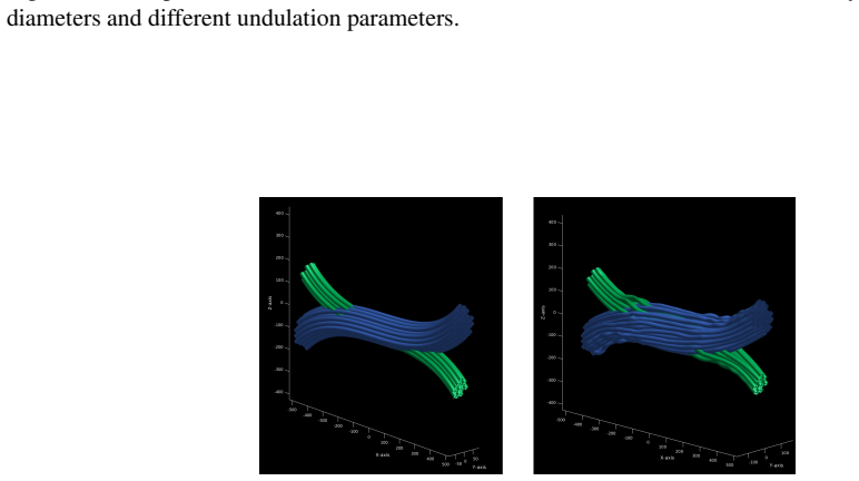
The argument rests on two mechanisms. The first is a bootstrapped error analysis: repeated Monte-Carlo runs at each particle and step count are compared with a high-quality reference, namely the analytical Gaussian-phase-distribution solution for restricted diffusion in cylinders for the intra-axonal signal and a 20-million-particle Monte-Carlo run for the extra-axonal signal, using the Relative Mean Absolute Error (RMAE) to turn raw signal deviations into convergence curves that set the recommended minima. The second is an energy-optimization substrate generator: an objective function penalizes overlap, curvature, and length of parametrized strands, and a modified cylinder-cylinder collision term lets those strands be subdivided into gamma-distributed diameters so that crossing bundles interdigitate without intersecting, preserving the volume fraction in the crossing area.
What would settle it
Recompute the extra-axonal signal for the same substrate with an independent finite-element or analytical effective-medium solver across the full acquisition protocol; if the reference signal disagrees by more than the reported 0.4–0.7% convergence margin, the recommended parameter minima are miscalibrated.
Extended reading notes
Core claim
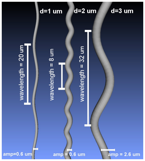
On its own terms, the paper establishes three quantitative claims. First, for the substrate and acquisition settings studied, simulations with fewer than $5\times10^5$ particles or $1\times10^4$ steps show a significant, avoidable variance between repeated runs in both the restricted intra-axonal signal and the hindered extra-axonal signal; the mean relative absolute error settles near 0.4–0.7% only above those values. Second, substrates built from fewer than 10,000 sampled cylinders produce extra-axonal radial signals whose directional symmetry is visibly broken and whose mean amplitude is biased, with the bias disappearing around a 200–230 µm voxel scale for the tested diameter distribution. Third, replacing straight cylinders with helically undulating axons shifts the apparent diameter recovered from a straight-cylinder model, severely for 1 µm axons, where the mis-estimation can exceed 300%. The paper also presents a framework for generating complex crossing substrates whose strands interdigitate without overlap, preserving volume in the crossing region and achieving an intra-axonal volume fraction above 48% even at low resolution.
Load-bearing premise
The load-bearing premise is that the 20-million-particle, $2\times10^4$-step extra-axonal run is itself an accurate reference; if that Monte-Carlo simulation carries a systematic bias from the fixed step size, substrate representation, or cylinder packing, then every error percentage and the recommended $5\times10^5$-particle, $1\times10^4$-step minimum inherit that bias.
Editorial extensions
If this is right
- Validations run below $5\times10^5$ particles and $1\times10^4$ steps should be treated as carrying unexplained run-to-run variance; reporting a single run is not enough.
- Extra-axonal signals from substrates with fewer than about 10,000 cylinders are not radially isotropic, so fitting procedures that assume axial symmetry inherit a systematic bias.
- Axon-diameter estimates from straight-cylinder models can be off by more than 300% for 1 µm undulating axons, meaning undulation is a confounding factor in diameter mapping.
- The proposed substrate generator can produce crossing-fibre ground truths with preserved volume and an intra-axonal volume fraction above 48% at coarse resolution, giving crossing-aware models a harder, more realistic test.
Reading between the lines
- A practical next step the paper does not take is to derive a scaling law for the particle and step minima as a function of diffusivity, diffusion time, and packing density, since the recommended values are tied to the specific protocol and substrate studied here.
- Because the extra-axonal reference is itself a Monte-Carlo run, an independent finite-element or effective-medium calculation on the same substrate would give a stronger absolute calibration of the recommended thresholds.
- The generator could be used to build controlled dose-response phantoms that vary undulation amplitude, crossing angle, and volume fraction independently, letting models be scored against known ground truth rather than inferred tissue properties.
- The observed substrate-size bias in the mean extra-axonal signal implies that shrinking the simulated voxel is not a neutral computational shortcut, and time-dependent diffusion metrics extracted from such simulations may inherit a false time-dependence.
Signed reviews
Editorial analysis
A structured set of objections, weighed in public.
Referee Report
Summary. This manuscript studies three methodological pitfalls in Monte-Carlo diffusion simulations (MCDS) used as ground truth for diffusion MRI: the number of particles and time steps, the intra-axonal representation (straight vs. undulating cylinders), and the size of the extra-axonal substrate. It reports that simulations with fewer than 5e5 particles and 1e4 steps show significant variability, and that substrates with fewer than 10,000 sampled cylinders induce a bias in the radial symmetry of the extra-axonal signal. It then presents a framework for generating complex crossing substrates that preserves volume in the crossing region and achieves high packing density, and it evaluates axon diameter estimation in such substrates. The paper also provides a complexity analysis of the simulator and makes the code and substrate data available.
Significance. If the central quantitative claims hold, the paper is practically important because many published MCDS-based ground-truth studies use parameter values below the proposed minima, and the paper identifies a concrete reproducibility risk. The bootstrap design in Section 3.1, with 50 repetitions per parameter combination, is a clear strength, as is the effort to validate the simulator against analytical solutions and an independent FEM approach. The proposed crossing-substrate framework is a useful step toward more realistic numerical phantoms, and the volume-preservation property is a genuine improvement over naive crossings. However, the paper's headline numbers (5e5 particles, 1e4 steps, 10,000 cylinders) are currently derived from visual inspection without a pre-specified tolerance, and the extra-axonal gold standard is itself a Monte-Carlo simulation whose systematic errors have not been independently bounded. These issues make the main recommendations not yet fully reproducible or falsifiable as stated, so the manuscript needs substantial revision before it can serve as a reliable reference for parameter choice.
major comments (4)
- [§4.1, §6] The central thresholds '5×10^5 particles' and '1×10^4 steps' are presented in the conclusions without a pre-specified acceptance criterion. In Figures 5 and 6 the RMAE is plotted against sample size, but no tolerance on the RMAE is defined and no confidence bounds are derived from the 50 repetitions; the reader cannot determine why 5×10^5 rather than 2×10^5 or 1×10^6 is the cut-off. In addition, when the number of steps is varied the reference uses the maximum value (2×10^4), so the RMAE for smaller step counts mixes the fixed-step discretization bias with Monte Carlo variance, and the text labels the combined effect 'variance'. Please define an explicit reproducibility criterion (e.g., a maximal acceptable RMAE with a confidence interval) and separate the discretization bias from the sampling variance, ideally by varying the step count in the gold-standard reference as well.
- [§3.1, §4.1] The extra-axonal gold standard is itself a Monte-Carlo simulation (20×10^6 particles, 2×10^4 steps), and the paper reports no comparison of this reference against an independent method beyond an assertion of convergence; the earlier FEM validation [38] is cited but not shown for this substrate and protocol. If the fixed-step integration or the cylinder packing carries a systematic bias, every RMAE in Section 4.1 inherits it, so the recommended minima could be miscalibrated. Please add either a convergence study for the extra-axonal gold standard with respect to step size (not just particle number) or a comparison with an independent numerical/analytical reference for the identical geometry.
- [§3.3, §4.3] The conclusion that substrates with fewer than 10,000 cylinders induce an 'important bias' is not supported by a quantitative threshold. The anisotropy metric (std/mean of the radial signal) is reported for one random substrate per size, with only sizes 100, 1,000, 10,000, 50,000, and 100,000 tested; the boundary 'less than 10,000' is an interpolation with no uncertainty. Please repeat the measurement over multiple random packing realizations, report the spread of the anisotropy metric, and state a pre-defined isotropy criterion (or at least a tolerance on the deviation from the large-substrate limit).
- [§3.2, §4.2] The diameter-fitting intervals in Figure 7 depend on the arbitrary threshold of '1% difference from the minimum fitting error'. Because the RMAE surface is strongly protocol-dependent (Figure 8), the reported intervals and the qualitative claim of 'considerable mis-estimation' are sensitive to this ad hoc choice. Please justify the 1% level (e.g., by linking it to the noise level or the known sensitivity of the protocol) or report how the intervals change when the threshold is varied.
minor comments (5)
- [§3.1] The list of tested particle numbers includes 2×10^6, but the preceding sentence says particles were varied from 1×10^3 to 1×10^6; please make the range consistent.
- [§3.2] The sentence 'choosing a the parameters that shows almost almost no variance' contains repeated words ('a the', 'almost almost') and a subject-verb agreement error; please correct it.
- [§3.3] 'This parameters where chooseng from the previous results' should read 'These parameters were chosen from the previous results'.
- [§4.1, Figure 5] The caption says 'RMAE of all the repetition' but should read 'RMAE of all the repetitions'; please also indicate which panels correspond to the intra- and extra-axonal cases in the text, since the figure has six panels.
- [§3.4] The optimization time (about 42 hours) and simulation time (less than 24 hours) are reported for a single run; please state whether these are wall-clock times and on what specific hardware configuration they were obtained, to make the computational claims reproducible.
Circularity Check
No significant circularity: thresholds and evaluations are empirical measurements against independent analytical and FEM references, not fitted re-statements of inputs.
full rationale
The paper's central quantitative claims are descriptive summaries of its own Monte-Carlo convergence and substrate-size experiments, not quantities derived from a fitted model that is then re-predicted. Intra-axonal signals are compared against the independent GPD analytical solution (Van Gelderen et al., [48]) and extra-axonal signals against a high-particle gold standard that was additionally checked against a finite-element method in [38]; even though [38] shares an author, it is an independent numerical method and the paper also reports its own convergence check at 1e6 particles/5e3 steps, so the citation is not load-bearing. The recommended thresholds (5e5 particles, 1e4 steps, 10,000 cylinders) are read off RMAE and anisotropy plots, with no equation forcing those numbers by construction; the absence of a formal threshold criterion is a reproducibility weakness, not a circularity. The undulation/diameter study simulates known geometries and fits them with a deliberately misspecified straight-cylinder GPD model, so the mis-estimation is a model-error measurement rather than an input re-stated as an output. The framework evaluation uses the same simulator but tests geometric properties and convergence, with no fitted parameter renamed as a prediction. No equation in the paper reduces to its own input, and no load-bearing claim rests on a self-citation chain.
Assumptions & free parameters
free parameters (2)
- Cost-function weights w_o, w_c, w_l =
not specified
- Diameter fitting threshold (1% above minimum RMAE) =
1%
assumptions (4)
- domain assumption The Gaussian Phase Distribution (GPD) analytical solution for restricted diffusion in straight cylinders is the correct reference for intra-axonal signals.
- domain assumption The cylinder packing algorithm from [22] produces an extra-axonal space representative of white matter.
- domain assumption The fixed-step-size random walk with r = sqrt(6D dt) accurately models Brownian motion in these restricted geometries.
- domain assumption The simulator itself is correct because it was validated against analytical solutions and a finite element method in [38].
Cite this review
Pith. "Pith review of Robust Monte-Carlo Simulations in Diffusion-MRI: Effect of the substrate complexity and parameter choice on the reproducibility of results." pith.science (2026). https://pith.science/paper/4LNC6ZPD
@misc{pith2026190811203,
  author       = {Pith},
  title        = {Pith review of: Robust Monte-Carlo Simulations in Diffusion-MRI: Effect of the substrate complexity and parameter choice on the reproducibility of results},
  year         = {2026},
  howpublished = {\url{https://pith.science/paper/4LNC6ZPD}},
  note         = {Machine review of arXiv:1908.11203}
}
read the original abstract
Monte-Carlo Diffusion Simulations (MCDS) have been used extensively as a ground truth tool for the validation of microstructure models for Diffusion-Weighted MRI. However, methodological pitfalls in the design of the biomimicking geometrical configurations and the simulation parameters can lead to approximation biases. Such pitfalls affect the reliability of the estimated signal, as well as its validity and reproducibility as ground truth data. In this work, we first present a set of experiments in order to study three critical pitfalls encountered in the design of MCDS in the literature, namely, the number of simulated particles and time steps, simplifications in the intra-axonal substrate representation, and the impact of the substrate's size on the signal stemming from the extra-axonal space. The results obtained show important changes in the simulated signals and the recovered microstructure features when changes in those parameters are introduced. Thereupon, driven by our findings from the first studies, we outline a general framework able to generate complex substrates. We show the framework's capability to overcome the aforementioned simplifications by generating a complex crossing substrate, which preserves the volume in the crossing area and achieves a high packing density. The results presented in this work,along with the simulator developed, pave the way towards more realistic and reproducible Monte-Carlo simulations for Diffusion-Weighted MRI.
Figures

Figures from the paper (10 more)


Reference graph
Works this paper leans on
-
[38]
J. Rafael-Patino, A. Ramirez-Manzanares, J. Pe˜na Acevedo, and H. Zhang. Validating particle dynamics in monte carlo diffusion simulation using the finite element method. Proc. 25th Scientific Meeting of the International Society for Magnetic Resonance in Medicine, page 1849, 2017
work page 2017
-
[1]
P. K. Agarwal, M. de Berg, J. Gudmundsson, M. Hammar, and H. J. Haverkort. Box-trees and r-trees with near-optimal query time. In Proceedings of the Seventeenth Annual Symposium on Computational Geometry, SCG ’01, pages 124–133, New York, NY , USA, 2001. ACM
work page 2001
-
[2]
D. C. Alexander, P. L. Hubbard, M. G. Hall, E. A. Moore, M. Ptito, G. J. Parker, and T. B. Dyrby. Orientationally invariant indices of axon diameter and density from diffusion mri. NeuroImage, 52(4):1374, 2010
work page 2010
-
[3]
D. F. M. Allison C. Bain, David I. Shreiber. Modeling of Microstructural Kinematics During Simple Elongation of Central Nervous System Tissue. J. Biomech Eng, (6), 2004
work page 2004
- [4]
-
[5]
G. T. Balls and L. R. Frank. A simulation environment for diffusion weighted mr experiments in complex media. Magnetic Resonance in Medicine, 62(3):771, 2009
work page 2009
-
[6]
R. Bammer. Basic principles of diffusion-weighted imaging. European Journal of Radiology, 45(3):169, 2003
work page 2003
-
[7]
V . R. Barlett, M. Hoyuelos, and H. O. Martin. Monte carlo simulation with fixed steplength for diffusion processes in nonhomogeneous media. Journal of Computational Physics, 239, 2013
work page 2013
Show all 52 references
-
[8]
G. T. Baxter and L. R. Frank. A computational model for diffusion weighted imaging of myelinated white matter. NeuroImage, page 204, 2013
2013
-
[9]
Benjamini, M
D. Benjamini, M. E. Komlosh, L. A. Holtzclaw, U. Nevo, and P. J. Basser. White matter microstructure from nonparametric axon diameter distribution mapping. Neuroimage, 135:333–344, 2016. 16
2016
-
[10]
Brabec, S
J. Brabec, S. Lasic, and M. Nilsson. Time-dependent diffusion in undulating structures: Impact on axon diameter estimation. arXiv, 2019
2019
-
[11]
L. M. Burcaw, E. Fieremans, and D. S. Novikov. Mesoscopic structure of neuronal tracts from time-dependent diffusion. NeuroImage, 114:15334406, 2015
2015
-
[12]
T. G. Close, J. D. Tournier, F. Calamante, L. A. Johnston, I. Mareels, and A. Connelly. A software tool to generate simulated white matter structures for the assessment of fibre-tracking algorithms. NeuroImage, 47(4):1288, 2009
2009
-
[13]
Daducci, E
A. Daducci, E. J. Canales-Rodr´ıguez, H. Zhang, T. B. Dyrby, D. C. Alexander, and J. P. Thiran. Accel- erated microstructure imaging via convex optimization (amico) from diffusion mri data. NeuroImage, 105:32, 2015
2015
-
[14]
Daducci, A
A. Daducci, A. D. Pal`u, A. Lemkaddem, and J. P. Thiran. Commit: Convex optimization modeling for microstructure informed tractography. IEEE Transactions on Medical Imaging, 34(1):246, 2015
2015
-
[15]
De Santis, D
S. De Santis, D. K. Jones, and A. Roebroeck. Including diffusion time dependence in the extra-axonal space improves in vivo estimates of axonal diameter and density in human white matter. NeuroImage, 130, 2016
2016
-
[16]
T. B. Dyrby, L. V . Sagaard, M. G. Hall, M. Ptito, and D. C. Alexander. Contrast and stability of the axon diameter index from microstructure imaging with diffusion mri. Magnetic Resonance in Medicine, 70(3):711, 2013
2013
-
[17]
Ferizi, T
U. Ferizi, T. Schneider, T. Witzel, L. L. Wald, H. Zhang, C. A. Wheeler-Kingshott, and D. C. Alexander. White matter compartment models for in vivo diffusion MRI at 300 mt/m. NeuroImage, 118:468–483, 2015
2015
-
[18]
Fieremans, Y
E. Fieremans, Y . D. Deene, S. Delputte, M. S.¨Ozdemir, Y . D’Asseler, J. Vlassenbroeck, K. Deblaere, E. Achten, and I. Lemahieu. Simulation and experimental verification of the diffusion in an anisotropic fiber phantom. Journal of Magnetic Resonance, 190(2):189, 2008
2008
-
[19]
Fieremans, D
E. Fieremans, D. S. Novikov, J. H. Jensen, and J. A. Helpern. Monte carlo study of a two-compartment exchange model of diffusion. NMR in Biomedicine, 23(7):711, 2010
2010
-
[20]
Garyfallidis, M
E. Garyfallidis, M. Brett, B. Amirbekian, A. Rokem, S. van der Walt, M. Descoteaux, and I. Nimmo- Smith. Dipy, a library for the analysis of diffusion MRI data. Frontiers in Neuroinformatics, 8, 2014
2014
-
[21]
Ginsburger, F
K. Ginsburger, F. Poupon, J. Beaujoin, D. Estournet, F. Matuschke, J.-F. Mangin, M. Axer, and C. Poupon. Improving the Realism of White Matter Numerical Phantoms: A Step toward a Better Understanding of the Influence of Structural Disorders in Diffusion MRI. Frontiers in Physic...
2018
-
[22]
M. G. Hall and D. C. Alexander. Convergence and parameter choice for monte-carlo simulations of diffusion mri. IEEE Transactions on Medical Imaging, 28(9):1354–1364, Sept 2009
2009
-
[23]
M. G. Hall, G. Nedjati-Gilani, and D. C. Alexander. Realistic voxel sizes and reduced signal variation in monte-carlo simulation for diffusion mr data synthesis. arXiv, 2017
2017
-
[24]
P. Haninec. Undulating course of nerve fibres and bands of Fontana in peripheral nerves of the rat. Anatomy and Embryology, 174(3):407–411, jan 1986
1986
-
[25]
Hrabe, S
J. Hrabe, S. Hrab˘etov´a, and K. Segeth. A Model of Effective Diffusion and Tortuosity in the Extracellular Space of the Brain. Biophys. J., 87(3):1606–1617, sep 2004
2004
-
[26]
M. Lin, H. He, G. Schifitto, and J. Zhong. Simulation of changes in diffusion related to different pathologies at cellular level after traumatic brain injury. Magnetic Resonance in Medicine, 76(1):290, 2016
2016
-
[27]
H. G. Lipinski. Monte carlo simulation of extracellular diffusion in brain tissues. Physics in Medicine and Biology, 35(3):441, 1990
1990
-
[28]
Lovas, N
G. Lovas, N. Szil´agyi, K. Majt´enyi, M. Palkovits, and S. Komoly. Axonal changes in chronic demyeli- nated cervical spinal cord plaques. Brain, 123:308, 2000
2000
-
[29]
C. H. Neuman. Spin echo of spins diffusing in a bounded medium. The Journal of Chemical Physics, 60(11):4508, 1974
1974
-
[30]
Nilsson, S
M. Nilsson, S. Lasiˇc, I. Drobnjak, D. Topgaard, and C. F. Westin. Resolution limit of cylinder diameter estimation by diffusion MRI: The impact of gradient waveform and orientation dispersion. NMR in Biomedicine, 2017
2017
-
[31]
Nilsson, J
M. Nilsson, J. Latt, F. Staahlberg, D. van Westen, and H. Hagslatt. The importance of axonal undulation in diffusion mr measurements: A monte carlo simulation study. NMR in Biomedicine, 25(5):795, 2012
2012
-
[32]
Nilsson, D
M. Nilsson, D. V . Westen, F. Staahlberg, P. C. Sundgren, and J. Latt. The role of tissue microstructure and water exchange in biophysical modelling of diffusion in white matter. Magnetic Resonance Materials in Physics, Biology and Medicine, 26(4):345, 2013
2013
-
[33]
D. S. Novikov, E. Fieremans, J. H. Jensen, and J. A. Helpern. Random walks with barriers. Nature Physics, 7(6):1004.2701, 2011
2011 arXiv
-
[34]
Panagiotaki, M
E. Panagiotaki, M. G. Hall, H. Zhang, B. Siow, M. F. Lythgoe, and D. C. Alexander. High-fidelity meshes from tissue samples for diffusion mri simulations. Medical Image Computing and Computer-Assisted Intervention – MICCAI 2010, page 404, 2010. 18
2010
-
[35]
Panagiotaki, T
E. Panagiotaki, T. Schneider, B. Siow, M. G. Hall, M. F. Lythgoe, and D. C. Alexander. Compartment models of the diffusion mr signal in brain white matter: A taxonomy and comparison. NeuroImage, 59(3):2241, 2012
2012
-
[36]
Plante and F
I. Plante and F. a Cucinotta. Monte-carlo simulation of particle diffusion in various geometries and application to chemistry and biology. Theory and Applications of Monte Carlo Simulations, page 193, 2013
2013
-
[37]
W. S. Price. Pulsed-field gradient nuclear magnetic resonance as a tool for studying translational diffusion: Part 1. basic theory. Concepts in Magnetic Resonance, 9(5):299, 1997
1997
-
[39]
Raffelt, J.-D
D. Raffelt, J.-D. Tournier, S. Rose, G. R. Ridgway, R. Henderson, S. Crozier, O. Salvado, and A. Con- nelly. Apparent fibre density: A novel measure for the analysis of diffusion-weighted magnetic resonance images. NeuroImage, 59(4):3976 – 3994, 2012
2012
-
[40]
Rensonnet, B
G. Rensonnet, B. Scherrer, G. Girard, A. Jankovski, S. K. Warfield, B. Macq, J.-P. Thiran, and M. Taquet. Towards microstructure fingerprinting: Estimation of tissue properties from a dictionary of Monte Carlo diffusion MRI simulations. NeuroImage, 184:964–980, 2019
2019
-
[41]
Rensonnet, B
G. Rensonnet, B. Scherrer, S. K. Warfield, B. Macq, and M. Taquet. Assessing the validity of the approximation of diffusion-weighted-MRI signals from crossing fascicles by sums of signals from single fascicles. Magnetic Resonance in Medicine, 79(4):2332–2345, 2018
2018
-
[42]
Sanguinetti and R
G. Sanguinetti and R. Deriche. Mapping average axon diameters under long diffusion time. In 2014 IEEE 11th International Symposium on Biomedical Imaging (ISBI), pages 242–245, April 2014
2014
-
[43]
C. E. Sexton, K. B. Walhovd, A. B. Storsve, C. K. Tamnes, L. T. Westlye, H. Johansen-Berg, and A. M. Fjell. Accelerated changes in white matter microstructure during aging: a longitudinal diffusion tensor imaging study. The Journal of neuroscience : the official journal of the ...
2014
-
[44]
Shacklock
M. Shacklock. Biomechanics of the nervous system breig revisited, 2007
2007
-
[45]
E. O. Stejskal and J. E. Tanner. Spin Diffusion Measurements: Spin Echoes in the Presence of a Time-Dependent Field Gradient. The Journal of Chemical Physics, 42(1):288–292, 1965
1965
-
[46]
Szafer, J
A. Szafer, J. Zhong, and J. C. Gore. Theoretical model for water diffusion in tissues. Magnetic Resonance in Medicine, 33(5):697, 1995. 19
1995
-
[47]
B. D. Trapp, J. Peterson, R. M. Ransohoff, R. Rudick, S. M¨ork, and L. B¨o. Axonal Transection in the Lesions of Multiple Sclerosis. New England Journal of Medicine, 338(5):278–285, 1998
1998
-
[48]
Van Gelderen, D
P. Van Gelderen, D. Des Pres, P. C. Van Zijl, and C. T. Moonen. Evaluation of Restricted Diffusion in Cylinders. Phosphocreatine in Rabbit Leg Muscle. Journal of Magnetic Resonance, Series B , 103(3):255–260, 1994
1994
-
[49]
J. M. V . Verth and L. M. Bishop. Chapter 12 - Intersection Testing. In J. M. V . Verth and L. M. Bishop, editors, Essential Mathematics for Games and Interactive Applications (Second Edition), pages 541–599. Morgan Kaufmann, Boston, second edi edition, 2008
2008
-
[50]
C.-H. Yeh, B. Schmitt, D. L. Bihan, J.-R. Li-Schlittgen, C.-P. Lin, and C. Poupon. Diffusion microscopist simulator: A general monte carlo simulation system for diffusion magnetic resonance imaging. PLoS ONE, 8(10):e76626, 2013
2013
-
[51]
Zhang, P
H. Zhang, P. L. Hubbard, G. J. M. Parker, and D. C. Alexander. Axon diameter mapping in the presence of orientation dispersion with diffusion MRI. Neuroimage, 56(3):1301–15, jun 2011
2011
-
[52]
Zhang, T
H. Zhang, T. Schneider, C. A. Wheeler-Kingshott, and D. C. Alexander. Noddi: Practical in vivo neurite orientation dispersion and density imaging of the human brain. NeuroImage, 61(4):1000, 2012. 20 Figure 1: Gamma distributed radii and corresponding intra-axonal diffusion sig...
2012
Reviewed August 14, 2026 · model on record in the stance chip above.
Discussion (0). Continue with ORCID to comment.