REVIEW 3 major objections 5 minor 88 references
Signaling gradients in surface dynamics as basis for planarian regeneration
T0 review · 3 major / 5 minor · reviewed 2026-08-14 · deepseek-v4-flash
Pith's one-line read The paper argues that polarity-preserving planarian regeneration is driven by wound-edge stem cells sensing the slope of a wnt-related gradient, not its level, and that a reaction–diffusion model built on this gradient-sensing boundary…
desk verdict The gradient-sensing mechanism is new and the reduction is solid, but the printed boundary terms have the wrong sign — the full model as written would regenerate tail at the anterior wound. read the letter →
The pith
A machine-rendered reading of the paper's core claim, the machinery that carries it, and where it could break.
The reading
What carries the argument
The load-bearing object is the dynamic (Wentzell) boundary compartment: a thin finite-size region at each body edge with its own concentrations and kinetics, coupled to the bulk through diffusive flux and containing an extra wound-healing source that differentiates stem cells according to the sign of $\partial_\nu w$ (Eqs. (3.8)–(3.15)). This boundary sensor provides the positive feedback—a small rise in boundary $w$ strengthens the gradient, which strengthens head/tail differentiation, which in turn raises boundary $w$—that makes the unpolarized trunk state unstable and starts regeneration. It also separates the model from Turing-style wavelength selection and from French-flag reading of absolute wnt levels; the reduced analysis distills the action to a scalar equation for $w$ whose stability boundaries predict when regeneration fails.
What would settle it
A concrete test: in an amputated fragment, image a wnt/β-catenin reporter in the first hours after cutting and compare the differentiation fate of cells at the two wounds with the signed slope of the reporter near each wound. If cells at a wound facing a flat or inverted residual gradient still reliably make the correct head or tail, the gradient-sensing mechanism is falsified. A model-level falsifier also exists: the linear stability calculation in Section 4.4 predicts that the unpolarized state is unstable only when the boundary feedback coefficients satisfy $\tau/\varepsilon > 1/\gamma + \sqrt{2}$ in the scalar reduction; measuring these rates and testing whether regeneration still occurs despite stability would settle the mechanism.
Extended reading notes
Core claim
On the paper's own terms, the central discovery is that polarity-preserving regeneration can be organized entirely by gradient sensing at body edges, without any cell reading the absolute level of a morphogen. In the model, a long-range wnt-related signal $w$ sits in a monotone gradient from head to tail. After a cut, the remaining fragment keeps a piece of that gradient, and stem cells in a thin boundary compartment at each wound differentiate into head or tail cells according to the sign of the normal derivative $\partial_\nu w$, through terms such as $\tau(1-h)(1-d)\chi^\varepsilon_{>\theta}(\partial_\nu w)$ for head and the mirror term for tail. The resulting dynamics regenerate correct poles from tiny trunk fragments and remain robust as the domain grows; the authors also show analytically that replacing the dynamic boundary conditions by instantaneous Robin-type conditions destroys regeneration, because the positive feedback between the boundary value of $w$ and the bulk gradient is lost.
Load-bearing premise
The load-bearing premise is that stem cells at a fresh wound can measure the sign (or small thresholded value) of the spatial slope of the wnt-related gradient and turn that into head-versus-tail differentiation; the paper postulates this sensing step rather than deriving it from a molecular mechanism or direct measurement.
Editorial extensions
If this is right
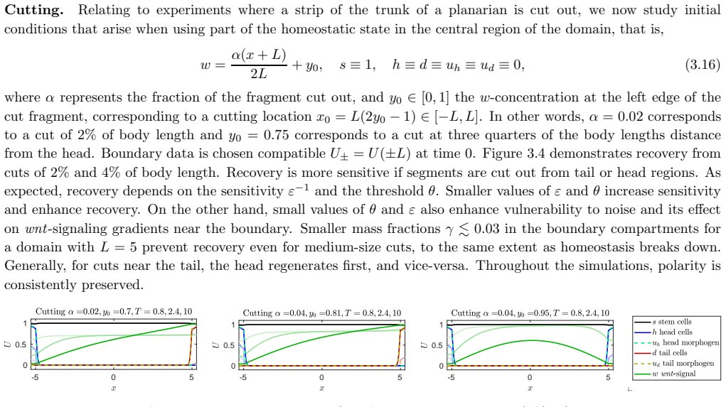
- A fragment cut from the trunk, head, or tail region regenerates head at the original head-facing edge and tail at the other, with polarity preserved across body sizes from $L=0.005$ to $L=40$ in simulations.
- Grafting reproduces the observed either-or outcome: donor head tissue placed near the host head merges and vanishes, while the same tissue placed far from the host head persists and can organize a second axis.
- Absolute levels of wnt signaling are not used for polarity in the early regeneration stages, so equally small fragments from different body regions behave alike despite very different local wnt concentrations.
- During uniform growth the pattern is maintained for moderate growth speeds; if dilution makes the wnt gradient shallower than the sensing threshold, the head is lost and a second tail replaces it.
- Regeneration begins as an instability of the gradientless trunk state, and near the boundary between recovery and failure the model predicts oscillatory wnt concentrations before the gradient collapses.
Reading between the lines
- A direct experimental test of the paper's mechanism would be to image wnt/β-catenin reporters at a fresh wound and ask whether differentiation direction correlates with the slope of the gradient on the two sides of the cut, rather than with the local concentration.
- Because the paper identifies a finite-size boundary compartment as mathematically necessary (dynamic rather than Robin conditions), it implies that a distinct 'pole' tissue with its own kinetics should be experimentally detectable at planarian body edges; failure to find such a compartment would undercut the model.
- The conservation of wnt signaling across planarians and hydra suggests the gradient-sensing boundary mechanism could be tested in hydra as well, where the same type of head/foot grafting experiments have been quantified.
- If the gradient-sensing rule is really generic, it might be implemented in synthetic tissues or organoids by engineering cells that measure a morphogen slope at an artificial boundary, providing an engineering route to polarity control.
Signed reviews
Editorial analysis
A structured set of objections, weighed in public.
Referee Report
Summary. The paper introduces a reaction-diffusion model for planarian regeneration along the anterior-posterior axis. The model tracks stem, head, and tail cell densities together with short-range head/tail signals and a long-range wnt-related signal w. Dynamics in the bulk are supplemented by Wentzell-type dynamic boundary conditions on a boundary compartment, where stem-cell differentiation into head or tail cells is switched by the sign of the normal derivative of w. The authors report numerical simulations of homeostasis, cutting, grafting, and growth, and claim correct reproduction of the main experiments with preservation of polarity over orders of magnitude in body size. The second half of the paper derives reduced two-species and scalar models, analyzes stability of the unpolarized state, and argues that dynamic boundary conditions are necessary for regeneration while Robin-type conditions fail. The paper also predicts oscillations near the threshold of regeneration failure.
Significance. If the central claim is correct, the model offers a minimal, analytically tractable mechanism for polarity-preserving regeneration that does not rely on Turing-type wavelength selection and that scales over large size changes. The explicit model reduction to an order-parameter equation and to a scalar equation, plus the stability analysis in Section 4.4, is a genuine strength: it identifies a concrete positive-feedback loop through boundary sensing and explains why dynamic boundary conditions are qualitatively different from instantaneous Robin conditions. The scalar model's oscillatory instability is a falsifiable prediction. However, the significance is conditional on resolving the sign inconsistency in the sensing terms and on demonstrating that the reported outcomes are robust rather than fine-tuned to near-critical parameter values. As written, the central claim is not supported by the printed equations.
major comments (3)
- [Section 3.2, Eqs. (3.13)-(3.15)] The signs in the gradient-sensing terms are inverted relative to the stated head/tail convention. In the healthy gradient and in the cutting initial condition (3.16), w increases from left to right, so w_x>0. At the left boundary x=-L the outward normal derivative is ∂ν w = -w_x <0, so the indicator χ^ε_{<-θ}(∂ν w) in Eq. (3.15) is active and χ^ε_{>θ}(∂ν w) in Eq. (3.14) is inactive. The written model therefore produces tail cells at the anterior wound and head cells at the posterior wound, which is the opposite of the experimental polarity the paper claims to reproduce. This is not a minor labeling issue: for a trunk fragment with h=d=0, the wrong Ψ term is unsuppressed and drives differentiation in the wrong direction. The reduced model in Eq. (4.5) uses the opposite sign convention (Ψ_c^± contains χ^ε_{>θ}(-∂ν w|±L) for the +1/head state), so the full and reduced systems are internally inconsistent. Either the full model in Section 3 has a sign typo that also affects the reported simulations, or the simulations were generated with a different system than the one printed. In either case, the central demonstration of Section 3.3 is not supported by the equations as written.
- [Sections 3.3 and 4.3] The paper repeatedly claims robust behavior for a wide range of parameter values, but the simulation parameters are explicitly chosen close to critical values. Section 3.3 states: 'Our choices of parameters are close to the critical values, where head and tail regions neither shrink nor expand.' Section 4.3 chooses κ=0.577, which is within 0.001 of the Maxwell point κ*=1/√3≈0.5774 identified in Section 4.2. For a bistable or tristable front system, operating at the Maxwell point makes the size and persistence of grafted head/tail regions highly sensitive to small parameter perturbations. The paper does not provide a systematic parameter scan, a phase diagram, or a basin-of-attraction study. Without such evidence, the qualitative statements about 'robustness' in Section 2.2 and the abstract cannot be distinguished from fine-tuning of the reported simulations.
- [Sections 1, 2.2, and 3.3] The abstract and introduction claim that the model 'correctly reproduces' cut and graft experiments and that it is the only reacting-diffusing-species model able to do so. The evidence consists of a small number of simulations with hand-picked initial conditions and no quantitative comparison to experimental data such as regeneration times, minimum fragment sizes, or the observed frequencies of two-headed versus two-tailed outcomes. The paper itself notes in Section 2.2 that the model cannot produce a bias between two-headed and two-tailed animals because head and tail are treated symmetrically. The claims should be softened to 'is consistent with the qualitative outcomes of selected experiments,' and the uniqueness claim should either be removed or supported by a systematic comparison with the models discussed in Section 2.4.
minor comments (5)
- [Eq. (3.15)] The second sensing function is labeled Ψ_w but should be Ψ_d, consistent with Eq. (3.12) and the surrounding text.
- [Title and formatting] The title contains a typo: 'planari an' should be 'planarian.' There are also repeated spacing errors in the text, e.g., 'Differential equations' and 'inhomegeneous Dirichlet boundary conditions' in Section 3.2.
- [Section 4.2] The text writes 'critical “Mawell” point' but should read 'Maxwell point.'
- [Section 3.3] The numerical simulations are not accompanied by code or data availability statements, and the parameter values in Table 1 are presented without a sensitivity analysis. Providing the simulation code would substantially improve reproducibility, especially given the sign inconsistency noted above.
- [Section 5] The statement that gradient sensing is 'necessary' to reproduce robust preservation of polarity is presented as a conclusion, but the paper only demonstrates sufficiency of the proposed mechanism. The wording should be adjusted to reflect that this is a conjecture supported by the reduced-model analysis, not a proven necessity.
Circularity Check
No circular derivation; the gradient-sensing mechanism is an openly stated modeling postulate, with one minor non-load-bearing self-citation. A printed sign error would reverse the claimed polarity, but that is a correctness issue, not circularity.
full rationale
The regeneration mechanism is introduced as an assumption, not inferred from the phenomena: 'We postulate that, as a key ingredient to robust regeneration and preservation of polarity, the production rate Ψ_h/d depends sensitively on the normal derivative of the long-range signal w' (Sec. 3.2). The cutting/grafting/growth simulations solve the full initial-boundary-value problem and exhibit non-obvious behaviors, including failure for small fragments, oscillations near the failure threshold, and instability of Robin-type relaxations, so the demonstrations are not logically identical to the inputs. The reduced model and linearization in Sec. 4.4 are additional analytic content, not a restatement of the boundary term. The only notable self-citation is [76] for the underlying cell-signal formulation and growth laws; it is background and not load-bearing, so at most a minor self-citation. Separately, under the standard outward-normal convention, Eqs. (3.14)-(3.15) assign head production to the posterior wound and tail production to the anterior wound, opposite to the paper's claim; this sign/typo discrepancy is a correctness issue that does not constitute circularity.
Assumptions & free parameters
free parameters (15)
- Ds =
1
- Dh =
1e-3
- Dd =
1e-3
- Duh and Dud =
1e-2 each
- Dw =
1
- ps and eta_s =
100 and 100
- ph, pd, eta_h, eta_d =
all 1
- r0, r1, r2, r3 =
18, 12, 6, 6
- pw =
10
- tau =
0.5
- theta and epsilon =
3 and 2e-3
- gamma =
0.3
- L =
10
- kappa (reduced model) =
0.577
- tau (scalar model) =
50
assumptions (7)
- domain assumption Cell densities and signals evolve as continuum reaction-diffusion fields on a one-dimensional interval x in [-L, L].
- ad hoc to paper The lumped variable w represents the full wnt-related signaling family and obeys degradation by head cells and saturated production by tail cells in Eq. (3.7).
- ad hoc to paper A finite-size boundary compartment with dynamic (Wentzell) boundary conditions (3.8)-(3.9) describes wound healing at body edges.
- ad hoc to paper Stem cells at boundary compartments sense the sign of the normal derivative of w and differentiate accordingly through the Psi functions in (3.13)-(3.15).
- domain assumption Quasi-steady-state reductions: stem cell density is constant s approximately 1 and short-range signals u_h and u_d are algebraically slaved to h and d (Section 4.1).
- domain assumption Head and tail cells have equal kinetics and the system is reflection-symmetric between h and d.
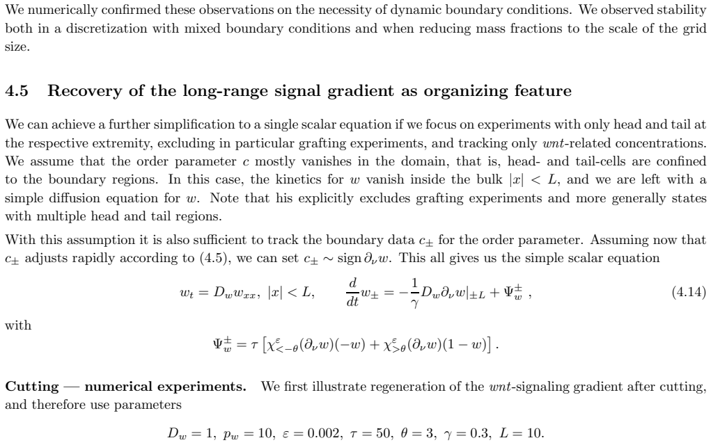
- ad hoc to paper For the scalar model, boundary order parameter c+ adjusts rapidly to sign of the normal derivative of w, excluding grafting and multiple head/tail regions.
invented entities (2)
-
Boundary compartment variables U+ with gradient-sensing differentiation kinetics Psi+
-
Lumped wnt-related signal w
independent evidence
Cite this review
Pith. "Pith review of Signaling gradients in surface dynamics as basis for planarian regeneration." pith.science (2026). https://pith.science/paper/A3TXL5SE
@misc{pith2026190804253,
  author       = {Pith},
  title        = {Pith review of: Signaling gradients in surface dynamics as basis for planarian regeneration},
  year         = {2026},
  howpublished = {\url{https://pith.science/paper/A3TXL5SE}},
  note         = {Machine review of arXiv:1908.04253}
}
read the original abstract
We introduce and analyze a mathematical model for the regeneration of planarian flatworms. This system of differential equations incorporates dynamics of head and tail cells which express positional control genes that in turn translate into localized signals that guide stem cell differentiation. Orientation and positional information is encoded in the dynamics of a long range wnt-related signaling gradient. We motivate our model in relation to experimental data and demonstrate how it correctly reproduces cut and graft experiments. In particular, our system improves on previous models by preserving polarity in regeneration, over orders of magnitude in body size during cutting experiments and growth phases. Our model relies on tristability in cell density dynamics, between head, trunk, and tail. In addition, key to polarity preservation in regeneration, our system includes sensitivity of cell differentiation to gradients of wnt-related signals %measured relative to the tissue surface. This process is particularly relevant in a small tissue layer close to wounds during their healing, and modeled here in a robust fashion through dynamic boundary conditions.
Figures

Figures from the paper (13 more)





Reference graph
Works this paper leans on
-
[1]
Achermann and T
J. Achermann and T. Sugiyama. Genetic analysis of developmenta l mechanisms in hydra: X. Morphogenetic potentials of a regeneration-deficient strain (reg-16). Developmental biology, 107(1):13–27, 1985
1985
-
[2]
Adell, F
T. Adell, F. Cebri` a, and E. Sal´ o. Gradients in planarian regenera tion and homeostasis. Cold Spring Harbor perspectives in biology, 2(1):a000505, 2010
2010
-
[3]
Almuedo-Castillo, A
M. Almuedo-Castillo, A. Bl¨ aßle, D. M¨ orsdorf, L. Marcon, G. H. Soh, K. W. Rogers, A. F. Schier, and P. M¨ uller. Scale-invariant patterning by size-dependent inhibition of nodal sig nalling. Nature Cell Biology, 20(9):1032–1042, 2018
2018
-
[4]
Almuedo-Castillo, M
M. Almuedo-Castillo, M. Sureda-G´ omez, and T. Adell. Wnt signaling in planarians: new answers to old questions. International Journal of Developmental Biology , 56(1-2-3):53–65, 2012
2012
-
[5]
W. Alt. Biased random walk models for chemotaxis and related diffus ion approximations. Journal of Mathe- matical Biology, 9(2):147–177, 1980
1980
-
[6]
Bagu˜ n` a
J. Bagu˜ n` a. Mitosis in the intact and regenerating planarian Dugesia mediterranea n. sp. II. Mitotic studies during regeneration, and a possible mechanism of blastema formation. Journal of Experimental Zoology , 195(1):65–79, 1976
1976
-
[7]
Bagu˜ n` a and R
J. Bagu˜ n` a and R. Romero. Quantitative analysis of cell types during growth, degrowt h and regeneration in the planarians Dugesia mediterranea and Dugesia tigrina . Springer, 1981
1981
-
[8]
Bagu˜ n` a and R
J. Bagu˜ n` a and R. Romero. Quantitative analysis of cell types d uring growth, degrowth and regeneration in the planarians Dugesia mediterranea and Dugesia tigrina. In The Biology of the Turbellaria , pages 181–194. Springer, 1981
1981
Show all 88 references
-
[9]
Bagu˜ n` a, R
J. Bagu˜ n` a, R. Romero, E. Sal´ o, J. Collet, C. Auladell, M. Ribas, M. Riutort, J. Garc ´ ıa-Fern` andez, F. Burgaya, and D. Bueno. Growth, degrowth and regeneration as developmen tal phenomena in adult freshwater planarians. In Experimental embryology in aquatic plants and...
1990
-
[10]
J. Bagu, S. Carranza, M. Pala, C. Ribera, G. Giribet, M. A. Arne do, M. Ribas, and M. Riutort. From morphology and karyology to molecules. new methods for taxonomical identifica tion of asexual populations of freshwater planarians. a tribute to professor mario benazzi. Italian ...
1999
-
[11]
C. R. Bardeen and F. Baetjer. The inhibitive action of the roent gen rays on regeneration in planarians. Journal of Experimental Zoology Part A: Ecological Genetics and Phy siology, 1(1):191–195, 1904. 25
1904
-
[12]
H. R. Bode. Head regeneration in Hydra. Developmental dynamics , 226(2):225–236, 2003
2003
-
[13]
Bowen, T
I. Bowen, T. Ryder, and J. Thompson. The fine structure of t he planarian Polycelis tenuis Iijima. Protoplasma, 79(1-2):1–17, 1974
1974
-
[14]
Cramer von Laue
C. Cramer von Laue. Untersuchungen zur dualen Funktion von β -Catenin im Wnt-Signalweg und der Cadherin- vermittelten Zelladh¨ asion bei Hydra. PhD thesis, Technische Universit¨ at, 2004
2004
-
[15]
Cusseddu, L
D. Cusseddu, L. Edelstein-Keshet, J. Mackenzie, S. Portet, and A. Madzvamuse. A coupled bulk-surface model for cell polarisation. Journal of Theoretical Biology , 2018
2018
-
[16]
De Vries, J
E. De Vries, J. Bagu˜ n` a, and I. Ball. Chromosomal polymorphis m in planarians (Turbellaria, Tricladida) and the plate tectonics of the western Mediterranean. Genetica, 62(3):187–191, 1984
1984
-
[17]
C. M. Elliott, T. Ranner, and C. Venkataraman. Coupled bulk-su rface free boundary problems arising from a mathematical model of receptor-ligand dynamics. SIAM J. Math. Anal. , 49(1):360–397, 2017
2017
-
[18]
P. C. Fife. Models for phase separation and their mathematics. Electron. J. Differential Equations , pages No. 48, 26, 2000
2000
-
[19]
Gagliardi, A
M. Gagliardi, A. Hernandez, I. J. McGough, and J.-P. Vincent. I nhibitors of endocytosis prevent Wnt/Wingless signalling by reducing the level of basal β -catenin/Armadillo. J Cell Sci , 127(22):4918–4926, 2014
2014
-
[20]
L. Gee, J. Hartig, L. Law, J. Wittlieb, K. Khalturin, T. C. Bosch, and H. R. Bode. β -catenin plays a central role in setting up the head organizer in hydra. Developmental biology, 340(1):116–124, 2010
2010
-
[21]
Gierer and H
A. Gierer and H. Meinhardt. A theory of biological pattern form ation. Kybernetik, 12(1):30–39, 1972
1972
-
[22]
K. A. Gurley, S. A. Elliott, O. Simakov, H. A. Schmidt, T. W. Holste in, and A. S. Alvarado. Expression of secreted Wnt pathway components reveals unexpected comple xity of the planarian amputation response. Developmental biology, 347(1):24–39, 2010
2010
-
[23]
H¨ arting and A
S. H¨ arting and A. Marciniak-Czochra. Spike patterns in a reac tion–diffusion ode model with Turing instability. Mathematical Methods in the Applied Sciences , 37(9):1377–1391, 2014
2014
-
[24]
H¨ arting, A
S. H¨ arting, A. Marciniak-Czochra, and I. Takagi. Stable patt erns with jump discontinuity in systems with Turing instability and hysteresis. Disc. Cont. Dyn. Sys. A. , 38:757–800, 2017
2017
-
[25]
Hobmayer, F
B. Hobmayer, F. Rentzsch, K. Kuhn, C. M. Happel, C. C. von La ue, P. Snyder, U. Rothb¨ acher, and T. W. Holstein. WNT signalling molecules act in axis formation in the diploblastic m etazoan Hydra. Nature, 407(6801):186–189, 2000
2000
-
[26]
Iijima, Y
M. Iijima, Y. E. Huang, and P. Devreotes. Temporal and Spatia l Regulation of Chemotaxis. Developmental Cell, 3:469–478, 2002
2002
-
[27]
Kakugawa, P
S. Kakugawa, P. F. Langton, M. Zebisch, S. A. Howell, T.-H. Cha ng, Y. Liu, T. Feizi, G. Bineva, N. O’Reilly, A. P. Snijders, et al. Notum deacylates Wnt proteins to suppress s ignalling activity. Nature, 519(7542):187–192, 2015
2015
-
[28]
Lengfeld, H
T. Lengfeld, H. Watanabe, O. Simakov, D. Lindgens, L. Gee, L. Law, H. A. Schmidt, S. ¨Ozbek, H. Bode, and T. W. Holstein. Multiple Wnts are involved in Hydra organizer formation and regeneration. Developmental biology, 330(1):186–199, 2009
2009
-
[29]
Levine and W.-J
H. Levine and W.-J. Rappel. Membrane-bound Turing patterns. Phys. Rev. E (3) , 72(6):061912, 5, 2005
2005
-
[30]
Y. Li, A. Marciniak-Czochra, I. Takagi, and B. Wu. Steady stat es of fitzhugh-nagumo system with non-diffusive activator and diffusive inhibitor. preprint, 2017. 26
2017
-
[31]
Y. Li, A. Marciniak-Czochra, I. Takagi, B. Wu, et al. Bifurcation analysis of a diffusion-ode model with Turing instability and hysteresis. Hiroshima Mathematical Journal , 47(2):217–247, 2017
2017
-
[32]
D. Lobo, W. S. Beane, and M. Levin. Modeling planarian regenera tion: a primer for reverse-engineering the worm. PLoS Comput Biol , 8(4):e1002481, 2012
2012
-
[33]
H. K. MacWilliams. Hydra transplantation phenomena and the mec hanism of hydra head regeneration: II. Properties of the head activation. Developmental biology, 96(1):239–257, 1983
1983
-
[34]
Marciniak-Czochra
A. Marciniak-Czochra. Receptor-based models with diffusion-d riven instability for pattern formation in hydra. Journal of Biological Systems , 11(03):293–324, 2003
2003
-
[35]
Marciniak-Czochra
A. Marciniak-Czochra. Receptor-based models with hysteres is for pattern formation in hydra. Mathematical biosciences, 199(1):97–119, 2006
2006
-
[36]
Marciniak-Czochra, S
A. Marciniak-Czochra, S. H¨ arting, G. Karch, and K. Suzuki. D ynamical spike solutions in a nonlocal model of pattern formation. Nonlinearity, 31(5):1757–1781, 2018
2018
-
[37]
Marciniak-Czochra, G
A. Marciniak-Czochra, G. Karch, and K. Suzuki. Instability of T uring patterns in reaction-diffusion-ode systems. Journal of Mathematical Biology , 74(3):583–618, 2017
2017
-
[38]
Marciniak-Czochra, G
A. Marciniak-Czochra, G. Karch, K. Suzuki, J. Zienkiewicz, et a l. Diffusion-driven blowup of nonnegative solutions to reaction-diffusion-ode systems. Differential and Integral Equations , 29(7/8):715–730, 2016
2016
-
[39]
Marciniak-Czochra, M
A. Marciniak-Czochra, M. Nakayama, I. Takagi, et al. Pattern formation in a diffusion-ode model with hysteresis. Differential and Integral Equations , 28(7/8):655–694, 2015
2015
-
[40]
Meinhardt
H. Meinhardt. Turing’s theory of morphogenesis of 1952 and th e subsequent discovery of the crucial role of local self-enhancement and long-range inhibition. Interface Focus, 2(4):407–416, 2012
1952
-
[41]
T. H. Morgan. Experimental studies of the regeneration of Pla naria maculata. Development Genes and Evolution , 7(2):364–397, 1898
-
[42]
T. H. Morgan. The control of heteromorphosis in Planaria macu lata. Archiv f¨ ur Entwicklungsmechanik der Organismen, 17(4):683–695, 1904
1904
-
[43]
T. H. Morgan. Polarity considered as a phenomenon of gradatio n of materials. Journal of Experimental Zoology , 2(4):495–506, 1905
1905
-
[44]
Y. Mori, A. Jilkine, and L. Edelstein-Keshet. Wave-pinning and ce ll polarity from a bistable reaction-diffusion system. Biophysical journal , 94(9):3684–3697, 2008
2008
-
[45]
J. D. Murray. Mathematical biology I: An introduction . Interdisciplinary Applied Mathematics, 17. Springer- Verlag, New York, 2002
2002
-
[46]
J. D. Murray. Mathematical biology II: Spatial models and biomedical app lications. Interdisciplinary Applied Mathematics, 18. Springer-Verlag, New York, 2003
2003
-
[47]
P. A. Newmark and A. S. Alvarado. Not your father’s planarian: a classic model enters the era of functional genomics. Nature Reviews Genetics , 3(3):210–219, 2002
2002
-
[48]
K. Noda. Reconstitution of dissociated cells of hydra. Zool. Mag., 80:99–101, 1971
1971
-
[49]
N¨ usslein-Volhard and E
C. N¨ usslein-Volhard and E. Wieschaus. Mutations affecting seg ment number and polarity in Drosophila. Nature, 287(5785):795–801, 1980. 27
1980
-
[50]
H. G. Othmer and E. Pate. Scale-invariance in reaction-diffusion models of spatial pattern formation. Proceedings of the National Academy of Sciences , 77(7):4180–4184, 1980
1980
-
[51]
Owlarn and K
S. Owlarn and K. Bartscherer. Go ahead, grow a head! A planar ian’s guide to anterior regeneration. Regener- ation, 3(3):139–155, 2016
2016
-
[52]
C. P. Petersen and P. W. Reddien. Smed- β catenin-1 is required for anteroposterior blastema polarity in plana rian regeneration. Science, 319(5861):327–330, 2008
2008
-
[53]
C. P. Petersen and P. W. Reddien. A wound-induced Wnt expres sion program controls planarian regeneration polarity. Proceedings of the National Academy of Sciences , 106(40):17061–17066, 2009
2009
-
[54]
C. P. Petersen and P. W. Reddien. Polarized notum activation at wounds inhibits Wnt function to promote planarian head regeneration. Science, 332(6031):852–855, 2011
2011
-
[55]
Philipp, R
I. Philipp, R. Aufschnaiter, S. ¨Ozbek, S. Pontasch, M. Jenewein, H. Watanabe, F. Rentzsch, T. W. Holstein, and B. Hobmayer. Wnt/ β -catenin and noncanonical Wnt signaling interact in tissue evaginatio n in the simple eumetazoan Hydra. Proceedings of the National Academy of Sc...
2009
-
[56]
Plickert, V
G. Plickert, V. Jacoby, U. Frank, W. A. M¨ uller, and O. Mokady. Wnt signaling in hydroid development: formation of the primary body axis in embryogenesis and its subsequ ent patterning. Developmental biology , 298(2):368–378, 2006
2006
-
[57]
Randolph
H. Randolph. The regeneration of the tail in Lumbriculus. Journal of Morphology , 7(3):317–344, 1892
-
[58]
Randolph
H. Randolph. Observations and experiments on regeneration in planarians. Development Genes and Evolution , 5(2):352–372, 1897
-
[59]
R¨ atz and M
A. R¨ atz and M. R¨ oger. Turing instabilities in a mathematical mod el for signaling networks. J. Math. Biol. , 65(6-7):1215–1244, 2012
2012
-
[60]
R¨ atz and M
A. R¨ atz and M. R¨ oger. Symmetry breaking in a bulk-surface r eaction-diffusion model for signalling networks. Nonlinearity, 27(8):1805–1827, 2014
2014
-
[61]
P. W. Reddien. Constitutive gene expression and the specificat ion of tissue identity in adult planarian biology. Trends in Genetics , 27(7):277–285, 2011
2011
-
[62]
P. W. Reddien and A. S. Alvarado. Fundamentals of planarian reg eneration. Annu. Rev. Cell Dev. Biol. , 20:725–757, 2004
2004
-
[63]
Reuter, M
H. Reuter, M. M¨ arz, M. C. Vogg, D. Eccles, L. Gr ´ ıfol-Bold´ u,D. Wehner, S. Owlarn, T. Adell, G. Weidinger, and K. Bartscherer. β -catenin-dependent control of positional information along the A P body axis in planarians involves a teashirt family member. Cell Reports , 10...
2015
-
[64]
J. C. Rink. Stem Cells, Patterning and Regeneration in Planarians: Sel f-Organization at the Organismal Scale , pages 57–172. Springer, New York, 2018
2018
-
[65]
Sal´ o and J
E. Sal´ o and J. Bagu˜ n` a. Regeneration and pattern formation in planarians. Development, 83(1):63–80, 1984
1984
-
[66]
Sal´ o and J
E. Sal´ o and J. Bagu˜ n` a. Cell movement in intact and regenerating planarians. Quantitation using chromosomal, nuclear and cytoplasmic markers. Development, 89(1):57–70, 1985
1985
-
[67]
F. V. Santos. Studies on transplantation in Planaria. Biological Bulletin , 57(3):188–197, 1929
1929
-
[68]
M. L. Scimone, S. W. Lapan, and P. W. Reddien. A forkhead tran scription factor is wound-induced at the planarian midline and required for anterior pole regeneration. PLoS Genet , 10(1):e1003999, 2014. 28
2014
-
[69]
Sherratt, P
J. Sherratt, P. Maini, W. J¨ ager, and W. M¨ uller. A receptor based model for pattern formation in hydra. Forma, 10(2):77–95, 1995
1995
-
[70]
Shimizu, Y
H. Shimizu, Y. Sawada, and T. Sugiyama. Minimum tissue size requir ed for hydra regeneration. Developmental biology, 155(2):287–296, 1993
1993
-
[71]
S. Shostak. Inhibitory gradients of head and foot regenerat ion in Hydra viridis. Developmental biology, 28(4):620– 635, 1972
1972
-
[72]
L. R. Smales and H. D. Blankespoor. The epidermis and sensory o rgans of Dugesia tigrina (Turbellaria: Tricla- dida). Cell and Tissue Research , 193(1):35–40, 1978
1978
-
[73]
St¨ uckemann, J
T. St¨ uckemann, J. P. Cleland, S. Werner, H. T.-K. Vu, R. Baye rsdorf, S.-Y. Liu, B. Friedrich, F. J¨ ulicher, and J. C. Rink. Antagonistic Self-Organizing Patterning Systems Contr ol Maintenance and Regeneration of the Anteroposterior Axis in Planarians. Developmental Cell ,...
2017
-
[74]
Sureda-G´ omez, J
M. Sureda-G´ omez, J. M. Mart ´ ın-Dur´ an, and T. Adell. Localization of planarian β -CATENIN-1 reveals multiple roles during anterior-posterior regeneration and organogenesis . Development, 143(22):4149–4160, 2016
2016
-
[75]
Tasaki, N
J. Tasaki, N. Shibata, O. Nishimura, K. Itomi, Y. Tabata, F. Son , N. Suzuki, R. Araki, M. Abe, K. Agata, et al. ERK signaling controls blastema cell differentiation during planarian re generation. Development, 138(12):2417– 2427, 2011
2011
-
[76]
Tenbrock
C. Tenbrock. Mathematical models for regeneration on the ex ample of planarians. PhD-Thesis, University of M¨ unster (WWU), Germany, 2017
2017
-
[77]
A. M. Turing. The chemical basis of morphogenesis. Philosophical Transactions of the Royal Society B , 1952
1952
-
[78]
D. M. Umulis. Analysis of dynamic morphogen scale invariance. Journal of The Royal Society Interface , 6(41):1179–1191, 2009
2009
-
[79]
D. M. Umulis and H. G. Othmer. Mechanisms of scaling in pattern fo rmation. Development, 140(24):4830–4843, 2013
2013
-
[80]
G. Webster. Studies on pattern regulation in hydra. Development, 16(1):123–141, 1966
1966
-
[81]
Wenemoser, S
D. Wenemoser, S. W. Lapan, A. W. Wilkinson, G. W. Bell, and P. W. R eddien. A molecular wound response program associated with regeneration initiation in planarians. Genes & Development , 26(9):988–1002, 2012
2012
-
[82]
Wenemoser and P
D. Wenemoser and P. W. Reddien. Planarian regeneration involve s distinct stem cell responses to wounds and tissue absence. Developmental biology, 344(2):979–991, 2010
2010
-
[83]
Werner, T
S. Werner, T. St¨ uckemann, M. Beir´ an Amigo, J. C. Rink, F. J¨ ulicher, and B. M. Friedrich. Scaling and regeneration of self-organized patterns. Phys. Rev. Lett. , 114:138101, Apr 2015
2015
-
[84]
O. K. Wilby and G. Webster. Experimental studies on axial polarit y in hydra. Development, 24(3):595–613, 1970
1970
-
[85]
J. N. Witchley, M. Mayer, D. E. Wagner, J. H. Owen, and P. W. Re ddien. Muscle cells provide instructions for planarian regeneration. Cell reports , 4(4):633–641, 2013
2013
-
[86]
L. Wolpert. Positional information and the spatial pattern of c ellular differentiation. Journal of Theoretical Biology, 25(1):1 – 47, 1969
1969
-
[87]
L. Wolpert. Positional Information and Pattern Formation in De velopment. Developmental Genetics , 1994
1994
-
[88]
Wurtzel, L
O. Wurtzel, L. E. Cote, A. Poirier, R. Satija, A. Regev, and P. W . Reddien. A generic and cell-type-specific wound response precedes regeneration in planarians. Developmental Cell , 35(5):632–645, 2015. 29
2015
Reviewed August 14, 2026 · model on record in the stance chip above.
Discussion (0). Continue with ORCID to comment.