REVIEW 3 major objections 5 minor 5 references
CT Field of View Extension Using Combined Channels Extension and Deep Learning Methods
T0 review · 3 major / 5 minor · reviewed 2026-08-14 · deepseek-v4-flash
Pith's one-line read CT scans can widen their field of view by stretching sinogram edges and cleaning the artifacts with a U-Net.
desk verdict Plausible industrial EFoV trick with an honest qualitative report, but the training description has a hole that needs fixing before the results can be believed. read the letter →
The pith
A machine-rendered reading of the paper's core claim, the machinery that carries it, and where it could break.
The reading
What carries the argument
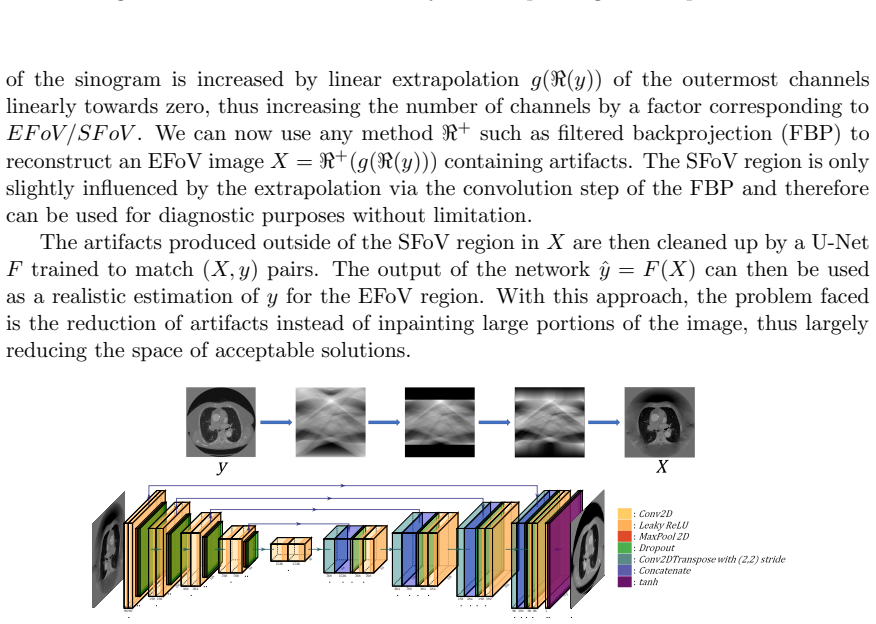
The machinery is a two-stage pipeline built around the Radon transform. A clinical slice is projected to a sinogram, the outermost channels are linearly extrapolated toward zero to match the desired EFoV/SFoV ratio, and filtered backprojection reconstructs an extended image whose outer band contains artifacts. A U-Net, a convolutional encoder-decoder with skip connections, is trained to map those artifact-laden extended images back to the original images, with the loss (mean squared error plus structural dissimilarity) evaluated only in the extended region. The SFoV portion of the network output is discarded and replaced by the original reconstruction, and the output's mean and standard deviation are matched to the input to soften the transition.
What would settle it
Acquire raw data from a body phantom wider than the scanner field of view, reconstruct it with the proposed pipeline and with HDFoV, and compare both against a ground-truth reconstruction of the same phantom made with a scanner that has a larger field of view; if the U-Net output is not closer to that ground truth than the plain linear extrapolation reconstruction in the extended band, the simulated-to-real transfer fails and the central claim does not hold.
Extended reading notes
Core claim
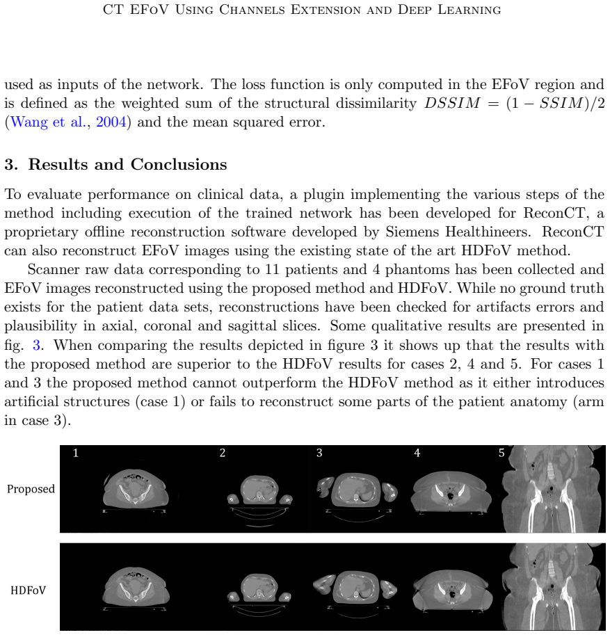
The central claim is that linear extrapolation of sinogram channels followed by deep-learning artifact removal in image space is an efficient and competitive way to extend the CT field of view. The paper's conclusion states that linear extrapolation followed by removal of artifacts in the reconstruction using a deep learning network proved to be an efficient method and shows potential to improve the quality of extended-field-of-view reconstructions. The extended region is not claimed to be diagnostic; it is meant to be sufficient for applications such as radiotherapy planning and imaging of obese patients. On the qualitative comparison with the existing HDFoV method, the proposed reconstructions look superior in cases 2, 4, and 5, but inferior in cases 1 and 3, where the method either introduces artificial structures or fails to reconstruct part of the patient anatomy.
Load-bearing premise
The whole result depends on the assumption that a network trained on artifacts created by stretching simulated projection data of clean CT slices will also clean up artifacts in real scanner measurements, which have noise and beam-hardening effects the simulation may not reproduce.
Editorial extensions
If this is right
- If the claim holds, the extrapolated region becomes usable for non-diagnostic clinical tasks such as radiotherapy planning and imaging of obese patients, where the standard scan field is simply too narrow.
- Because the method is one linear extrapolation followed by a single network pass, it offers a computationally cheaper alternative to iterative extended-field-of-view reconstructions.
- Framing the problem as artifact reduction rather than image inpainting restricts the space of possible outputs, which should make reconstructed anatomy more plausible than direct extrapolation alone.
- The method does not dominate HDFoV in every case: on the five displayed comparisons it is judged superior in three and inferior in two, so it should be considered a selective improvement rather than a universal replacement.
Reading between the lines
- A natural extension the paper does not test is varying the noise level and beam-hardening model in the simulated training data; a controlled study could map how much realism the artifact generator needs before the network transfers to raw scanner data.
- The failure cases suggest the network is weakest on compact high-contrast anatomy (the missed arm in one case) and strongest on smooth soft-tissue extensions; a phantom study varying tissue contrast in the truncated region could confirm that pattern and guide retraining.
- The same two-step idea, linear or model-based extension of incomplete measurements followed by an artifact-removal network, could be transferred to other incomplete-data CT problems such as limited-angle or sparse-view scanning, with the artifact generator replaced by the corresponding forward model.
Signed reviews
Editorial analysis
A structured set of objections, weighed in public.
Referee Report
Summary. The paper proposes a two-step method for extending the CT field of view. First, the sinogram channels are linearly extrapolated outward, and an EFoV image is reconstructed with FBP, producing characteristic artifacts outside the SFoV. Second, a U-Net is trained to remove these artifacts in image space; the SFoV region is copied from the input, and the network output is statistically matched to the input distribution. The method is evaluated qualitatively on clinical raw data from 11 patients and 4 phantoms, comparing the proposed approach to a prior HDFoV method, with the authors reporting favorable results in three of five illustrated cases. The central claim is that linear extrapolation plus deep-learning artifact removal is an efficient and potentially superior EFoV reconstruction strategy.
Significance. If the central claim holds, the method offers a computationally inexpensive alternative to iterative EFoV reconstruction, which is clinically relevant for radiotherapy planning and imaging of large patients. The combination of a deterministic sinogram-extension step with a learned artifact-reduction step is a sensible division of labor, and the use of a U-Net in image space keeps the runtime low. The paper is an extended abstract, so the contribution is at the proof-of-concept level. The authors are transparent about the qualitative nature of the evaluation and explicitly acknowledge two cases where the proposed method does not outperform HDFoV. However, the current manuscript lacks the training-data details needed to reproduce the method, and the evaluation does not provide quantitative or statistical support for the superiority claim.
major comments (3)
- [Section 2, training data paragraph] The description of the training data construction is ambiguous in a way that is load-bearing for the entire method. The text states that 'a collection of clinical CT scan slices y are projected to their Radon transform to obtain sinograms' and that the loss is 'only computed in the EFoV region.' If y is a standard SFoV reconstruction, then the EFoV region of y is identically zero, and the optimal network output there is zero, which would make the reported EFoV anatomy (e.g., the arm in case 3) impossible. The authors must clarify whether y is reconstructed with a field of view larger than the simulated SFoV, or whether the sinogram is first truncated/cropped to a smaller SFoV before the linear extrapolation is applied. Without an explicit statement of how nonzero anatomy enters the EFoV part of the training targets, the method description is incomplete and the results cannot be interpreted or reproduced.
- [Section 3, results] The evaluation is entirely qualitative. The authors report that 11 patient and 4 phantom datasets were reconstructed, yet the only evidence presented is five displayed cases, of which three are judged favorable to the proposed method and two are unfavorable. There are no quantitative error metrics (e.g., RMSE, SSIM, or mean absolute error in the EFoV region), even though the phantom datasets could provide ground truth, and no statistical test of the claim that the proposed method is 'superior' to HDFoV in cases 2, 4, and 5. The conclusion that linear extrapolation plus deep learning 'proved to be an efficient method' and 'shows potential to improve quality' is not supported by the evidence shown. The authors should provide quantitative comparisons and, ideally, a reader study or a scoring protocol that substantiates the superiority claim.
- [Section 2, domain transfer] The network is trained on simulated artifacts generated by linearly extrapolating Radon transforms of clinical CT slices, but it is applied to real scanner raw data in Section 3. The manuscript does not describe whether the training simulation accounts for the actual noise, beam hardening, and truncation characteristics of the scanner, nor does it report the parameters used for the simulated extrapolation (e.g., the extrapolation slope, the amount of padding, or the EFoV/SFoV ratio). If the simulated artifact structure differs from the real artifacts, the U-Net may not remove artifacts on actual scans, and the reported clinical results would not be reproducible. This is a correctness risk that should be addressed by specifying the simulation settings and by showing, at minimum, that the artifact statistics in training match those in the test reconstructions.
minor comments (5)
- [Section 2, loss function] The loss is defined as a weighted sum of DSSIM and MSE, but the weights are not reported. Since the relative weighting is a free parameter that can materially affect the trade-off between structural fidelity and pixel accuracy, it should be stated explicitly.
- [Section 2, SFoV replacement and normalization] The order of operations between replacing the SFoV region with X and the statistical adaptation (mean/variance matching) is not made clear. The sentence 'In order to improve the final SFoV/EFoV transition, the network output is adapted to the input distribution' suggests the adaptation is applied before or after the replacement, but the exact sequence should be spelled out.
- [Figure 2] Figure 2 is difficult to interpret: the caption says 'Data generation pipeline and network architecture,' but the figure appears to mix the U-Net structure with the sinogram-to-image pipeline, and the numbers are not annotated. A separate schematic of the training pipeline and a standard U-Net diagram would improve readability.
- [Section 3, clinical evaluation protocol] The paper states that volumes were 'checked for consistency and plausibility' but does not describe who performed the checks, how many slices were reviewed, or which criteria were used to judge 'superior' results. A brief description of the evaluation protocol would increase confidence in the qualitative comparison.
- [General] The abstract and Section 1 mention that the proposed method delivers 'improved results' compared to existing methods, but the introduction cites no quantitative comparison and the body only offers a qualitative comparison to HDFoV. The wording should be aligned with the actual evidence presented.
Circularity Check
No significant circularity: the U-Net is trained on simulated forward-degraded data and evaluated against an independent HDFoV baseline; no fitted parameter is relabeled as a prediction.
full rationale
The paper's pipeline is g(y) -> X -> F(X) -> y_hat. The network is trained on pairs (X,y) where y are clinical slices and X is formed by projecting y, linearly extrapolating the sinogram, and backprojecting. This is a standard supervised artifact-removal setup: the target y is not derived from the network output, and the network is not used to fit a parameter that is then renamed a prediction. The only self-citations (Bruder et al. 2008, Kunze et al. 2007) are prior Siemens EFoV reconstruction techniques used as comparisons or background; they are not invoked as proof of correctness of the proposed method, and the paper presents qualitative comparisons against the HDFoV method. There is no uniqueness theorem imported from the authors' prior work, and no ansatz is smuggled in via citation. The reader's noted ambiguity about whether training slices y contain anatomy outside the simulated SFoV is a description or reproducibility gap, not a circular reduction: even if the training-data construction were under-specified, computing the loss in the EFoV region would not make the network output equal to its input by construction. Therefore no circularity is found and the score is 0.
Assumptions & free parameters
free parameters (1)
- Loss weighting between DSSIM and MSE =
Not stated
assumptions (5)
- standard math Radon transform and filtered backprojection adequately model the CT imaging process.
- domain assumption Linear extrapolation of outer sinogram channels toward zero yields artifacts that a U-Net can learn to remove.
- domain assumption The SFoV region is only slightly influenced by the extrapolation and can be used for diagnosis without limitation.
- domain assumption Networks trained on simulated projections of clinical slices transfer to real scanner raw data.
- ad hoc to paper Qualitative visual assessment on selected slices is a sufficient evaluation of EFoV quality.
Cite this review
Pith. "Pith review of CT Field of View Extension Using Combined Channels Extension and Deep Learning Methods." pith.science (2026). https://pith.science/paper/ACP2FBLQ
@misc{pith2026190809529,
  author       = {Pith},
  title        = {Pith review of: CT Field of View Extension Using Combined Channels Extension and Deep Learning Methods},
  year         = {2026},
  howpublished = {\url{https://pith.science/paper/ACP2FBLQ}},
  note         = {Machine review of arXiv:1908.09529}
}
read the original abstract
This paper proposes a method to extend the field of view of computed tomography images. In a first step, the field of view is increased by extrapolating linearly the outer channels in the sinogram space. The modified sinogram is then used to reconstruct extended field of view (EFoV) images containing artifacts due to the channels extension. In a second step, those artifacts are reduced by a deep learning network in image space. The proposed method has been evaluated on a collection of clinical scans. The resulting volumes have been checked for consistency and plausibility and compared to an existing state of the art EFoV method.
Figures

Reference graph
Works this paper leans on
-
[1]
H. Bruder, C. Suess, and K. Stierstorfer. Efficient extended field of view (efov) reconstruction techniques for multi-slice helical ct. 6913: 0 81--, 04 2008. doi:10.1117/12.768477
- [2]
-
[3]
H. Kunze, W. Härer, and K. Stierstorfer. Iterative extended field of view reconstruction. Proceedings of SPIE - The International Society for Optical Engineering, 03 2007. doi:10.1117/12.707336
-
[4]
O. Ronneberger, P.Fischer, and T. Brox. U-net: Convolutional networks for biomedical image segmentation. In Medical Image Computing and Computer-Assisted Intervention (MICCAI), volume 9351 of LNCS, pages 234--241. Springer, 2015. URL http://lmb.informatik.uni-freiburg.de/Publications/2015/RFB15a. (available on arXiv:1505.04597 [cs.CV])
arXiv 2015
-
[5]
Z. Wang, A. Bovik, H. Sheikh, and E. Simoncelli. Image quality assessment: From error visibility to structural similarity. IEEE TRANSACTIONS ON IMAGE PROCESSING, 13 0 (4): 0 600--612, 2004
work page 2004
Reviewed August 14, 2026 · model on record in the stance chip above.
Discussion (0). Continue with ORCID to comment.