REVIEW 1 major objections 6 minor 1 references
Generation of abdominal synthetic CTs from 0.35T MR images using generative adversarial networks for MR-only liver radiotherapy
T0 review · 1 major / 6 minor · reviewed 2026-08-14 · deepseek-v4-flash
Pith's one-line read GANs convert low-field MRI into synthetic CT for liver radiotherapy
desk verdict A competent feasibility study of GAN-based sCT from low-field MR for liver radiotherapy, undermined a bit by unvalidated deformable registration as ground truth. read the letter →
The pith
A machine-rendered reading of the paper's core claim, the machinery that carries it, and where it could break.
The reading
What carries the argument
The load-bearing mechanism is the generative adversarial network, specifically two variants: a conditional GAN (cGAN) trained on paired MR and deformed-CT slices, and a cycle-consistent GAN (cycleGAN) trained on unpaired slices through cycle-consistency losses that translate MR to CT and back. Each generator is a convolutional neural network mapping 0.35T TrueFISP MR slices into synthetic CT slices; each discriminator tries to distinguish real from generated images. The synthetic CT then supplies Hounsfield units to the treatment planning system's Monte Carlo dose calculation, so the quantity that carries the argument is the learned map from MR intensity patterns to electron-density-equivalent HU values.
What would settle it
Take one liver patient with a native breath-hold CT that can be aligned to the MR without deformable registration, generate the sCT, and recalculate the clinical plan; if the 2%, 2 mm gamma pass rate against the native-CT dose drops below 95% or the PTV dose deviation exceeds 1%, the MR-only dose calculation claim would be violated for that protocol.
Extended reading notes
Core claim
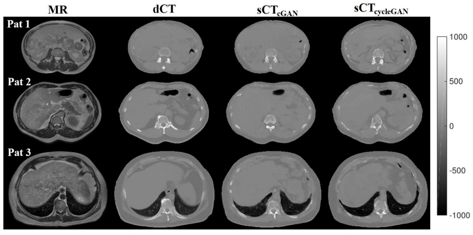
The paper claims that abdominal synthetic CT images generated by two generative adversarial networks, cGAN and cycleGAN, from 0.35T MR images are accurate enough for dose calculation in liver radiotherapy. Against the deformed CT reference, the networks produce mean absolute errors of 89.8 HU (cGAN) and 94.1 HU (cycleGAN); on eight liver plans, both deliver average gamma passing rates higher than 95% at the 2%, 2 mm criterion and higher than 99% at 3%, 3 mm, with mean dose and DVH deviations within ±0.6% of prescribed dose for the PTV and within ±0.15% for evaluated organs. The authors conclude that MR-only liver radiotherapy is achievable with these models, and that cGAN is somewhat more accurate than cycleGAN in this cohort.
Load-bearing premise
The reference images used for training and evaluation are CTs that were deformably registered to the MR images, and the paper does not report how accurate that registration was; the reported HU and gamma errors are measured against these registered images, not against an independently verified ground truth.
Editorial extensions
If this is right
- Both cGAN and cycleGAN produce abdominal sCTs whose HU errors are comparable to previously reported abdominal sCT results from high-field MRI, suggesting the low-field source is not a barrier.
- Dose recalculations on eight liver plans meet the 2%, 2 mm gamma criterion on average, so these models could replace the planning CT in the MR-only workflow for similar patients.
- With sCT generation under about 10 seconds per patient, the method is fast enough to sit inside an online adaptive treatment session without stretching it.
- Because cycleGAN does not require paired MR-CT training data, it can be trained on larger unpaired clinical datasets, potentially improving generalization.
Reading between the lines
- If the deformable registration that creates the dCT reference has errors comparable to the reported MAE, the true HU accuracy against native CT could be worse than the quoted ~90 HU; an independent rigidly co-registered comparison would resolve this.
- Both models had visible difficulty reproducing ribs; this may matter more for plans whose beams traverse the chest wall than for the liver plans tested here, and could be checked with a rib-tissue phantom or a thoracic cohort.
- The cGAN's small advantages over cycleGAN may shrink or reverse with more training data, since cycleGAN's unpaired training can exploit routine scans that lack a co-registered CT.
Signed reviews
Editorial analysis
A structured set of objections, weighed in public.
Referee Report
Summary. This manuscript investigates the generation of synthetic CT (sCT) images from 0.35T MR images using two generative adversarial networks, cGAN and cycleGAN, for MR-only liver radiotherapy. A retrospective cohort of 12 abdominal cancer patients (8 liver, 4 non-liver) was used. CT images were deformably registered to the MR images to create deformed CT (dCT) images, which served as the reference for training and evaluation. The models were trained on transverse slices with four-fold cross-validation, and sCT quality was assessed via MAE and PSNR inside the body contour. Dose calculation accuracy was evaluated on 8 liver patients by recalculating clinical plans on sCT and comparing dose distributions to dCT-based plans using gamma analysis and DVH metrics. The results show average MAE of 89.8 HU (cGAN) and 94.1 HU (cycleGAN), gamma passing rates above 95% (2%, 2 mm) and 99% (3%, 3 mm), and average dose metric deviations within ±0.6% for the PTV and ±0.15% for OARs. The authors conclude that both models achieve accurate dose calculation and are promising for MR-only liver radiotherapy, with cGAN appearing slightly more accurate than cycleGAN.
Significance. If validated, this work would be the first demonstration of deep learning-based sCT generation from low-field (0.35T) MR images for abdominal sites, specifically liver, with potential direct application to MR-guided online adaptive radiotherapy. The study has notable strengths: it uses a held-out cross-validation scheme, performs dosimetric evaluation with gamma analysis and clinically relevant DVH metrics on actual treatment plans, and directly compares two popular GAN architectures under identical data conditions. The reported sCT generation time of about 6 s per patient is practically attractive for online adaptive workflows. However, the evaluation relies entirely on a dCT reference created by deformable registration whose accuracy is not reported, and the sample size for dosimetric conclusions is small (n=8) with no statistical testing. These issues currently limit the strength of the feasibility claim, though the methodology is appropriate and the results are promising as a preliminary feasibility study.
major comments (1)
- [Section 2.1, 2.4, Eqs. (1)-(2), Tables 3-4] The claim that cGAN outperforms cycleGAN (abstract, Section 4) is not supported by statistical evidence. The differences in MAE (89.8 HU vs 94.1 HU) and gamma passing rates (e.g., 97.4% vs 95.6% for D ≥ 90% at 2%, 2 mm) are based on only 12 patients for HU metrics and 8 for dosimetric metrics, with large standard deviations. No confidence intervals, hypothesis tests, or non-inferiority analyses are provided. Given the small cohort, these differences could easily be within sampling variability. The authors should either provide appropriate statistical comparisons or temper the comparative conclusion to a descriptive observation that warrants future investigation with larger cohorts.
minor comments (6)
- [Abstract] There is a typo: "sCTcyleGAN" should be "sCTcycleGAN".
- [Eq. (2)] The PSNR formula uses a constant of 4095, but the text states the networks process 16-bit single-channel images; 4095 is the maximum value for a 12-bit range. Please clarify the bit depth used for the images and ensure consistency.
- [Table 2] The column header "PNSR" is a typo; it should be "PSNR".
- [Section 2.2] The sentence describing cycleGAN's cycle consistency is confusing: "convert generated sMR (sCT) slices back to cycleCT (cycleMR) slices" should be rephrased to clearly indicate that the cycle loss computes L1 distance between the original CT/MR and the cycled CT/MR slices.
- [Section 2.3] The four-fold cross-validation uses groups of 3 patients for training and 1 for testing, but the paper does not specify whether the groups were stratified by tumor site or dose fractionation. Given the heterogeneous cohort, a stratified split would be more robust; please specify or justify the random split.
- [Section 3] Figure 3 shows dose distribution comparisons for only one patient; while representative, providing dose difference maps for an additional patient with larger differences (e.g., the cycleGAN patient with 1.0% PTV mean dose deviation) would strengthen visual evidence of the variability.
Circularity Check
No significant circularity: the reported sCT metrics are held-out cross-validation results against a standard dCT reference, and self-citations are only contextual.
full rationale
The derivation chain is self-contained. Both GANs are trained on MR/dCT slice pairs with four-fold cross-validation, and all reported MAE, PSNR, gamma, and DVH comparisons are computed on held-out patient folds, so the results are not fitted values or training losses. Although Eq. (1) uses the same L1-style distance that appears in the cGAN objective, the numbers in Table 2 are test-fold averages, not the optimized training objective. The dosimetric evaluation is an independent empirical comparison: clinical plans optimized on dCT are copied to sCT and recalculated, and the resulting distributions are compared by gamma and DVH metrics. The dCT itself is created by deformable CT-to-MR registration in the MRIdian system; this makes dCT a surrogate reference rather than the original CT, which is a legitimate accuracy concern about the ground truth, but it does not make the sCT prediction logically equivalent to its input. The only self-citation (Fu et al 2019) is a contextual comparison in the introduction and is not load-bearing for the present results. No equation reduces to its own input, no fitted parameter is relabeled as a prediction, and no uniqueness or ansatz is imported from prior work to force the outcome. The authors' own statements about small patient numbers and future validation are statistical-power limitations, not circularity.
Assumptions & free parameters
free parameters (4)
- L1 loss weight =
100
- Training epochs =
200
- MR intensity clip =
[0, 99th percentile]
- dCT HU clip =
[-1000, 1200] HU
assumptions (4)
- domain assumption dCT is a valid surrogate for the true CT in dose calculation
- domain assumption The deformable registration between CT and MR is accurate
- domain assumption GAN architectures from prior work transfer to 0.35T abdominal MR
- domain assumption The 12-patient cohort is representative of the abdominal cancer population
Cite this review
Pith. "Pith review of Generation of abdominal synthetic CTs from 0.35T MR images using generative adversarial networks for MR-only liver radiotherapy." pith.science (2026). https://pith.science/paper/CE67EH6I
@misc{pith2026190804809,
  author       = {Pith},
  title        = {Pith review of: Generation of abdominal synthetic CTs from 0.35T MR images using generative adversarial networks for MR-only liver radiotherapy},
  year         = {2026},
  howpublished = {\url{https://pith.science/paper/CE67EH6I}},
  note         = {Machine review of arXiv:1908.04809}
}
read the original abstract
Electron density maps must be accurately estimated to achieve valid dose calculation in MR-only radiotherapy. The goal of this study is to assess whether two deep learning models, the conditional generative adversarial network (cGAN) and the cycle-consistent generative adversarial network (cycleGAN), can generate accurate abdominal synthetic CT (sCT) images from 0.35T MR images for MR-only liver radiotherapy. A retrospective study was performed using CT images and 0.35T MR images of 12 patients with liver (n=8) and non-liver abdominal (n=4) cancer. CT images were deformably registered to the corresponding MR images to generate deformed CT (dCT) images for treatment planning. Both cGAN and cycleGAN were trained using MR and dCT transverse slices. Four-fold cross-validation testing was conducted to generate sCT images for all patients. The HU prediction accuracy was evaluated by voxel-wise similarity metric between each dCT and sCT image for all 12 patients. dCT-based and sCT-based dose distributions were compared using gamma and dose-volume histogram (DVH) metric analysis for 8 liver patients. sCTcycleGAN achieved the average mean absolute error (MAE) of 94.1 HU, while sCTcGAN achieved 89.8 HU. In both models, the average gamma passing rates within all volumes of interest were higher than 95% using a 2%, 2 mm criterion, and 99% using a 3%, 3 mm criterion. The average differences in the mean dose and DVH metrics were within +/-0.6% for the planning target volume and within +/-0.15% for evaluated organs in both models. Results demonstrated that abdominal sCT images generated by both cGAN and cycleGAN achieved accurate dose calculation for 8 liver radiotherapy plans. sCTcGAN images had smaller average MAE and achieved better dose calculation accuracy than sCTcyleGAN images. More abdominal patients will be enrolled in the future to further evaluate two models.
Figures


Reference graph
Works this paper leans on
-
[1]
Abadi M, Agarwal A, Barham P, Brevdo E, Chen Z, Citro C, Corrado G S, Davis A, Dean J and Devin M 2016 Tensorflow: Large-scale machine learning on heterogeneous distributed systems arXiv Prepr. arXiv1603.04467 Andreasen D, Edmund J M, Zografos V, Menze B H and Van Leemput K 2016 Computed tomography synthesis from magnetic resonance images in the pelvis us...
arXiv 2016
Reviewed August 14, 2026 · model on record in the stance chip above.
Discussion (0). Continue with ORCID to comment.