MERMAID-v1 PET Scanner Prototype: Initial Characterization and First Zebrafish Scans
Pith reviewed 2026-06-26 21:42 UTC · model grok-4.3
The pith
A two-head PET prototype reaches 0.7 mm resolution and produces the first ex- and in-vivo zebrafish images showing tracer uptake in brain and eyes.
A machine-rendered reading of the paper's core claim, the machinery that carries it, and where it could break.
Core claim
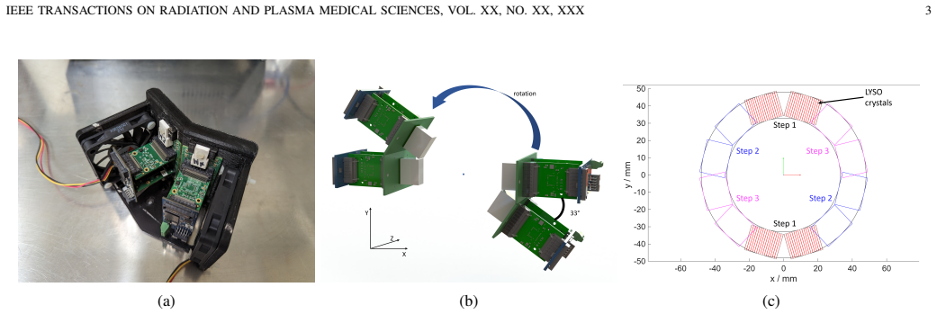
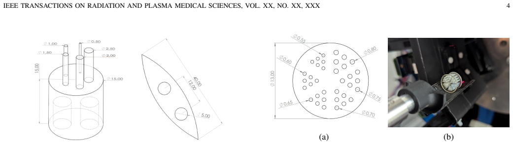
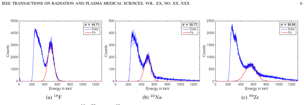
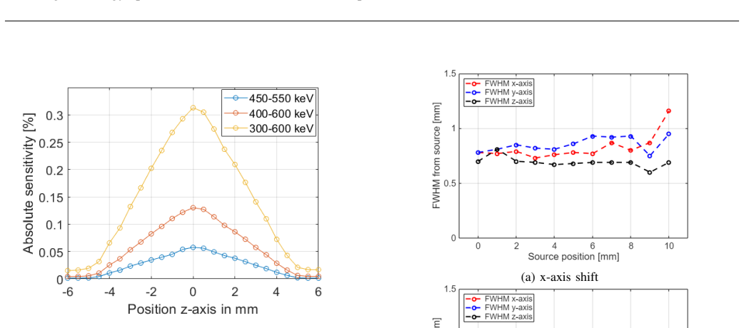
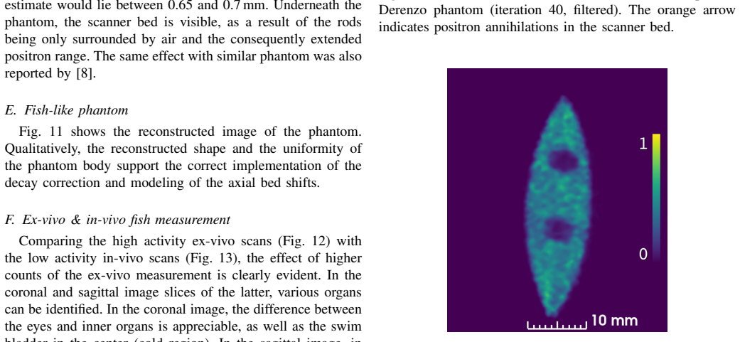
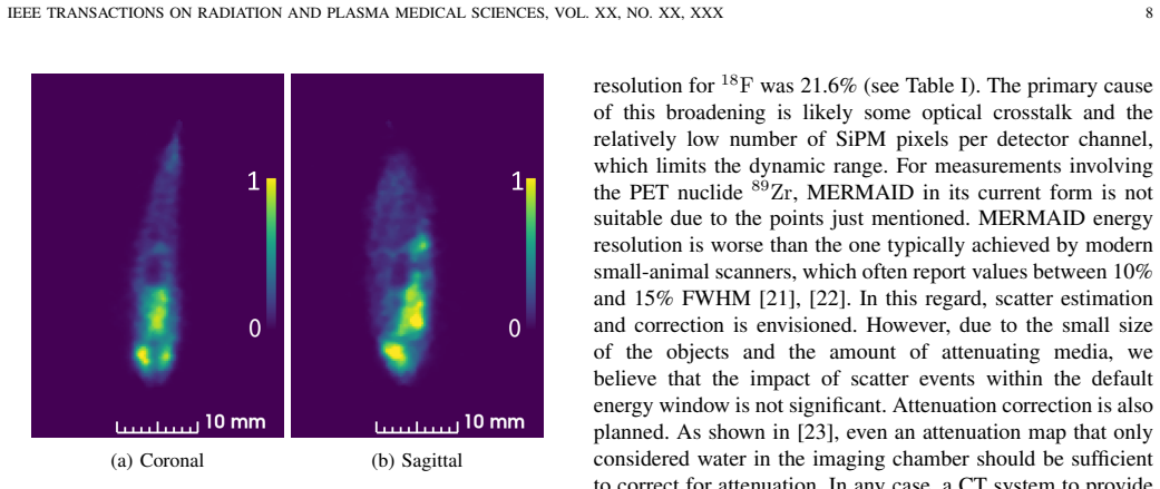
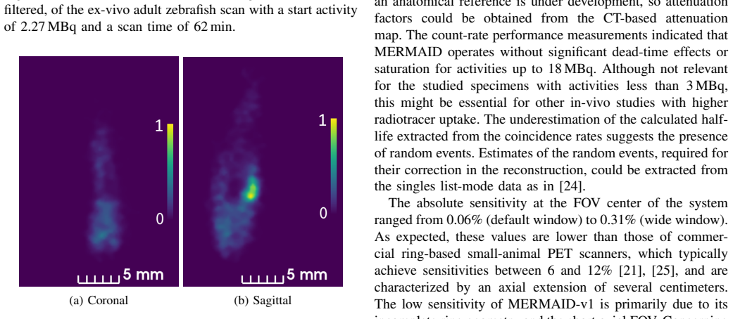
MERMAID-v1 is a prototype PET scanner designed for biomedical research on adult zebrafish. Its two-head configuration was characterized with average energy resolution of 21.6% FWHM at 511 keV, absolute sensitivity between 0.06% and 0.31% depending on energy window, and averaged spatial resolution of 0.77 mm transaxially and 0.66 mm axially in the central FOV. Dedicated reconstruction software models the parallax effect. Phantom images from a downscaled NEMA IQ phantom and 3D-printed Derenzo phantom indicate 0.7-0.8 mm resolution despite absent depth-of-interaction information. The first ex- and in-vivo scans of adult zebrafish successfully detected tracer uptake in organs including the brain
What carries the argument
The two-head detector configuration paired with parallax-aware reconstruction software that produces images from coincidence data acquired in a water-filled chamber.
If this is right
- The scanner supports PET imaging of living anesthetized zebrafish inside a water-filled chamber at low injected activities.
- Detectable uptake in small organs such as brain and eyes is possible with the current configuration.
- Future addition of scatter, attenuation, and efficiency corrections plus expansion to more detector heads will improve quantitative accuracy and image quality.
- The approach opens the door to integrating complementary modalities such as CT on the same platform.
Where Pith is reading between the lines
- The resolution achieved suggests the method could be extended to study functional changes in zebrafish disease models over time.
- Similar dedicated scanners might be adapted for other small aquatic vertebrates used in biomedical research.
- Qualitative uptake maps may suffice for initial screening studies even before full quantitative corrections are available.
Load-bearing premise
That the limited two-head setup without depth-of-interaction, scatter, attenuation, or efficiency corrections can still generate interpretable images of zebrafish organs.
What would settle it
Repeated in-vivo zebrafish scans that show no detectable tracer uptake in brain or eyes, or phantom measurements that yield spatial resolution substantially coarser than 0.8 mm FWHM.
Figures

read the original abstract
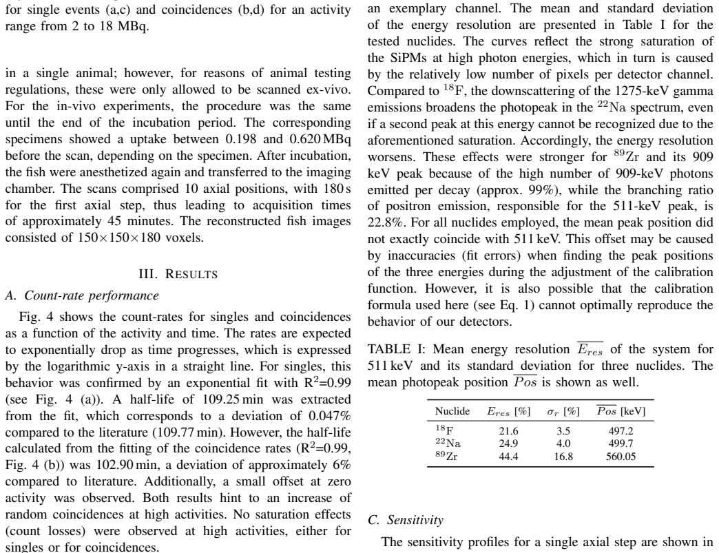
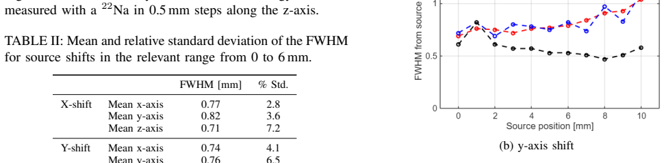
MERMAID-v1 is a prototype PET scanner designed to support biomedical research involving adult zebrafish and similar species. The current experimental setup has been characterized, and scans of various phantoms, as well as adult zebrafish have been conducted. A dedicated reconstruction software was implemented, including accurate modeling of the parallax effect. The average energy resolution was 21.6% (FWHM at 511keV), with no significant dead-time effects observed for activities up to 18MBq. The absolute sensitivity at the center of the field of view (FOV) ranged from 0.06% to 0.31%, depending on the energy window (from 450-550 to 300-600keV), reflecting the limitations of the current two-head configuration. In the central 12mm of the transaxial FOV, the averaged spatial resolution is approximately 0.77mm (FWHM) transaxially and 0.66mm axially, as evaluated using a point source. Image quality was assessed using a downscaled NEMA-inspired IQ phantom and a 3D-printed Derenzo phantom. The reconstructed images suggest a spatial resolution around 0.7mm - 0.8mm, despite the lack of depth-of-interaction information. The first ex- and in-vivo PET scans of adult zebrafish were successfully performed, showing detectable tracer uptake in organs such as the brain and eyes despite low initial activity levels. These results confirm MERMAID-v1 capability to obtain useful results from the acquired data from living, anesthetized fish in a water-filled imaging chamber. While no scatter, attenuation, or efficiency corrections have yet been implemented, this work establishes a working proof-of-concept for dedicated PET imaging of small aquatic vertebrates. Future developments will focus on developing correction techniques, expanding the detector array, and integrating complementary modalities such as CT.
Editorial analysis
A structured set of objections, weighed in public.
Referee Report
Summary. The manuscript reports the design and initial characterization of the MERMAID-v1 two-head PET prototype for adult zebrafish imaging. It provides measured performance metrics (21.6% energy resolution at 511 keV, sensitivity 0.06–0.31% depending on energy window, ~0.77 mm transaxial / 0.66 mm axial FWHM resolution from point source) and phantom-based image quality results suggesting 0.7–0.8 mm resolution. The central claim is that the first ex- and in-vivo zebrafish scans demonstrate detectable tracer uptake in organs such as brain and eyes, establishing a working proof-of-concept for dedicated PET of small aquatic vertebrates despite the absence of DOI, scatter, attenuation, and efficiency corrections.
Significance. If the zebrafish image interpretations are validated, the work would provide the first experimental demonstration of organ-level PET imaging in living adult zebrafish, opening a new niche for biomedical research on aquatic vertebrate models. The direct experimental measurements of resolution and sensitivity constitute a strength; however, the low sensitivity and lack of corrections mean the immediate biological utility remains limited until further development.
major comments (2)
- [Zebrafish scans] Abstract and zebrafish scans section: the claim that the reconstructed images show 'detectable tracer uptake in organs such as the brain and eyes' and 'confirm capability to obtain useful results' is load-bearing for the proof-of-concept but is supported only by qualitative description. With the two-head geometry (sensitivity 0.06–0.31%), no DOI, and no scatter/attenuation/efficiency corrections applied, quantitative metrics (e.g., contrast-to-noise ratio, ROI values, or comparison to tracer-free controls) are required to demonstrate that focal signals are not reconstruction artifacts amplified by the parallax modeling or noise.
- [Image quality assessment] Image quality assessment section: the Derenzo and downscaled NEMA-inspired phantom results are reported to suggest 0.7–0.8 mm resolution, yet no contrast recovery coefficients, recovery coefficients for hot/cold rods, or false-positive rate estimates in water-filled low-activity chambers are provided. These omissions directly affect whether the same reconstruction pipeline can be trusted to distinguish true organ uptake from artifacts in the zebrafish water chamber.
minor comments (2)
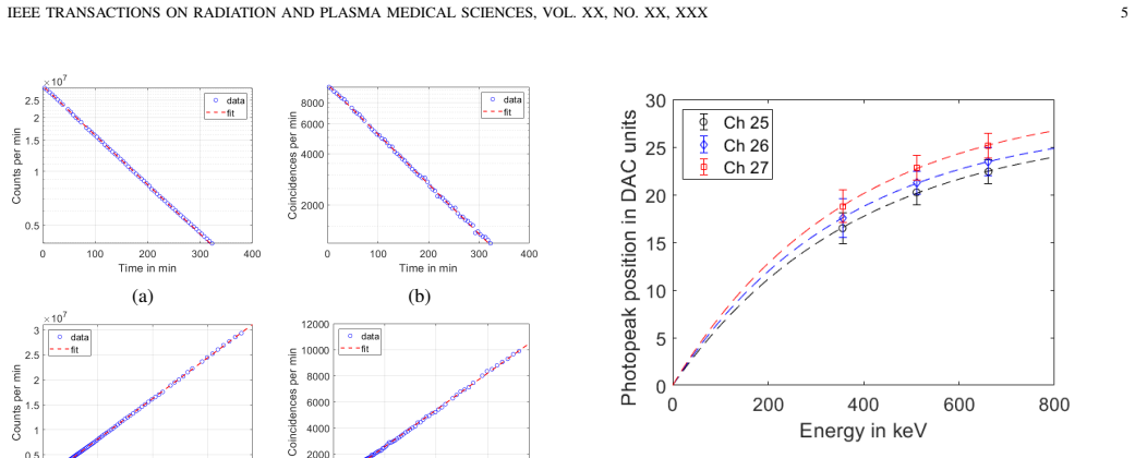
- [Performance characterization] The abstract states 'no significant dead-time effects observed for activities up to 18 MBq' but does not specify the metric or count-rate curve used to reach this conclusion.
- [Sensitivity measurements] The energy window dependence of sensitivity is given as a range but the exact windows and corresponding measured values should be tabulated for reproducibility.
Simulated Author's Rebuttal
We thank the referee for the constructive and detailed feedback on our manuscript. We address each major comment below and indicate the revisions we will make.
read point-by-point responses
-
Referee: [Zebrafish scans] Abstract and zebrafish scans section: the claim that the reconstructed images show 'detectable tracer uptake in organs such as the brain and eyes' and 'confirm capability to obtain useful results' is load-bearing for the proof-of-concept but is supported only by qualitative description. With the two-head geometry (sensitivity 0.06–0.31%), no DOI, and no scatter/attenuation/efficiency corrections applied, quantitative metrics (e.g., contrast-to-noise ratio, ROI values, or comparison to tracer-free controls) are required to demonstrate that focal signals are not reconstruction artifacts amplified by the parallax modeling or noise.
Authors: We agree that quantitative metrics would provide stronger support for the organ-level uptake claims given the prototype limitations. In the revised manuscript we will add ROI values and background estimates from the zebrafish images to compute contrast-to-noise ratios. We will also include any available tracer-free control data for comparison; if such controls were not acquired, we will explicitly note this as a limitation while retaining the proof-of-concept framing. revision: partial
-
Referee: [Image quality assessment] Image quality assessment section: the Derenzo and downscaled NEMA-inspired phantom results are reported to suggest 0.7–0.8 mm resolution, yet no contrast recovery coefficients, recovery coefficients for hot/cold rods, or false-positive rate estimates in water-filled low-activity chambers are provided. These omissions directly affect whether the same reconstruction pipeline can be trusted to distinguish true organ uptake from artifacts in the zebrafish water chamber.
Authors: We acknowledge that reporting contrast recovery coefficients and rod recovery coefficients would strengthen validation of the reconstruction pipeline. We will compute and include these metrics for both phantoms in the revised manuscript, along with any feasible estimates of activity in low-activity chambers, to better demonstrate that the observed zebrafish signals are consistent with the phantom performance. revision: yes
Circularity Check
No circularity: experimental measurements only
full rationale
The paper reports direct empirical measurements of energy resolution (21.6% FWHM), sensitivity (0.06–0.31%), spatial resolution (~0.77 mm transaxial from point source), and image quality from Derenzo/NEMA phantoms plus zebrafish scans. No derivations, predictions, or fitted parameters are presented as independent results; the reconstruction is described as implemented software with parallax modeling but contains no equations that reduce to self-inputs. No self-citation chains or ansatzes are invoked as load-bearing for any claim. The work is self-contained against external benchmarks of scanner characterization.
Axiom & Free-Parameter Ledger
axioms (2)
- standard math Positron annihilation produces two 511 keV photons emitted approximately 180 degrees apart.
- domain assumption The energy resolution, sensitivity, and spatial resolution measurements accurately reflect detector performance without major unaccounted systematic errors from the two-head geometry.
Reference graph
Works this paper leans on
-
[1]
Zebrafish: Development of a Vertebrate Model Organism,
J. R. Meyers, “Zebrafish: Development of a Vertebrate Model Organism,”Current Protocols Essential Laboratory Techniques, vol. 16, no. 1, May 2018. [Online]. Available: http://dx.doi.org/10.1002/cpet.19
-
[2]
Utilization of zebrafish as a model system in medical research,
S. Liu, “Utilization of zebrafish as a model system in medical research,” BIO Integration, vol. 3, no. 4, p. 188–192, 2022
2022
-
[3]
The use of zebrafish (Danio rerio) as biomedical models,
T. Teameet al., “The use of zebrafish (Danio rerio) as biomedical models,”Animal Frontiers, vol. 9, no. 3, p. 68–77, Jun 2019. [Online]. Available: http://dx.doi.org/10.1093/af/vfz020
-
[4]
PET/CT Technology in Adult Zebrafish: A Pilot Study Toward Live Longitudinal Imaging,
C. Tucker, R. Collins, M. A. Denvir, and W. A. McDougald, “PET/CT Technology in Adult Zebrafish: A Pilot Study Toward Live Longitudinal Imaging,”Frontiers in Medicine, vol. 8, Oct. 2021. [Online]. Available: https://doi.org/10.3389/fmed.2021.725548
-
[5]
Developing a Practical Approach to imaging PET/CT of Zebrafish,
E. Snay, M. Dang, F. Fahey, and L. Zon, “Developing a Practical Approach to imaging PET/CT of Zebrafish,”Journal of Nuclear Medicine, vol. 58, no. supplement 1, pp. 1127–1127, 2017. [Online]. Available: https://jnm.snmjournals.org/content/58/supplement 1/1127
2017
-
[6]
Dedicated Chamber for Multimodal In Vivo Imaging of Adult Zebrafish,
S. Seeger, M. Zvolsk ´y, S. Melikov, M. Frerkes, and M. Rafecas, “Dedicated Chamber for Multimodal In Vivo Imaging of Adult Zebrafish,”Zebrafish, vol. 19, no. 2, pp. 67–70, Apr 2022. [Online]. Available: https://doi.org/10.1089/zeb.2021.0066
-
[7]
3d printed radioactive phantoms for Positron Emission Tomography,
S. Seeger, E. Elmoujarkach, N. M ¨oller, C. Schmidt, and M. Rafecas, “3d printed radioactive phantoms for Positron Emission Tomography,” Transactions on Additive Manufacturing Meets Medicine, vol. 4, no. S1, 2022. [Online]. Available: https://www.journals.infinite-science. de/index.php/ammm/article/view/640
2022
-
[8]
Dedicated 3D Printed Radioactive Phantoms With 18F-FDG for Ultra-High Resolution PET,
E. Elmoujarkachet al., “Dedicated 3D Printed Radioactive Phantoms With 18F-FDG for Ultra-High Resolution PET,”IEEE Transactions on Radiation and Plasma Medical Sciences, 2024
2024
-
[9]
MER- MAID - A PET Prototype for Small Aquatic Animal Imaging,
M. Zvolsky, S. Seeger, M. Schaar, C. Schmidt, and M. Rafecas, “MER- MAID - A PET Prototype for Small Aquatic Animal Imaging,” in2019 IEEE Nuclear Science Symposium and Medical Imaging Conference (NSS/MIC). IEEE, Oct 2019
2019
-
[10]
Characterisation of the Upgraded MERMAID Prototype, a PET/CT Device for Small Aquatic Animals,
S. Seeger, H. V o, A. Bolke, and M. Rafecas, “Characterisation of the Upgraded MERMAID Prototype, a PET/CT Device for Small Aquatic Animals,” in2022 IEEE Nuclear Science Symposium and Medical Imaging Conference (NSS/MIC). IEEE, Nov. 2022, p. 1–2
2022
-
[11]
First Images from MERMAID, a Small Aquatic Animal PET Scanner Prototype,
S. Seeger, H. V o, C. Florack, J. Werner, C. Schmidt, and M. Rafecas, “First Images from MERMAID, a Small Aquatic Animal PET Scanner Prototype,” in2023 IEEE Nuclear Science Symposium, Medical Imaging Conference and International Symposium on Room-Temperature Semi- conductor Detectors (NSS MIC RTSD), 2023, pp. 1–1. IEEE TRANSACTIONS ON RADIATION AND PLASMA...
2023
-
[12]
First in-Vivo Tests of Zebrafish with Mermaid, a PET Prototype for Small Aquatic Animals,
S. Seegeret al., “First in-Vivo Tests of Zebrafish with Mermaid, a PET Prototype for Small Aquatic Animals,” in2025 IEEE Nuclear Science Symposium (NSS), Medical Imaging Conference (MIC) and Room Temperature Semiconductor Detector Conference (RTSD), 2025, pp. 1–1
2025
-
[13]
NEMA Standards Publication NU 4 – 2008 Performance Measurements of Small Animal Positron Emission Tomographs,
“NEMA Standards Publication NU 4 – 2008 Performance Measurements of Small Animal Positron Emission Tomographs,” National Electrical Manufacturers Association, Rosslyn, V A, Tech. Rep., 2008
2008
-
[14]
Experimental results with TOFPET2 ASIC for time-of-flight applications,
R. Bugalhoet al., “Experimental results with TOFPET2 ASIC for time-of-flight applications,”Nuclear Instruments and Methods in Physics Research Section A: Accelerators, Spectrometers, Detectors and Associated Equipment, vol. 912, pp. 195–198, 2018, new Developments In Photodetection 2017. [Online]. Available: https: //www.sciencedirect.com/science/article/...
2018
-
[15]
Application of Silicon Photomultipliers to Positron Emission Tomography,
E. Roncali and S. R. Cherry, “Application of Silicon Photomultipliers to Positron Emission Tomography,”Annals of Biomedical Engineering, vol. 39, no. 4, pp. 1358–1377, Apr 2011. [Online]. Available: https://doi.org/10.1007/s10439-011-0266-9
-
[16]
Simulated one- pass list-mode: an approach to on-the-fly system matrix calculation,
J. E. Gillam, P. Solevi, J. F. Oliver, and M. Rafecas, “Simulated one- pass list-mode: an approach to on-the-fly system matrix calculation,” Physics in Medicine and Biology, vol. 58, no. 7, p. 2377, Mar. 2013. [Online]. Available: https://dx.doi.org/10.1088/0031-9155/58/7/2377
-
[17]
Dedicated prostate DOI-TOF-PET based on the ProVision detection concept,
H. P. V o, T. Williams, K. Doroud, C. Williams, and M. Rafecas, “Dedicated prostate DOI-TOF-PET based on the ProVision detection concept,”Physics in Medicine & Biology, vol. 70, no. 18, p. 185001, 2025
2025
-
[18]
Development and Characterization of 3D Printed Radioactive Phantoms for High Resolution PET,
E. Elmoujarkach, S. Seeger, N. M ¨oller, C. Schmidt, and M. Rafecas, “Development and Characterization of 3D Printed Radioactive Phantoms for High Resolution PET,” in2022 IEEE Nuclear Science Symposium and Medical Imaging Conference (NSS/MIC), 2022, pp. 1–2
2022
-
[19]
Submillimeter-Resolution PET for High- Sensitivity Mouse Brain Imaging,
H. G. Kanget al., “Submillimeter-Resolution PET for High- Sensitivity Mouse Brain Imaging,”Journal of Nuclear Medicine, vol. 64, no. 6, pp. 978–985, 2023. [Online]. Available: https: //jnm.snmjournals.org/content/64/6/978
2023
-
[20]
Comments on the NEMA NU 4-2008 Standard on Performance Measurement of Small Animal Positron Emission Tomographs,
P. Hallen, D. Schug, and V . Schulz, “Comments on the NEMA NU 4-2008 Standard on Performance Measurement of Small Animal Positron Emission Tomographs,”EJNMMI Physics, vol. 7, no. 1, Feb
2008
-
[21]
Available: http://dx.doi.org/10.1186/s40658-020-0279-2
[Online]. Available: http://dx.doi.org/10.1186/s40658-020-0279-2
-
[22]
S. Krishnamoorthy, E. Blankemeyer, P. Mollet, S. Surti, R. Van Holen, and J. S. Karp, “Performance evaluation of the molecubesβ-cube—a high spatial resolution and high sensitivity small animal pet scanner utilizing monolithic lyso scintillation detectors,”Physics in Medicine and Biology, vol. 63, no. 15, p. 155013, Jul. 2018. [Online]. Available: http://d...
-
[23]
Depth of interaction resolution measurements for a high resolution PET detector using position sensitive avalanche photodiodes,
Y . Yanget al., “Depth of interaction resolution measurements for a high resolution PET detector using position sensitive avalanche photodiodes,” Physics in Medicine and Biology, vol. 51, no. 9, p. 2131–2142, Apr
-
[24]
Available: http://dx.doi.org/10.1088/0031-9155/51/9/001
[Online]. Available: http://dx.doi.org/10.1088/0031-9155/51/9/001
-
[25]
Development of a digital zebrafish phantom and its application to dedicated small-fish PET,
M. Zvolsky, M. Schaar, S. Seeger, S. Rakers, and M. Rafecas, “Development of a digital zebrafish phantom and its application to dedicated small-fish PET,”Physics in Medicine and Biology, vol. 67, no. 17, Aug 2022. [Online]. Available: http://dx.doi.org/10. 1088/1361-6560/ac71ee
2022
-
[26]
Modelling random coincidences in positron emission tomography by using singles and prompts: a comparison study,
J. F. Oliver and M. Rafecas, “Modelling random coincidences in positron emission tomography by using singles and prompts: a comparison study,”PloS one, vol. 11, no. 9, p. e0162096, 2016
2016
-
[27]
Spatial Resolution and Sensitivity of the Inveon Small-Animal PET Scanner,
E. P. Visseret al., “Spatial Resolution and Sensitivity of the Inveon Small-Animal PET Scanner,”Journal of Nuclear Medicine, vol. 50, no. 1, p. 139–147, Jan. 2009. [Online]. Available: http://dx.doi.org/10.2967/jnumed.108.055152
-
[28]
H. Wanget al., “Performance evaluation of the ACTIVE 7 MAX benchtop preclinical PET scanner in accordance with the NEMA NU 4-2008 standard,”EJNMMI Physics, vol. 13, no. 1, Dec. 2025. [Online]. Available: http://dx.doi.org/10.1186/s40658-025-00813-9
-
[29]
Inclusion of inter-crystal scattering in pet: Analytical models and dedicated reconstruction,
J. Roser, H. P. V o, R. Kantorek, S. Seeger, and M. Rafecas, “Inclusion of inter-crystal scattering in pet: Analytical models and dedicated reconstruction,”IEEE Transactions on Radiation and Plasma Medical Sciences, pp. 1–1, 2026
2026
-
[30]
Performance evaluation of the nanoScan® P123S total-body PET,
D. R ´etiet al., “Performance evaluation of the nanoScan® P123S total-body PET,”EJNMMI Physics, vol. 13, no. 1, Dec. 2025. [Online]. Available: http://dx.doi.org/10.1186/s40658-025-00817-5
discussion (0)
Sign in with ORCID, Apple, or X to comment. Anyone can read and Pith papers without signing in.