REVIEW 3 major objections 6 minor 26 references
Advanced Plaque Modeling for Atherosclerosis Detection Using Molecular Communication
T0 review · 3 major / 6 minor · reviewed 2026-08-12 · deepseek-v4-flash
Pith's one-line read This paper argues that plaque-induced changes in blood flow and nanoparticle arrival times form a measurable molecular-communication signature for detecting atherosclerosis non-invasively.
desk verdict Solid analytical extension of the authors' prior MC plaque work, with correct new CIR derivations; the quantitative claims for severe stenosis are weakened by an acknowledged but likely violated laminar-flow assumption. read the letter →
The pith
A machine-rendered reading of the paper's core claim, the machinery that carries it, and where it could break.
The reading
What carries the argument
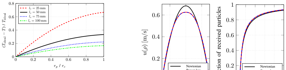
The central object is the channel impulse response $h(t)$, the fraction of released nanoparticles that have crossed the receiver plane at time $t$. The argument is carried by three interlocking pieces: the flow-dominated-regime CIR derivation adapted from pipe-flow molecular communication, with closed-form expressions for Newtonian, power-law, and Herschel-Bulkley fluids; the Venturi-effect model that treats the plaque as a piecewise radial constriction and predicts traversal-time reduction $T=\int_0^{l_c} dx/u(x)$; and the OpenFOAM MPPIC particle simulation driven by a realistic carotid pulsatile flow profile with particle release at peak systole, early diastole, and late diastole. The simulation supplies what the analytical models cannot: the lasting asymmetric flow profile downstream of the plaque.
What would settle it
Run a turbulence-resolving or experimentally validated simulation of 50-nm SPIONs through a 75% stenosis at peak systole with the same carotid waveform and compare the first-arrival traversal speedup and full CIR against the unobstructed case; if the roughly 43% speedup or the clean separation of CIRs by plaque radius is not reproduced, the proposed detection signature does not survive realistic high-systole flow.
Extended reading notes
Core claim
In the paper's own terms, the discovery is that plaque-induced stenosis leaves a detectable fingerprint in the molecular communication channel impulse response. Using a CFD simulation with a non-Newtonian Casson blood model, a human-carotid pulsatile inlet waveform, and superparamagnetic iron-oxide nanoparticles as information carriers, the authors show that the fraction of received particles over time separates by plaque size: first-arrival traversal speed increases by 5.5% for $r_p=0.25\,r_c$, 33% for $r_p=0.5\,r_c$, and 43% for $r_p=0.75\,r_c$, close to the 15%, 30%, and 43% predicted by their Venturi model. They further derive closed-form channel impulse responses for power-law and Herschel-Bulkley non-Newtonian models, show that the two models nearly coincide in this parameter regime, and demonstrate that the simulated flow profile stays skewed and asymmetric downstream of the plaque, in line with in-vivo observations. The intended upshot is that flow-profile shape and CIR changes are usable indicators for early, non-invasive plaque detection.
Load-bearing premise
The result rests on the assumption that flow remains laminar even at peak systole through a 75% stenosis; turbulence would change the arrival statistics and could erase the speedup signature.
Editorial extensions
If this is right
- A plaque that narrows the vessel to 75% of its normal radius can speed up nanoparticle traversal by about 43%, making traversal time a direct physical indicator of stenosis severity.
- Particle release should be synchronized with peak systole for the fastest transport through an obstructed vessel, while early-diastole release produces the slowest arrivals.
- The largest separation between plaque and no-plaque channel impulse responses occurs at peak systole, making that phase the most useful detection window for future IoBNT sensors.
- Analytical non-Newtonian CIRs and the Venturi traversal-time estimate give quick approximations that match simulation well for larger plaques, even though full CFD is needed to capture the skewed downstream flow profile.
Reading between the lines
- An implicit design consequence is that an IoBNT detector must either control or measure the release phase: at small plaque sizes, the shift between systole and diastole release can be as large as the plaque-induced shift, so phase-unaware measurements could confound plaque size estimation.
- The Venturi traversal-time formula could be inverted: given a measured speedup and known channel length, a device could estimate the effective plaque radius without imaging.
- A natural extension would test wall compliance via fluid-solid interaction; the published dataset already includes a first FSI trial, so the signature's robustness to moving walls is checkable.
- If turbulence modeling is added, the same simulation pipeline could quantify whether the peak-systole detection window remains the best or shifts to another phase.
Editorial analysis
A structured set of objections, weighed in public.
Referee Report
Summary. The paper models a 50 mm carotid-artery segment as a molecular communication channel with a circular transmitter, a passive cross-sectional receiver, and an idealized annular plaque. It derives closed-form channel impulse responses (CIRs) for Newtonian, power-law, and Herschel-Bulkley fluids in the flow-dominated regime, adds a Venturi-based travel-time reduction model, and compares the analytical velocity profiles with OpenFOAM simulations. It then imposes a human carotid pulsatile inlet velocity profile and simulates particle release at peak systole, early diastole, and late diastole for four plaque sizes. The main reported results are that the plaque accelerates traversal by 5.5%, 33%, and 43% for rp = 0.25, 0.5, and 0.75 times the vessel radius, that peak-systole release gives the earliest arrivals, and that these CIR changes could serve as a non-invasive plaque-detection metric.
Significance. If the results hold, the paper offers a physically plausible and novel sensing signature for molecular-communication-based plaque detection: plaque-induced changes in flow profile and CIR, including Venturi-style speedups and sensitivity to the cardiac phase at release. The analytical CIR derivations in Section III-A are transparent and reduce correctly to the Newtonian limit, the Venturi model gives a simple falsifiable prediction, and the authors explicitly compare analytical and simulated profiles rather than hiding the mismatch. The release of the simulation dataset [7] is a genuine reproducibility strength. However, the most severe stenosis case is very likely outside the laminar regime on which all simulations rest, and the simulation results are presented without uncertainty quantification, so the quantitative claims currently outrun the evidence.
major comments (3)
- [§II, §IV-B, Fig. 6] The load-bearing assumption that flow remains laminar in all scenarios is almost certainly violated for the most severe stenosis. With rc = 3.0 mm and rp = 0.75×rc, the lumen radius at the throat is 0.75 mm; conservation of mass gives an average throat velocity of about 5.5 m/s at the stated mean inlet speed of 34.2 cm/s, and the pulsatile peak in Fig. 2 is several times higher. With blood density 1050 kg/m³ and viscosity near 4×10⁻³ Pa·s, the throat Reynolds number is of order 10³–10⁴, far above the transitional threshold for pipe flow. A laminar solver cannot capture flow separation, recirculation, or turbulent mixing in this regime, all of which directly alter particle residence times and CIR shape. Since the 43% speedup and the peak-systole fastest-arrival claim for rp = 0.75×rc rest on these simulations, the central quantitative claim for severe stenosis is not supported. The authors should either restrict the conclusions to stenosis levels where the flow remains laminar, or add a transitional/turbulence-resolving validation for the worst case.
- [§IV-B, Fig. 6] The speedups of 5.5%, 33%, and 43% are quoted to two significant figures, yet Fig. 6 shows single curves with no error bars, confidence intervals, or repetition count, and the text does not define the "traversal speed" metric (first-arrival time, median arrival, or shift in the full CIR). Without this information, a reader cannot distinguish a genuine plaque-induced channel change from sampling noise or from the choice of metric. Please state whether the runs are deterministic, report the number of repetitions and the dispersion across them, and define exactly how the traversal speed was computed.
- [§III-B, Eq. (15)] The piecewise radius function in Eq. (15) is inconsistent with the definition of rp in §II. rp is defined as the radial expansion of the plaque, so the lumen radius in Region 3 should be rc − rp, not rp; this is also the only interpretation consistent with the factor r_c²/r(x)² = 1/0.75² = 1.78 quoted for rp = 0.25×rc in Fig. 5. As written, Eq. (15) makes Eq. (16) and the derived 15%/30%/43% Venturi predictions irreproducible. Please correct the notation (for example, introduce r_lumen = rc − rp) and specify the integration domain for each region.
minor comments (6)
- [§III-A3, Eq. (14)] The condition "for t ≥ d/u0t" is dimensionally wrong; it should be t ≥ d/u0. In addition, the symbol a in the formula is never defined and should be set to a = rc.
- [§IV-B] The text says that particles "arrive later at the TX" for release time tED; the receiver is at the RX, not the TX, so this should read "arrive later at the RX."
- [Fig. 5 caption] The caption says the power-law and Herschel-Bulkley models have been fitted to the simulated profile at x = 0.01 m, but the fitted parameter values and the fitting procedure are not reported; please provide them.
- [§III-B, Eq. (15)] The domain intervals for the four regions in the piecewise radius function are not stated; please define them explicitly in terms of lc, lp,outer, and lp,inner.
- [§II and §III] The simulations are said to use the Casson model while the analytical comparison uses power-law and Herschel-Bulkley models; please clarify whether the same rheological parameters were used in the pulsatile simulations and how the Casson parameters relate to the analytical model parameters.
- [Fig. 6] The text mentions an "irregularity in the upper third of the PS plot curve" but does not explain its physical origin; please annotate or discuss this feature.
Circularity Check
No circular derivation found: the analytical CIRs and Venturi predictions are derived from constitutive flow equations and geometry, not from the simulation outputs, and the self-citations are used as method/baseline references rather than as the target result.
full rationale
The paper's central claim—that plaque size and release phase within the cardiac cycle alter the MC channel—is supported by OpenFOAM simulations plus independently derived analytical models. The Newtonian, power-law, and Herschel-Bulkley CIRs (Eqs. (3), (8), (14)) are obtained by integrating the assumed velocity profiles with a delta-distributed initial condition, following the derivation in Wicke et al. [20]; no simulation output is inserted into these equations. The Venturi traversal-time model in Eq. (16) is a geometric conservation-of-mass calculation with no fitted parameters. The comparison in Fig. 5 fits only local non-Newtonian parameters for visualization and does not feed the central detection claim. The speedup percentages in Section IV-B come from the particle simulations and are compared with, not fitted to, the Venturi prediction; agreement for larger plaques is an independent check rather than a reduction to the model's own inputs. Self-citations [6], [7], and [25] supply the prior baseline scenario, the released dataset, and the solver method; none is invoked as an external theorem that forces the result. The laminar-flow assumption is a substantive modeling limitation affecting realism, but it is not a circularity. Thus no load-bearing step reduces to its own inputs by construction; the minor self-citations are not load-bearing.
Assumptions & free parameters
free parameters (1)
- Non-Newtonian model fit parameters for Fig. 5 =
unstated
assumptions (5)
- domain assumption Laminar flow in all simulated scenarios, including pulsatile inlet through stenoses.
- domain assumption Flow-dominated transport regime with negligible diffusion (alpha = D lc / (uavg rc^2) = 1.8e-8 << 1).
- domain assumption Human carotid pulsatile flow profile and release times (tPS, tED, tLD) from literature are representative.
- domain assumption MPPIC/OpenFOAM solver accurately models particle transport in blood with Casson rheology.
- standard math Closed-form Hagen-Poiseuille solutions for power-law and Herschel-Bulkley fluids in a straight pipe apply to the idealized channel.
Cite this review
Pith. "Pith review of Advanced Plaque Modeling for Atherosclerosis Detection Using Molecular Communication." pith.science (2026). https://pith.science/paper/IKMSVJJ5
@misc{pith2026241113241,
  author       = {Pith},
  title        = {Pith review of: Advanced Plaque Modeling for Atherosclerosis Detection Using Molecular Communication},
  year         = {2026},
  howpublished = {\url{https://pith.science/paper/IKMSVJJ5}},
  note         = {Machine review of arXiv:2411.13241}
}
read the original abstract
As one of the most prevalent diseases worldwide, plaque formation in human arteries, known as atherosclerosis, is the focus of many research efforts. Previously, molecular communication (MC) models have been proposed to capture and analyze the natural processes inside the human body and to support the development of diagnosis and treatment methods. In the future, synthetic MC networks are envisioned to span the human body as part of the Internet of Bio-Nano Things (IoBNT), turning blood vessels into physical communication channels. By observing and characterizing changes in these channels, MC networks could play an active role in detecting diseases like atherosclerosis. In this paper, building on previous preliminary work for simulating an MC scenario in a plaque-obstructed blood vessel, we evaluate different analytical models for non-Newtonian flow and derive associated channel impulse responses (CIRs). Additionally, we add the crucial factor of flow pulsatility to our simulation model and investigate the effect of the systole-diastole cycle on the received particles across the plaque channel. We observe a significant influence of the plaque on the channel in terms of the flow profile and CIR across different emission times in the cycle. These metrics could act as crucial indicators for early non-invasive plaque detection in advanced future MC methods.
Figures


Figures from the paper (1 more)

Reference graph
Works this paper leans on
-
[6]
A Molecular Communication Perspective on Detecting Arterial Plaque Formation,
P. Hofmann, S. Schmidt, A. Wietfeld, P. Zhou, J. Fuchtmann, F. H. P. Fitzek, and W. Kellerer, “A Molecular Communication Perspective on Detecting Arterial Plaque Formation,” IEEE Trans. Mol. Biol. Multi- Scale Commun., vol. 10, p. 458–463, Sept. 2024
work page 2024
-
[7]
Dataset for Advanced Plaque Modeling for Atherosclerosis Detection using Molecular Communication
P. Hofmann, A. Wietfeld, J. Fuchtmann, P. Zhou, R. Zheng, J. Cabrera, F. H. Fitzek, and W. Kellerer, “Dataset for Advanced Plaque Modeling for Atherosclerosis Detection using Molecular Communication.” https://ieee-dataport.org/documents/dataset-advanced-plaque-modeling- atherosclerosis-detection-using-molecular-communication, Nov. 2024
work page 2024
-
[25]
OpenFOAM Simulation of Microfluidic Molecular Communications: Method and Experimental Validation,
P. Hofmann, P. Zhou, C. Lee, M. Reisslein, F. H. Fitzek, and C.-B. Chae, “OpenFOAM Simulation of Microfluidic Molecular Communications: Method and Experimental Validation,” IEEE Access, Aug. 2024. Early access
work page 2024
-
[1]
The Internet of Bio-Nano Things,
I. F. Akyildiz, M. Pierobon, S. Balasubramaniam, and Y . Koucheryavy, “The Internet of Bio-Nano Things,” IEEE Commun. Mag., vol. 53, Mar. 2015
work page 2015
-
[2]
V . R. Preedy and R. R. Watson, eds., Handbook of Disease Burdens and Quality of Life Measures . New York, NY: Springer, 2010
work page 2010
-
[3]
P. Song, Z. Fang, H. Wang, Y . Cai, K. Rahimi, Y . Zhu, F. G. R. Fowkes, F. J. Fowkes, and I. Rudan, “Global and regional prevalence, burden, and risk factors for carotid atherosclerosis: A systematic review, meta- analysis, and modelling study,” The Lancet Global Health , vol. 8, May 2020
work page 2020
-
[4]
Diagnosis and management of carotid atherosclerosis,
A. Thapar, I. H. Jenkins, A. Mehta, and A. H. Davies, “Diagnosis and management of carotid atherosclerosis,” BMJ, vol. 346, Mar. 2013
work page 2013
-
[5]
Modeling CD40-Based Molecular Communications in Blood Vessels,
L. Felicetti, M. Femminella, G. Reali, P. Gresele, M. Malvestiti, and J. N. Daigle, “Modeling CD40-Based Molecular Communications in Blood Vessels,” IEEE Trans. on NanoBioscience , vol. 13, pp. 230–243, Sept. 2014
work page 2014
Show all 26 references
-
[8]
Carotid Diameter and Blood Flow Velocities in Cerebral Circulation in Hypertensive Patients,
L. A. Ferrara, M. Mancini, R. Iannuzzi, T. Marotta, I. Gaeta, F. Pasanisi, A. Postiglione, and L. Guida, “Carotid Diameter and Blood Flow Velocities in Cerebral Circulation in Hypertensive Patients,” Stroke, vol. 26, Mar. 1995
1995
-
[9]
The measurement of blood density and its meaning,
T. Kenner, “The measurement of blood density and its meaning,” Basic Research in Cardiology, vol. 84, Mar. 1989
1989
-
[10]
OpenFOAM v6 User Guide - 7.3 Trans- port/Rheology Models,
Chris Greenshields, “OpenFOAM v6 User Guide - 7.3 Trans- port/Rheology Models,” July 2018
2018
-
[11]
Numerical Studies of Blood Flow in Healthy, Stenosed, and Stented Carotid Arteries,
M. Gay and L. T. Zhang, “Numerical Studies of Blood Flow in Healthy, Stenosed, and Stented Carotid Arteries,” International Journal for Numerical Methods in Fluids , vol. 61, p. 453–472, Nov. 2008
2008
-
[12]
Characterization of Common Carotid Artery Blood-Flow Waveforms in Normal Human Subjects,
D. W. Holdsworth, C. J. D. Norley, R. Frayne, D. A. Steinman, and B. K. Rutt, “Characterization of Common Carotid Artery Blood-Flow Waveforms in Normal Human Subjects,” Physiological Measurement , vol. 20, p. 219–240, Jan. 1999
1999
-
[13]
Mathematical Modeling of Micro-/Nanoparticles Transport in Blood Vessels: A Review,
S.-S. Ashrafmansouri, M. Nasr Esfahany, and S.-S. Ashrafmansouri, “Mathematical Modeling of Micro-/Nanoparticles Transport in Blood Vessels: A Review,” Korean Journal of Chemical Engineering , vol. 41, pp. 1273–1305, May 2024
2024
-
[14]
A review on non-Newtonian fluid models for multi-layered blood rheology in constricted arteries,
S. A. Wajihah and D. S. Sankar, “A review on non-Newtonian fluid models for multi-layered blood rheology in constricted arteries,” Archive of Appl. Mech. , vol. 93, pp. 1771–1796, May 2023
2023
-
[15]
Predicting laminar–turbulent transition in Poiseuille pipe flow for non-Newtonian fluids,
B. G ¨uzel, I. Frigaard, and D. M. Martinez, “Predicting laminar–turbulent transition in Poiseuille pipe flow for non-Newtonian fluids,” Chemical Eng. Science, vol. 64, pp. 254–264, Jan. 2009
2009
-
[16]
White, Fluid Mechanics
F. White, Fluid Mechanics. WCB/McGraw-Hill, 1999
1999
-
[17]
Channel modeling for diffusive molecular communication—A tutorial review,
V . Jamali, A. Ahmadzadeh, W. Wicke, A. Noel, and R. Schober, “Channel modeling for diffusive molecular communication—A tutorial review,” Proc. IEEE, vol. 107, June 2019
2019
-
[18]
Efficient drug-delivery using magnetic nanoparticles–biodistribution and therapeutic effects in tumour bearing rabbits,
R. Tietze, S. Lyer, S. D ¨urr, T. Struffert, T. Engelhorn, M. Schwarz, E. Eckert, T. G ¨oen, S. Vasylyev, W. Peukert, F. Wiekhorst, L. Trahms, A. D ¨orfler, and C. Alexiou, “Efficient drug-delivery using magnetic nanoparticles–biodistribution and therapeutic effects in tumour ...
2013
-
[19]
Channel parameter studies of a molecular communication testbed with biocompatible infor- mation carriers: Methods and data,
M. Bartunik, J. Teller, G. Fischer, and J. Kirchner, “Channel parameter studies of a molecular communication testbed with biocompatible infor- mation carriers: Methods and data,” IEEE Trans. Mol. Biol. Multi-Scale Commun., vol. 9, Oct. 2023
2023
-
[20]
Ex- perimental System for Molecular Communication in Pipe Flow With Magnetic Nanoparticles,
W. Wicke, H. Unterweger, J. Kirchner, L. Brand, A. Ahmadzadeh, D. Ahmed, V . Jamali, C. Alexiou, G. Fischer, and R. Schober, “Ex- perimental System for Molecular Communication in Pipe Flow With Magnetic Nanoparticles,” IEEE Trans. Mol. Biol. Multi-Scale Commun., vol. 8, pp. 56...
2022
-
[21]
Schaschke, A Dictionary of Chemical Engineering
C. Schaschke, A Dictionary of Chemical Engineering . OUP Oxford, 2014
2014
-
[22]
The Rheology of Blood Flow in a Branched Arterial System,
S. S. Shibeshi and W. E. Collins, “The Rheology of Blood Flow in a Branched Arterial System,” Appl. Rheo., vol. 15, no. 6, p. 398, 2005
2005
-
[23]
Rheology of Human Blood, near and at Zero Flow,
E. Merrill, E. Gilliland, G. Cokelet, H. Shin, A. Britten, and R. Wells, “Rheology of Human Blood, near and at Zero Flow,” Biophysical Journal, vol. 3, pp. 199–213, May 1963
1963
-
[24]
In vivo three-dimensional blood velocity profile shapes in the human common, internal, and external carotid arteries,
A. V . Kamenskiy, Y . A. Dzenis, J. N. Mactaggart, A. S. Desyatova, and I. I. Pipinos, “In vivo three-dimensional blood velocity profile shapes in the human common, internal, and external carotid arteries,” J. Vasc. Surg., vol. 54, pp. 1011–1020, Oct. 2011
2011
-
[26]
An open-source finite volume toolbox for solid mechanics and fluid-solid interaction simulations,
P. Cardiff, A. Karac, P. D. Jaeger, H. Jasak, J. Nagy, A. Ivankovi´c, and ˇZ. Tukovi´c, “An open-source finite volume toolbox for solid mechanics and fluid-solid interaction simulations,” arXiv: Numerical Analysis , pp. 1– 45, Sept. 2018
2018
Reviewed August 12, 2026 · model on record in the stance chip above.
Discussion (0). Continue with ORCID to comment.