A Signal Extraction Approach for Remote Heart Rate Variability Assessment Using Proxy Measure in a Driving Simulator
Pith reviewed 2026-06-30 12:15 UTC · model grok-4.3
The pith
Remote photoplethysmography extracts pulse rate and heart rate variability from facial videos in a driving simulator with errors low enough to match electrocardiography statistics.
A machine-rendered reading of the paper's core claim, the machinery that carries it, and where it could break.
Core claim
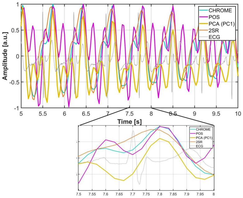
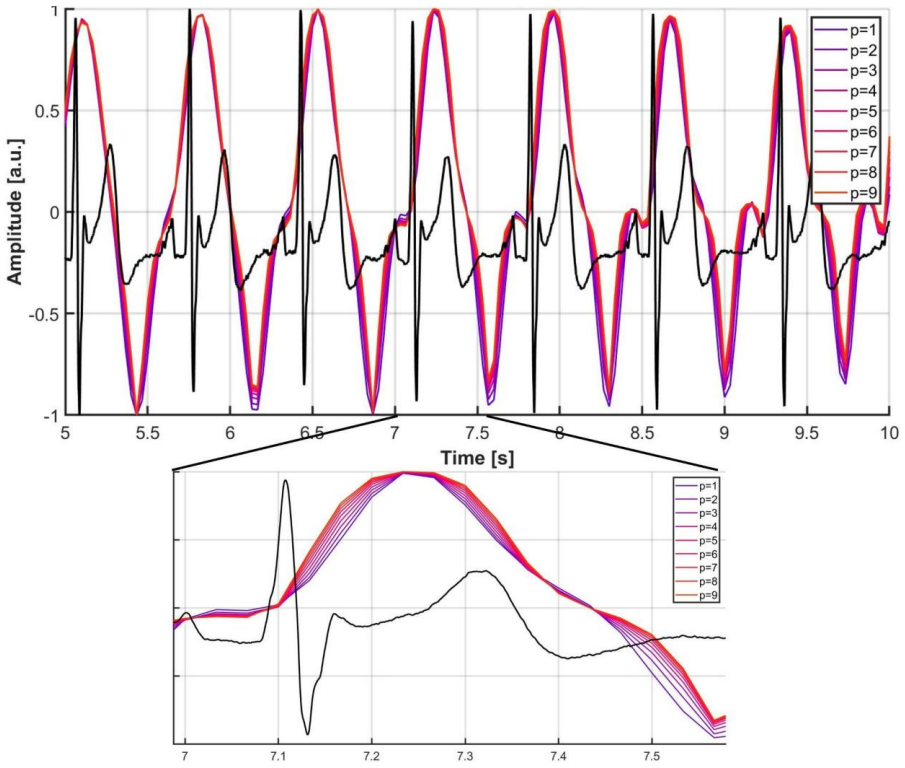
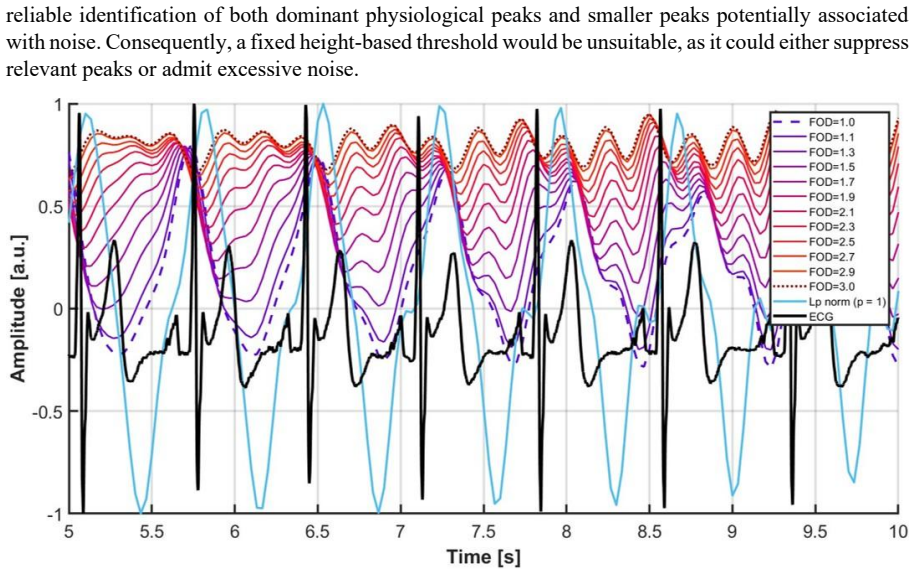
In recordings from 29 participants, the 2SR algorithm combined with fractional-order derivative enhancement and 20 superpixel regions yields a mean absolute error of 1.92 bpm for pulse rate against ECG, while the best configurations reach 0.061 s MAE for SDNN and 0.081 s for RMSSD; inter-beat interval detection reaches an F1 score of 0.93, and all rPPG-derived parameters reproduce the statistical structure of the reference ECG across both baseline and driving conditions, with CHROM recommended for HRV and Lp norm at p around 6-7 as an effective enhancer.
What carries the argument
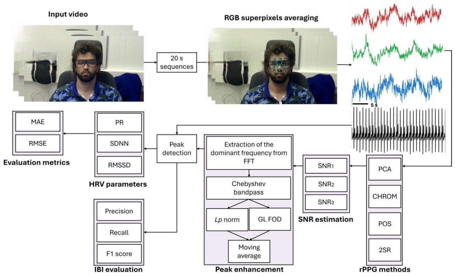
Application of spatial subspace rotation or chrominance methods to 10 or 20 superpixel facial regions, sharpened by Lp-norm or fractional-order derivative peak enhancement, followed by signal-to-noise ratio filtering of 20-second segments to reduce motion artifacts.
If this is right
- 2SR with FOD and 20 superpixels gives the lowest pulse rate error, while CHROM with Lp norm performs best for HRV parameters.
- Optimal settings cluster around p=6-7 for the Lp norm and fractional orders of 1.0-1.4.
- All tested configurations reproduce the reference ECG statistical structure for both baseline and driving conditions.
- FOD requires caution because it can introduce slow changes in the rPPG waveform shape.
Where Pith is reading between the lines
- The same pipeline could support fatigue or stress monitoring in real vehicles if motion patterns remain comparable to the simulator.
- Extending the SNR quality filter to variable-length segments might reduce data loss in longer recordings.
- Combining the recommended CHROM configuration with other facial or vehicle sensors could test robustness beyond isolated video input.
Load-bearing premise
The signal-to-noise ratio assessment on 20-second segments removes motion artifacts without discarding valid physiological signals or creating selection bias in the retained data.
What would settle it
A new dataset of driving simulator recordings where rPPG-derived SDNN and RMSSD values show statistically different distributions from simultaneous ECG values after identical processing would falsify the reproduction of statistical structure.
Figures
read the original abstract
This study evaluates remote Photopletismography (rPPG) algorithms, Spatial Subspace Rotation (2SR), Chrominance-based method (CHROM), Plane-Orthogonal-to-Skin (POS), and Principal Component Analysis (PCA), applied to selected superpixel-based facial regions (with target counts of 10 and 20 regions) for monitoring in a driving simulator. Two novel peak enhancement approaches, based on the Lp norm and Fractional-Order Derivative (FOD), are introduced to enable robust Heart Rate Variability (HRV) estimation. A signal-to-noise ratio-based quality assessment of 20 s segments serves as a data cleaning mechanism to mitigate motion artifacts inherent to dynamic recording conditions. In a sample of 29 participants recorded during baseline and driving simulation conditions, Pulse Rate (PR) is calculated with clinically acceptable accuracy across configurations (validated against simultaneous Electrocardiography (ECG) recordings), achieving the lowest Mean Absolute Error (MAE) of 1.92 bpm (sd = 1.72) using 2SR with FOD and 20 superpixel regions. The best-case MAE reached 0.061 s for Standard Deviation of Normal-to-Normal intervals (SDNN) and 0.081 s for Root Mean Square of Successive Differences (RMSSD), with inter-beat interval detection yielding an F1 score of 0.93. Optimal parameters clustered around p = 6-7 for Lp norm and fractional orders of 1.0-1.4. All rPPG-derived parameters reproduced the statistical structure of the reference ECG across conditions and configurations. Caution is advised when using FOD due to slow changes in the rPPG waveform. Overall, 2SR is recommended for PR, while CHROM for HRV estimation, using Lp norm with 20 superpixels, providing clear methodological guidance for rPPG monitoring in driving simulators
Editorial analysis
A structured set of objections, weighed in public.
Referee Report
Summary. The paper evaluates rPPG algorithms (2SR, CHROM, POS, PCA) on superpixel facial regions (10 or 20) for remote PR and HRV monitoring in a driving simulator. It introduces Lp-norm and fractional-order derivative (FOD) peak enhancement, applies SNR-based quality assessment to 20 s segments for motion-artifact mitigation, and validates against simultaneous ECG in 29 participants. It reports lowest MAE of 1.92 bpm (sd=1.72) for PR using 2SR+FOD+20 regions, best-case MAEs of 0.061 s (SDNN) and 0.081 s (RMSSD), F1=0.93 for IBI detection, reproduction of ECG statistical structure, optimal parameters p=6-7 and fractional order 1.0-1.4, and recommends 2SR for PR and CHROM for HRV with Lp norm and 20 superpixels.
Significance. If the central claims hold, the work supplies concrete, directly validated performance numbers for rPPG under realistic motion, together with explicit parameter ranges and configuration recommendations that could guide practical deployment in simulators or vehicles. The simultaneous ECG reference and reporting of both PR and HRV metrics (SDNN, RMSSD) plus F1 scores constitute a clear strength.
major comments (2)
- [Abstract / Methods (SNR quality assessment)] Abstract (data-cleaning paragraph) and Methods (SNR quality assessment): the manuscript states that an SNR-based gate on 20 s segments is used to remove motion artifacts but supplies no quantitative check that the retained segments preserve the original distribution of HRV statistics. No Kolmogorov-Smirnov test, paired comparison of SDNN/RMSSD before vs. after filtering, or per-condition discard fraction is reported. Because the headline claim that “all rPPG-derived parameters reproduced the statistical structure of the reference ECG” is made on the filtered data only, this omission is load-bearing for the validity of the reproduction result.
- [Results (parameter optimization)] Results (parameter-optimization paragraph): the reported optimal ranges (p = 6-7, fractional order 1.0-1.4) are presented without stating whether they were obtained by participant-wise cross-validation, nested validation, or post-hoc selection on the pooled data. If the latter, the quoted “best-case” MAEs may be optimistically biased and the recommendation of specific configurations requires re-evaluation.
minor comments (2)
- [Abstract] Abstract: the phrase “clinically acceptable accuracy” is used without citing the specific clinical tolerance thresholds (e.g., <5 bpm or <10 % error) against which the 1.92 bpm MAE is judged.
- [Methods] Notation: Lp norm order p and fractional order are introduced without an explicit equation or reference to the precise definition employed (e.g., the fractional derivative operator).
Simulated Author's Rebuttal
We thank the referee for the constructive comments, which highlight important aspects of methodological transparency. We address each major comment below and will revise the manuscript to incorporate the requested clarifications and additional analyses.
read point-by-point responses
-
Referee: [Abstract / Methods (SNR quality assessment)] Abstract (data-cleaning paragraph) and Methods (SNR quality assessment): the manuscript states that an SNR-based gate on 20 s segments is used to remove motion artifacts but supplies no quantitative check that the retained segments preserve the original distribution of HRV statistics. No Kolmogorov-Smirnov test, paired comparison of SDNN/RMSSD before vs. after filtering, or per-condition discard fraction is reported. Because the headline claim that “all rPPG-derived parameters reproduced the statistical structure of the reference ECG” is made on the filtered data only, this omission is load-bearing for the validity of the reproduction result.
Authors: We agree that quantitative verification of the SNR filtering's impact on HRV distributions is necessary to support the reproduction claim. In the revised manuscript, we will add a Kolmogorov-Smirnov test comparing SDNN and RMSSD distributions before versus after the SNR gate, paired comparisons where appropriate, and the per-condition fraction of discarded segments. These additions will be reported in the Methods and Results sections. revision: yes
-
Referee: [Results (parameter optimization)] Results (parameter-optimization paragraph): the reported optimal ranges (p = 6-7, fractional order 1.0-1.4) are presented without stating whether they were obtained by participant-wise cross-validation, nested validation, or post-hoc selection on the pooled data. If the latter, the quoted “best-case” MAEs may be optimistically biased and the recommendation of specific configurations requires re-evaluation.
Authors: The reported ranges were identified via grid search on the pooled dataset. We acknowledge this can introduce optimistic bias in the best-case MAEs. In the revision, we will explicitly describe the optimization procedure, note the potential for bias as a limitation, and add a participant-wise cross-validation analysis to re-evaluate and confirm the robustness of the recommended configurations (p=6-7, fractional order 1.0-1.4). revision: yes
Circularity Check
No significant circularity; results are direct empirical comparisons to ECG reference
full rationale
The paper's central claims consist of MAE, SDNN/RMSSD errors, and F1 scores obtained by comparing rPPG-derived quantities (from 2SR/CHROM/POS/PCA with Lp-norm or FOD peak enhancement) against simultaneous ECG recordings on the same 20 s segments. These metrics are computed externally and do not reduce, by any equation in the paper, to quantities defined by the paper's own fitted parameters or preprocessing choices. The SNR-based segment filter is a data-cleaning step whose effect on the retained distribution is not itself used to define the reported accuracies. No self-citation chains, uniqueness theorems, or ansatzes imported from prior author work appear in the provided text; the derivation chain is standard signal processing followed by independent validation against an external reference.
Axiom & Free-Parameter Ledger
free parameters (2)
- Lp norm order p =
6-7
- fractional order =
1.0-1.4
axioms (2)
- domain assumption Facial video color changes can serve as a proxy for cardiac pulse despite head motion in a driving simulator
- domain assumption SNR-based segment selection removes motion artifacts without systematically biasing HRV statistics
Reference graph
Works this paper leans on
-
[1]
Cao, R., Azimi, I., Sarhaddi, F., Niela -Vilen, H., Axelin, A., Liljeberg, P., & Rahmani, A. M. (2022). Accuracy assessment of oura ring nocturnal heart rate and heart rate variability in comparison with electrocardiography in time and frequency domains: co mprehensive analysis. Journal of Medical Internet Research , 24(1), e27487. https://doi.org/10.2196/27487
-
[2]
Dudarev, V.; Barral, O.; Zhang, C.; Davis, G.; Enns, J.T. On the reliability of wearable technology: A tutorial on measuring heart rate and heart rate variability in the wild. Sensors 2023, 23, 5863. https://doi.org/10.3390/s23135863
-
[3]
Barka, R. E., & Politis, I. (2024). Driving into the future: A scoping review of smartwatch use for real -time driver monitoring. Transportation Research Interdisciplinary Perspectives , 25, 101098. https://doi.org/10.1016/j.trip.2024.101098
-
[4]
Prucnal, M. A., Polak, A. G., & Kazienko, P. (2025). Improving the quality of pulse rate variability derived from wearable devices using adaptive, spectrum and nonlinear filtering. Biomedical Signal Processing and Control, 102, 107336. https://doi.org/10.1016/j.bspc.2024.107336
-
[5]
S., Hegarty-Craver, M., Boyce, M
Gaur, P., Temple, D. S., Hegarty-Craver, M., Boyce, M. D., Holt, J. R., Wenger, M. F., ... & Dausch, D. E. (2024). Continuous monitoring of heart rate variability in free -living conditions using wearable sensors: Exploratory observational study. JMIR Formative Research, 8, e53977. https://doi.org/10.2196/53977
-
[6]
Brookhuis, K. A., & De Waard, D. (2010). Monitoring drivers’ mental workload in driving simulators using physiological measures. Accident Analysis & Prevention, 42(3), 898-903. https://doi.org/10.1016/j.aap.2009.06.001
-
[7]
Xiao, H., Liu, T., Sun, Y., Li, Y., Zhao, S., & Avolio, A. (2024). Remote photoplethysmography for heart rate measurement: A review. Biomedical Signal Processing and Control , 88, 105608. https://doi.org/10.1016/j.bspc.2023.105608
-
[8]
Sakib, S., Hasan, Z., & Roy, N. (2025). A state‐of‐the‐art survey of remote photoplethysmography for contactless health parameters sensing. Wiley Interdisciplinary Reviews: Data Mining and Knowledge Discovery, 15(3), e70039. https://doi.org/10.1002/widm.70039
-
[9]
Seo, H., Kim, S., & Lee, E. C. (2025). Estimation of respiratory signals from remote photoplethysmography of RGB facial videos. Electronics, 14(11), 2152. https://doi.org/10.3390/electronics14112152
-
[10]
Measuring pulse rate with a webcam
Lewandowska, M.; Nowak, J. Measuring pulse rate with a webcam. J. Med. Imaging Health Inform . 2012, 2, 87 –
2012
-
[11]
https://doi.org/10.1166/jmihi.2012.1064
-
[12]
Validation of heart rate extraction using video imaging on a built -in camera system of a smartphone
Kwon, S.; Kim, H.; Park, K.S. Validation of heart rate extraction using video imaging on a built -in camera system of a smartphone. In Proceedings of the 2012 Annual International Conference of the IEEE Engineering in Medicine and Biology Society, San Diego, CA, USA, 28 August –1 September 2012; IEEE: Piscataway, NJ, USA, 2012; pp. 2174–2177. https://doi....
-
[13]
Non -contact, automated cardiac pulse measurements using video imaging and blind source separation
Poh, M.Z.; McDuff, D.J.; Picard, R.W. Non -contact, automated cardiac pulse measurements using video imaging and blind source separation. Opt. Express 2010, 18, 10762–10774. https://doi.org/10.1364/OE.18.010762
-
[14]
Ernst, H.; Scherpf, M.; Malberg, H.; Schmidt, M. Optimal color channel combination across skin tones for remote heart rate measurement in camera -based photoplethysmography. Biomedical Signal Processing and Control 2021, 68, 102644. https://doi.org/10.1016/j.bspc.2021.102644. 23
-
[15]
Renner, P.; Gleichauf, J.; Winkelmann, S. Non -Contact In-Car Monitoring of Heart Rate: Evaluating the Eulerian Video Magnification Algorithm in a Driving Simulator Study. In Proceedings of the Mensch und Computer 2024 , Karlsruhe, Germany, 1–4 September 2024; pp. 651–654. https://doi.org/10.1145/3670653.3677493
-
[16]
Dasari, A., Prakash, S. K. A., Jeni, L. A., & Tucker, C. S. (2021). Evaluation of biases in remote photoplethysmography methods. NPJ digital medicine, 4(1), 91. https://doi.org/10.1038/s41746-021-00462-z
-
[17]
Wang, W., Den Brinker, A. C., Stuijk, S., & De Haan, G. (2016). Algorithmic principles of remote PPG. IEEE Transactions on Biomedical Engineering, 64(7), 1479-1491. https://doi.org/10.1109/TBME.2016.2609282
-
[18]
Elgendi, M., Martinelli, I., & Menon, C. (2024). Optimal signal quality index for remote photoplethysmogram sensing. npj Biosensing, 1, 5. https://doi.org/10.1038/s44328-024-00002-1
-
[19]
De Haan, G., & Jeanne, V. (2013). Robust pulse rate from chrominance -based rPPG. IEEE Transactions on Biomedical Engineering, 60(10), 2878-2886. https://doi.org/10.1109/TBME.2013.2266196
-
[20]
Wang, W., Stuijk, S., & De Haan, G. (2015). A novel algorithm for remote photoplethysmography: Spatial subspace rotation. IEEE Transactions on Biomedical Engineering , 63(9), 1974 -1984. https://doi.org/10.1109/TBME.2015.2508602
-
[21]
Eulerian video magnification for revealing subtle changes in the world
Wu, H.Y.; Rubinstein, M.; Shih, E.; Guttag, J.; Durand, F.; Freeman, W. Eulerian video magnification for revealing subtle changes in the world. ACM Trans. Graph. (TOG) 2012, 31, 1–8. https://doi.org/10.1145/2185520.2185561
-
[22]
Pulse rate assessment: Eulerian video magnification vs
Miljković, N.; Trifunovi´c, D. Pulse rate assessment: Eulerian video magnification vs. electrocardiography recordings. In Proceedings of the 12th Symposium on Neural Network Applications in Electrical Engineering (NEUREL), Belgrade, Serbia, 25 –27 November 2014; IEEE: Piscataway, NJ, USA, 2012; pp. 17 –20. https://doi.org/10.1109/NEUREL.2014.7011447
-
[23]
D., Stojmenova Pečečnik, K., Sodnik, J., & Miljković, N
Nešković, Đ. D., Stojmenova Pečečnik, K., Sodnik, J., & Miljković, N. (2025). Contactless pulse rate assessment: Results and insights for application in driving simulators. Applied Sciences , 15(17), 9512. https://doi.org/10.3390/app15179512
-
[24]
Nagar, S., Hasegawa-Johnson, M., Beiser, D. G., & Ahuja, N. (2024). R2I-rPPG: A robust region of interest selection method for remote photoplethysmography to extract heart rate. arXiv preprint arXiv:2410.15851 . https://doi.org/10.48550/arXiv.2410.15851
-
[25]
Pai, A., Veeraraghavan, A., & Sabharwal, A. (2021). HRVCam: robust camera -based measurement of heart rate variability. Journal of biomedical optics, 26(2), 022707-022707. https://doi.org/10.1117/1.JBO.26.2.022707
-
[26]
McDuff, D. (2023). Camera measurement of physiological vital signs. ACM Computing Surveys , 55(9), 1 -40. https://doi.org/10.1145/3558518
-
[27]
Bobbia, S., Luguern, D., Benezeth, Y., Nakamura, K., Gomez, R., & Dubois, J. (2018). Real -time temporal superpixels for unsupervised remote photoplethysmography. In Proceedings of the IEEE Conference on Computer Vision and Pattern Recognition Workshops, pp. 1341-1348
2018
-
[28]
Medarević, J., Miljković, N., Stojmenova Pečečnik, K., & Sodnik, J. (2025). Distress detection in VR environment using Empatica E4 wristband and Bittium Faros 360. Frontiers in Physiology , 16, 1480018. https://doi.org/10.3389/fphys.2025.1480018
-
[29]
Yu, S. G., Kim, S. E., Kim, N. H., Suh, K. H., & Lee, E. C. (2021). Pulse rate variability analysis using remote photoplethysmography signals. Sensors, 21(18), 6241. https://doi.org/10.3390/s21186241
-
[30]
Shaffer F., Ginsberg J. P. (2017). An overview of heart rate variability metrics and norms. Frontiers in Public Health 5, 258. https://doi.org/10.3389/fpubh.2017.00258
-
[31]
Finžgar, M., & Podržaj, P. (2020). Feasibility of assessing ultra -short-term pulse rate variability from video recordings. PeerJ, 8, e8342. https://doi.org/10.7717/peerj.8342
-
[32]
Pander, T., Przybyła, T., & Czabański, R. (2013). An application of the Lp -norm in robust weighted averaging of biomedical signals. Journal of Medical Informatics & Technologies, 22
2013
-
[33]
Tanasković, I., & Miljković, N. (2023). A new algorithm for fetal heart rate detection: Fractional order calculus approach. Medical Engineering & Physics, 118, 104007. https://doi.org/10.1016/j.medengphy.2023.104007
-
[34]
Miljković, N., Popović, N., Djordjević, O., Konstantinović, L., & Šekara, T. B. (2017). ECG artifact cancellation in surface EMG signals by fractional order calculus application. Computer Methods and Programs in Biomedicine , 140, 259-264. https://doi.org/10.1016/j.cmpb.2016.12.017
-
[35]
Design of head -up display interfaces for automated vehicles
Stojmenova Pečečnik, K., Tomažič, S., Sodnik, J., (2023). Design of head -up display interfaces for automated vehicles. International Journal of Human -Computer Studies. 177, 103060. https://doi.org/10.1016/j.ijhcs.2023.103060
-
[36]
Nešković D. Đ, Miljković N , (2026). Supplementary Material for Manuscript A Signal Extraction Approach for Remote Heart Rate Variability Assessment Using Proxy Measure in a Driving Simulator , Zenodo https://doi.org/10.5281/zenodo.20354210
-
[37]
Li, P., Benezeth, Y., Nakamura, K., Gomez, R., & Yang, F. (2019). Model-based region of interest segmentation for remote photoplethysmography. In the 14th International Conference on Computer Vision Theory and Applications , pp. 383-388, SCITEPRESS-Science and Technology Publications. https://dx.doi.org/10.5220/0007389803830388
-
[38]
Benezeth, Y., Bobbia, S., Nakamura, K., Gomez, R., & Dubois, J. (2019). Probabilistic signal quality metric for reduced complexity unsupervised remote photoplethysmography. In 2019 13th International Symposium on Medical Information and Communication Technology (ISMICT) , pp. 1 -5, IEEE. https://doi.org/10.1109/ISMICT.2019.8744004. 24
-
[39]
Huang, Y., Huang, D., Huang, J., Lu, H., He, M., & Wang, W. (2023, July). Camera wavelength selection for multi- wavelength pulse transit time based blood pressure monitoring. In 2023 45th Annual International Conference of the IEEE Engineering in Medicine & Biology Society (EMBC) (pp. 1 -5). IEEE. https://doi.org/10.1109/EMBC40787.2023.10340068
-
[40]
Saritas, T., Greber, R., Venema, B., Puelles, V. G., Ernst, S., Blazek, V., ... & Schlieper, G. (2019). Non -invasive evaluation of coronary heart disease in patients with chronic kidney disease using photoplethysmography. Clinical kidney journal, 12(4), 538-545. https://doi.org/10.1093/ckj/sfy135
-
[41]
Xi, L., Wu, X., Chen, W., Wang, J., & Zhao, C. (2022). Weighted combination and singular spectrum analysis based remote photoplethysmography pulse extraction in low -light environments. Medical engineering & physics , 105, 103822. https://doi.org/10.1016/j.medengphy.2022.103822
-
[42]
Lin, Y. C., & Lin, Y. H. (2017). A study of color illumination effect on the SNR of rPPG signals. In 2017 39th Annual International Conference of the IEEE Engineering in Medicine and Biology Society (EMBC), pp. 4301-4304, IEEE. https://doi.org/10.1109/EMBC.2017.8037807
-
[43]
Pan, J., & Tompkins, W. J. (1985). A real -time QRS detection algorithm. IEEE Tansactions on Biomedical Engineering, (3), 230-236. https://doi.org/10.1109/TBME.1985.325532
-
[44]
Guler, S., Golparvar, A., Ozturk, O., Dogan, H., & Yapici, M. K. (2023). Optimal digital filter selection for remote photoplethysmography (rPPG) signal conditioning. Biomedical Physics & Engineering Express , 9(2), 027001. https://iopscience.iop.org/article/10.1088/2057-1976/acaf8a/meta
-
[45]
Liao, G., Lu, H., Shan, C., & Wang, W. (2023). Plethysmographic waveform features and hemodynamic features for camera-based blood pressure estimation. IEEE Transactions on Instrumentation and Measurement , 73, 1-14. https://doi.org/10.1109/TIM.2023.3338714
-
[46]
B., Ben-David, K., Morris, M., Wittels, H
Kantrowitz, A. B., Ben-David, K., Morris, M., Wittels, H. L., Wishon, M. J., McDonald, S. M., ... & Wittels, S. H. (2025). Pulse rate variability is not the same as heart rate variability: Findings from a large, diverse clinical population study. Frontiers in Physiology, 16, 1630032. https://doi.org/10.3389/fphys.2025.1630032
-
[47]
Schäfer, A., & Vagedes, J. (2013). How accurate is pulse rate variability as an estimate of heart rate variability?: A review on studies comparing photoplethysmographic technology with an electrocardiogram. International Journal of Cardiology, 166(1), 15-29. https://doi.org/10.1016/j.ijcard.2012.03.119
-
[48]
Allen, J., & Murray, A. (2002). Age -related changes in peripheral pulse timing characteristics at the ears, fingers and toes. Journal of Human Hypertension, 16(10), 711-717. https://doi.org/10.1038/sj.jhh.1001478
-
[49]
Allen, J., & Murray, A. (2000). Variability of photoplethysmography peripheral pulse measurements at the ears, thumbs and toes. IEE Proceedings -Science, Measurement and Technology , 147(6), 403 -407. https://doi.org/10.1049/ip-smt:20000846
-
[50]
Hsieh, F., & Chen, T. L. (2025). A novel R -peak detection algorithm. IEEE Access , 13, 210351 -210359. https://doi.org/10.1109/ACCESS.2025.3643153
-
[51]
AAMI, A., & EC57, A. A. M. I. (2008). Testing and reporting performance results of cardiac rhythm and st segment measurement algorithms. American National Standards Institute, Arlington, VA, USA, 43
2008
-
[52]
A., Sidorov, I
Kamshilin, A. A., Sidorov, I. S., Babayan, L., Volynsky, M. A., Giniatullin, R., & Mamontov, O. V. (2016). Accurate measurement of the pulse wave delay with imaging photoplethysmography. Biomedical optics express, 7(12), 5138-
2016
-
[53]
https://doi.org/10.1364/BOE.7.005138
-
[54]
Fariha, M. A. Z., Ikeura, R., Hayakawa, S., & Tsutsumi, S. (2020, June). Analysis of Pan -Tompkins algorithm performance with noisy ECG signals. In Journal of Physics: Conference Series (Vol. 1532, No. 1, p. 012022). IOP Publishing. https://iopscience.iop.org/article/10.1088/1742-6596/1532/1/012022/meta
- [55]
-
[56]
Robust confidence intervals for effect sizes: A comparative study of Cohen’s d and Cliff’s delta under non -normality and heterogeneous variances
Hess, M.R.; Kromrey, J.D. Robust confidence intervals for effect sizes: A comparative study of Cohen’s d and Cliff’s delta under non -normality and heterogeneous variances. In Proceedings of the Annual Meeting of the American Educational Research Association, San Diego, CA, USA, 12–16 April 2004; Volume 1
2004
-
[57]
Talukdar, D., De Deus, L. F., & Sehgal, N. (2023). The Evaluation of Remote Monitoring Technology Across Participants With Different Skin Tones. Cureus, 15(9). https://doi.org/10.7759/cureus.45075
-
[58]
Wang, K., Wei, Y., Tang, J., Wang, Y., Li, Z., Tong, M., ... & Zhao, Z. (2024, December). Camera -based hrv prediction for remote learning environments. In 2024 IEEE Smart World Congress (SWC) (pp. 1165-1173). IEEE. https://doi.org/10.1109/SWC62898.2024.00185
-
[59]
Huang, R. Y., & Dung, L. R. (2016). Measurement of heart rate variability using off -the-shelf smart phones. Biomedical Engineering Online, 15(1), 11. https://doi.org/10.1186/s12938-016-0127-8
-
[60]
Odinaev, I., Wong, K. L., Chin, J. W., Goyal, R., Chan, T. T., & So, R. H. (2023). Robust heart rate variability measurement from facial videos. Bioengineering, 10(7), 851. https://doi.org/10.3390/bioengineering10070851
-
[61]
Unifying frame rate and temporal dilations for improved remote pulse detection
Speth, J.; Vance, N.; Flynn, P.; Bowyer, K.; Czajka, A. Unifying frame rate and temporal dilations for improved remote pulse detection. Comput. Vis. Image Underst . 2021, 210, 103246. https://doi.org/10.1016/j.cviu.2021.103246
-
[62]
Unsupervised skin tissue segmentation for remote photoplethysmography
Bobbia, S.; Macwan, R.; Benezeth, Y.; Mansouri, A.; Dubois, J. Unsupervised skin tissue segmentation for remote photoplethysmography. Pattern Recognition Letter 2019, 124, 82–90. https://doi.org/10.1016/j.patrec.2017.10.017. 25
-
[63]
van Putten, L. D., Ahmed, A., & Wegerif, S. (2025). Remote photoplethysmography for contactless pulse rate monitoring: algorithm development and accuracy assessment. Physiological Measurement , 46(11), 115004. https://iopscience.iop.org/article/10.1088/1361-6579/ae1804/meta
-
[64]
Sun, Z., Junttila, J., Tulppo, M., Seppänen, T., & Li, X. (2022). Non -contact atrial fibrillation detection from face videos by learning systolic peaks. IEEE Journal of Biomedical and Health Informatics , 26(9), 4587 -4598. https://doi.org/10.1109/JBHI.2022.3193117
-
[65]
H., Landesberg, A., & Behar, J
Kotzen, K., Charlton, P. H., Landesberg, A., & Behar, J. A. (2021, September). Benchmarking photoplethysmography peak detection algorithms using the electrocardiogram signal as a reference. In 2021 Computing in Cardiology (CinC) (Vol. 48, pp. 1-4). IEEE. https://doi.org/10.23919/CinC53138.2021.9662889
-
[66]
Boccignone, G., Conte, D., Cuculo, V., d’Amelio, A., Grossi, G., & Lanzarotti, R. (2020). An open framework for remote-PPG methods and their assessment. Ieee Access , 8, 216083 -216103. https://doi.org/10.1109/ACCESS.2020.3040936
-
[67]
Spiegelberg, J., & Rusz, J. (2017). Can we use PCA to detect small signals in noisy data?. Ultramicroscopy, 172, 40-46. https://doi.org/10.1016/j.ultramic.2016.10.008
-
[68]
Caroppo, A., Manni, A., Rescio, G., Siciliano, P., & Leone, A. (2024). Vital signs estimation in elderly using camera- based photoplethysmography. Multimedia Tools and Applications , 83(24), 65363 -65386. https://doi.org/10.1007/s11042-023-18053-3
-
[69]
Comparison of Apple watch vs KardiaMobile: A tale of two devices
Lee, C.; Lee, C.; Fernando, C.; Chow, C.M. Comparison of Apple watch vs KardiaMobile: A tale of two devices. CJC Open 2022, 4, 939–945. https://doi.org/10.1016/j.cjco.2022.07.011
-
[70]
J., Chahal, T., Stinchcombe, A., Mullen, N., Weaver, B., & Bédard, M
Johnson, M. J., Chahal, T., Stinchcombe, A., Mullen, N., Weaver, B., & Bédard, M. (2011). Physiological responses to simulated and on -road driving. International journal of Psychophysiology , 81(3), 203 -208. https://doi.org/10.1016/j.ijpsycho.2011.06.012
-
[71]
Bondarenko, M., Menon, C., & Elgendi , M. (2025). The role of face regions in remote photoplethysmography for contactless heart rate monitoring. npj Digital Medicine, 8(1), 479. https://doi.org/10.1038/s41746-025-01814-9
-
[72]
Kiddle, A., Barham, H., Wegerif, S., & Petronzio, C. (2023). Dynamic region of interest selection in remote photoplethysmography: Proof -of-concept study. JMIR Formative Research , 7, e44575. https://formative.jmir.org/2023/1/e44575
2023
-
[73]
Y., Lee, K., & Sohn, C
Kim, D. Y., Lee, K., & Sohn, C. B. (2021). Assessment of roi selection for facial video-based rPPG. Sensors, 21(23),
2021
-
[74]
https://doi.org/10.3390/s21237923
-
[75]
Premkumar, S., & Hemanth, D. J. (2022, August). Intelligent remote photoplethysmography-based methods for heart rate estimation from face videos: A survey. Informatics, 9(3), 57. MDPI. https://doi.org/10.3390/informatics9030057
-
[76]
Soares, S., Ferreira, S., & Couto, A. (2020). Driving simulator experiments to study drowsiness: A systematic review. Traffic Injury Prevention, 21(1), 29-37. https://doi.org/10.1080/15389588.2019.1706088
-
[77]
Lohani, M., Payne, B. R., & Strayer, D. L. (2019). A review of psychophysiological measures to assess cognitive states in real-world driving. Frontiers in Human Neuroscience, 13, 57. https://doi.org/10.3389/fnhum.2019.00057
-
[78]
Rasche, S., Trumpp, A., Waldow, T., Gaetjen, F., Plötze, K., Wedekind, D., ... & Zaunseder, S. (2016). Camera - based photoplethysmography in critical care patients. Clinical Hemorheology and Microcirculation , 64(1), 77-90. https://doi.org/10.3233/CH-162048
-
[79]
P., Kaminer, K., Nussinovitch, M., Segev, S., Volovitz, B., & Nussinovitch, N
Nussinovitch, U., Elishkevitz, K. P., Kaminer, K., Nussinovitch, M., Segev, S., Volovitz, B., & Nussinovitch, N. (2011). The efficiency of 10‐second resting heart rate for the evaluation of short‐term heart rate variability indices. Pacing and Clinical Electrophysiology, 34(11), 1498-1502. https://doi.org/10.1111/j.1540-8159.2011.03178.x
-
[80]
Developed with the Special Contribution of the European Heart Rhythm Association (EHRA), Endorsed by the European Association for Cardio -Thoracic Surgery (EACTS), Authors/Task Force Members, Camm, A. J., Kirchhof, P., Lip, G. Y., ... & Zupan, I. (2010). Guidelines for the management of atrial fibrillation: the Task Force for the Management of Atrial Fibr...
discussion (0)
Sign in with ORCID, Apple, or X to comment. Anyone can read and Pith papers without signing in.