REVIEW 2 major objections 5 minor 85 references
A Multiphase Model of Growth Factor-Regulated Atherosclerotic Cap Formation
T0 review · 2 major / 5 minor · reviewed 2026-08-14 · deepseek-v4-flash
Pith's one-line read A multiphase model of atherosclerotic cap formation claims that a stable collagenous fibrous cap can be built by a small population of smooth muscle cells, with TGF-β the decisive regulatory factor.
desk verdict Solid model paper with a genuinely new analytical result; the quantitative validation is partly calibrated to the comparison data, but the qualitative TGF-beta story is independently supported and worth referee time. read the letter →
The pith
A machine-rendered reading of the paper's core claim, the machinery that carries it, and where it could break.
The reading
What carries the argument
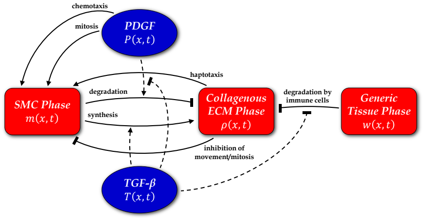
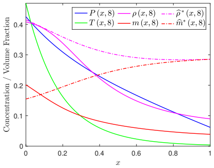
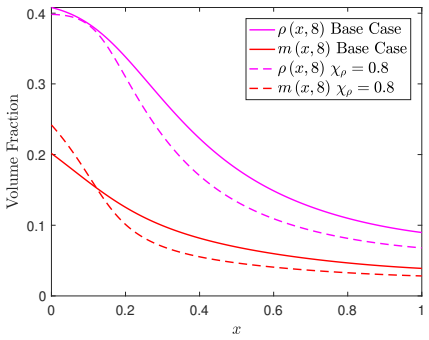
The load-bearing object is the reduced three-phase model, equations (39)–(42): volume fractions $m$ (SMC), $\rho$ (collagenous ECM), and $w=1-m-\rho$ (generic tissue), with quasi-steady reaction–diffusion equations for PDGF $P$ and TGF-$\beta$ $T$, and non-standard boundary conditions in which SMC influx at the medial edge is proportional to the PDGF gradient times a fixed medial SMC fraction. At a fixed location with fixed local growth-factor concentrations $P^*$, $T^*$, the ECM equation reduces to a quadratic in $\rho^*$ whose admissible root is the negative branch; differentiating that root yields the closed-form optimal SMC fraction $\hat{m}^*$ and maximum ECM fraction $\hat{\rho}^*$ in terms of $\mu$ and $\lambda$. The fact that $\rho^*(m^*)$ is flat near its maximum is what lets a small SMC population produce near-maximal cap collagen, and it also explains why haptotaxis (the $\chi_\rho$ coupling in $\psi$) changes cap shape but not cap density.
What would settle it
Measure the time course of SMC and collagen content in fibrous caps of ApoE-deficient mice after TGF-β blockade: the model predicts cap collagen falls roughly 40% while SMC content rises, whereas the cited TGF-β blockade experiments report about 50% collagen loss with no change in SMC content; the SMC trajectory is the distinguishing observable.
Extended reading notes
Core claim
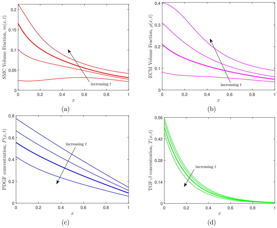
The paper's central claim is that the collagenous fibrous cap that stabilises an atherosclerotic plaque can be produced and maintained by a surprisingly small population of vascular smooth muscle cells, provided TGF-β is present; TGF-β is the dominant control of cap density because it simultaneously stimulates collagen synthesis and inhibits both SMC-mediated and immune-cell-mediated degradation. In the analytical steady-state reduction, the ECM volume fraction $\rho^*$ at a fixed plaque location depends biphasically on the SMC volume fraction $m^*$: the optimal SMC fraction $\hat{m}^* = \frac{1+\sqrt{\lambda}}{1+\mu+\lambda+2\sqrt{\lambda}}$ maximises the ECM fraction $\hat{\rho}^* = \frac{\mu}{1+\mu+\lambda+2\sqrt{\lambda}}$, where $\mu$ is the ratio of SMC-driven synthesis to immune-cell-driven degradation and $\lambda$ is the ratio of SMC-driven degradation to immune-cell-driven degradation. With the model's parameter values the optimum lies at a small SMC fraction, below 15–20% even at moderate TGF-β, and the $\rho^*(m^*)$ curve is flat around the maximum, so cap ECM stays near maximal across a wide range of SMC densities. The base-case simulation ends with about 8.6% SMC and 21.4% collagen, matching ApoE mouse measurements, and removing TGF-β influx cuts cap collagen by roughly 40% while raising SMC numbers.
Load-bearing premise
Quantitative predictions stand on parameter values chosen, where direct measurements were unavailable, to produce biologically realistic results, so the numerical agreement with the ApoE mouse data is not a fully independent test.
Editorial extensions
If this is right
- If the model is right, preserving TGF-β signalling is the most direct route to cap stability, because TGF-β both raises collagen synthesis and suppresses two separate degradation pathways.
- Cap collagen density is not a monotone function of SMC density: beyond the optimal SMC fraction, extra SMCs reduce ECM by occupying space and degrading collagen, so SMC-rich caps can be thinner than SMC-poor ones.
- A moderate excess of SMCs above the optimum makes the cap robust to later SMC loss or falling TGF-β, whereas a cap formed below the optimum degrades quickly under the same perturbations.
- The model predicts that lowering PDGF influx delays, but does not prevent, cap formation; a sparse SMC population still builds a substantial cap, just more slowly.
Reading between the lines
- Editorial inference: the flatness of $\rho^*(m^*)$ around its maximum implies a threshold-like robustness rule—cap thickness stays near-maximal until SMC density falls below a fairly sharp lower bound, after which degradation accelerates; quantifying that threshold against in vivo SMC-density data would be a direct test.
- Editorial inference: transplanting the same steady-state machinery to human arteries, which have resident intimal SMCs absent in mice, would likely push plaque SMC densities above the optimum and make human caps depend more on TGF-β responsiveness than on SMC number.
- Editorial inference: the paper's observation that cholesterol loading attenuates cellular TGF-β responsiveness translates in this model into a lower $\mu$ and higher $\lambda$, which lowers the maximal ECM ceiling; a quantitative simulation of that coupling is a natural extension the paper leaves implicit.
Signed reviews
Editorial analysis
A structured set of objections, weighed in public.
Referee Report
Summary. The paper develops a one-dimensional multiphase PDE model of fibrous cap formation in atherosclerosis. It tracks SMC volume fraction, collagenous ECM volume fraction, a generic tissue phase, and quasi-steady PDGF and TGF-beta concentrations, with non-standard boundary conditions modeling endothelial growth-factor influx and SMC influx at the medial boundary. After nondimensionalization, the authors derive a local steady-state ECM relation, yielding explicit formulas for the SMC fraction that maximizes ECM at a point (Eqs. 54-55). Numerical simulations with base-case parameters reproduce the temporal pattern of SMC and collagen accumulation in ApoE mouse plaques, and sensitivity studies probe the roles of SMC haptotaxis and growth-factor influx. The paper claims that a relatively small SMC population can form a stable cap and that TGF-beta is critical for effective cap formation and maintenance.
Significance. If the quantitative claims are robust, this would be a valuable mechanistic contribution to atherosclerosis modeling: it combines multiphase mechanics with growth-factor-regulated ECM remodeling and provides explicit analytical expressions for the optimal SMC fraction. The qualitative TGF-beta result is supported by independent experimental studies (Mallat et al.; Lutgens et al.), and the local steady-state analysis is a useful interpretative tool. Strengths include the explicit analytical reduction (Eqs. 50-55), the detailed parameterization from in vitro and in vivo data, and the systematic sensitivity studies over alpha_P, alpha_T, and chi_rho. However, the central quantitative claim about small SMC populations rests on unmeasured baseline ECM parameters whose values are calibrated to the same dataset later used for validation; this needs to be addressed before the claim is fully supported.
major comments (2)
- [Section 2.6 and Section 3.2.1] The base-case parameter rs is set 'sufficiently large to allow plaque collagen to accumulate on a timescale similar to that reported in Reifenberg et al. (2012)' (Section 2.6, Table 1), and Section 3.2.1 then presents the agreement of total SMC and ECM fractions (8.6% and 21.4% versus 7% and 24%) as quantitative validation using the same Reifenberg et al. data. Because rs, rd, and beta_rho are not measured, the agreement is partly built into the parameter choice. Please add a global sensitivity analysis over rs, rd, and beta_rho (and ideally beta_m, rm, and the growth-factor uptake rates) and report the resulting ranges of steady-state SMC/ECM totals and of the cap-region V_m and V_rho, so that the 'small SMC population' conclusion can be checked against parameter uncertainty.
- [Section 3.1, Eqs. (54)-(55), and Figure 4] The claim that a relatively small SMC volume fraction (0.1-0.2) maximizes ECM deposition is read off Figure 4 using base-case values. Equation (54) shows that m_hat depends on mu=R_s/B_rho and lambda=R_d/B_rho, and the optimum shifts upward as mu decreases; since mu is proportional to rs/beta_rho, the 'small SMC' conclusion is not robust without specifying the uncertainty in these baseline rates. I recommend either plotting m_hat and rho_hat over the plausible ranges of (rs, rd, beta_rho) or explicitly restricting the claim to the base-case parameter set.
minor comments (5)
- [Abstract] The final sentence, 'an important step towards the development of a comprehensive in silico plaque', is missing a noun; it should read 'comprehensive in silico plaque model'.
- [Section 2.1.3] Immediately after Eq. (17), 'immune cell ECM degration' should be 'degradation'.
- [Section 3.1, Eq. (50)] The assertion that the discriminant is 'trivially' strictly positive for all admissible parameter values is not demonstrated; a short derivation or reference would help.
- [Section 3.2.1, Figure 6 caption] The statement that time points for the ECM panel do not correspond exactly to the other panels is vague; please list the actual time values used for each panel or clarify the convention.
- [Section 4.6] The reported sensitivity simulation with beta_rho=1.5 (32% vs 38% ECM in the cap region) is not shown; either include it in the results or mark it consistently as 'results not shown'.
Circularity Check
Quantitative validation partly circular: baseline ECM synthesis rate rs is tuned to the Reifenberg et al. collagen-accumulation timescale, which is then cited as independent confirmation of the simulated ECM/SMC dynamics; qualitative TGF-beta and small-SMC-population findings retain independent support.
-
fitted input called prediction
[Section 2.6 (Model Parameterisation) and Section 3.2.1 (Base Case Simulation)]
"we assume that the value of rs must be sufficiently large to allow plaque collagen to accumulate on a timescale similar to that reported in Reifenberg et al. (2012)... Based on these assumptions, we estimate rs = 1.8, rd = 1.5 and βρ = 0.75. ... The results in Figures 6a and 6b are qualitatively and quantitatively consistent with the experimental observations of Reifenberg et al. (2012)."
The baseline ECM synthesis rate rs directly controls collagen accumulation in Eq. (40). In Section 2.6, rs is explicitly chosen so that plaque collagen accumulates on the timescale reported by Reifenberg et al. (2012). Section 3.2.1 then cites that same study as independent confirmation that the simulated SMC/ECM time courses and totals (8.6% and 21.4% versus 7% and 24%) are 'quantitatively consistent'. The agreement with Reifenberg et al. is therefore partly enforced by construction: the calibration target and the validation dataset are one and the same.
full rationale
The central analytical result, Eq. (54), is derived internally from the model equations rather than imported from a self-citation, and the qualitative finding that a relatively small SMC population can generate a stable ECM-rich cap is an emergent consequence of the model structure, not an input fitted to data. The TGF-beta-criticality conclusion is independently supported both by the alpha_T = 0 simulation and by external experimental blockade studies (Mallat et al. 2001; Lutgens et al. 2002). There is also a self-citation to Watson et al. (2018) for parameter values and the multiphase framework, but that citation is not the load-bearing justification for any new claim, since the current model adds new biology and the parameter choices are usually traceable to independent experimental references. The genuine circularity is limited to the quantitative validation step involving Reifenberg et al.: rs was set to reproduce that study's collagen-accumulation timescale, and the same study is later invoked to certify quantitative agreement. The resulting score of 4 reflects this partial calibration-validation circularity while acknowledging that the paper's main qualitative insights are independently grounded and not forced by self-citation.
Assumptions & free parameters
free parameters (10)
- rs =
1.8
- rd =
1.5
- beta_rho =
0.75
- alpha_P =
0.7
- alpha_T =
2.5
- chi_rho =
0.3
- mM =
0.01
- eta_P =
2.5
- eta_T =
2.5
- sigma_P and sigma_T =
4
assumptions (9)
- standard math Conservation of mass and momentum for continuous phases
- domain assumption All phases have equal, constant density
- domain assumption ECM phase is a rigid scaffold with zero velocity
- domain assumption Growth factors diffuse quasi-statically, with flux modulated by w
- domain assumption Fixed domain, no intimal growth; mass influx balanced by efflux
- domain assumption Constitutive forms for extra pressures Lambda(P) and psi(m,rho) from Byrne-Owen and Lemon et al.
- domain assumption No voids: m+rho+w=1; all phases strictly positive
- domain assumption SMC entry across the IEL is purely chemotactic, proportional to dLambda/dP times the PDGF gradient
- ad hoc to paper Unmeasured parameters are chosen to ensure biologically realistic results
Cite this review
Pith. "Pith review of A Multiphase Model of Growth Factor-Regulated Atherosclerotic Cap Formation." pith.science (2026). https://pith.science/paper/MVEEY7K3
@misc{pith2026190802889,
  author       = {Pith},
  title        = {Pith review of: A Multiphase Model of Growth Factor-Regulated Atherosclerotic Cap Formation},
  year         = {2026},
  howpublished = {\url{https://pith.science/paper/MVEEY7K3}},
  note         = {Machine review of arXiv:1908.02889}
}
abstract
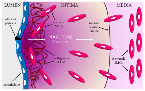
Atherosclerosis is characterised by the growth of fatty plaques in the inner (intimal) layer of the artery wall. In mature plaques, vascular smooth muscle cells (SMCs) are recruited from the adjacent medial layer to deposit a cap of fibrous collagen over the fatty plaque core. The fibrous cap isolates the thrombogenic content of the plaque from the bloodstream and prevents the formation of blood clots that cause myocardial infarction or stroke. Despite the important protective role of the cap, the mechanisms that regulate cap formation and maintenance are not well understood. It remains unclear why certain caps become stable, while others become vulnerable to rupture. We develop a multiphase PDE model with non-standard boundary conditions to investigate collagen cap formation by SMCs in response to growth factor signals from the endothelium. Diffusible platelet-derived growth factor (PDGF) stimulates SMC migration, proliferation and collagen degradation, while diffusible transforming growth factor (TGF)-$\beta$ stimulates SMC collagen synthesis and inhibits collagen degradation. The model SMCs respond haptotactically to gradients in the collagen phase and have reduced rates of migration and proliferation in dense collagenous tissue. The model, which is parameterised using a range of in vivo and in vitro experimental data, reproduces several observations from studies of plaque growth in atherosclerosis-prone mice. Numerical simulations and model analysis demonstrate that a stable cap can be formed by a relatively small SMC population and emphasise the critical role of TGF-$\beta$ in effective cap formation and maintenance. These findings provide unique insight into the cellular and biochemical mechanisms that may lead to plaque destabilisation and rupture. This work represents an important step towards the development of a comprehensive in silico plaque.
Figures

Figures from the paper (11 more)





Reference graph
Works this paper leans on
-
[1]
Adiguzel, P
E. Adiguzel, P. J. Ahmad, C. Franco, and M. P. Bendeck. Collagens in the progression and complications of atherosclerosis. Vasc. Med., 14: 0 73--89, 2009
2009
-
[2]
Ahamed, N
J. Ahamed, N. Burg, K. Yoshinaga, C. A. Janczak, D. B. Rifkin, and B. S. Coller. In vitro and in vivo evidence for shear-induced activation of latent transforming growth factor- 1. Blood, 112: 0 3650--3660, 2008
2008
-
[3]
M. R. Alexander and G. K. Owens. Epigenetic control of smooth muscle cell differentiation and phenotypic switching in vascular development and disease. Annu. Rev. Physiol., 74: 0 13--40, 2012
2012
-
[4]
Astanin and L
S. Astanin and L. Preziosi. Multiphase models of tumour growth. In A. Angelis, M. A. J. Chaplain, and N. Bellomo, editors, Selected Topics in Cancer Modeling: Genesis, Evolution, Immune Competition, and Therapy. Modeling and Simulation in Science, Engineering and Technology, pages 223--253. Birkh \"a user, Boston, 2008
2008
-
[5]
M. R. Bennett, S. Sinha, and G. K. Owens. Vascular smooth muscle cells in atherosclerosis. Circ. Res., 118: 0 692--702, 2016
2016
-
[6]
R. Bhui and H. N. Hayenga. An agent-based model of leukocyte transendothelial migration during atherogenesis. PLoS Comput. Biol., 13: 0 e1005523, 2017
work page 2017
-
[7]
V. Borrelli, L. di Marzo , P. Sapienza, M. Colasanti, E. Moroni, and A. Cavallaro. Role of platelet-derived growth factor and transforming growth factor _1 in the regulation of metalloproteinase expressions. Surgery, 140: 0 454--463, 2006
work page 2006
- [8]
Show all 85 references
-
[9]
Budu-Grajdeanu, R
P. Budu-Grajdeanu, R. C. Schugart, A. Friedman, C. Valentine, A. K. Agarwal, and B. H. Rovin. A mathematical model of venous neointimal hyperplasia formation. Theor. Biol. Med. Model., 5: 0 2, 2008
2008
-
[10]
M. A. K. Bulelzai and J. L. A. Dubbeldam. Long time evolution of atherosclerotic plaques. J. Theor. Biol., 297: 0 1--10, 2012
2012
-
[11]
H. M. Byrne and M. R. Owen. A new interpretation of the K eller- S egel model based on multiphase modelling. J. Math. Biol., 49: 0 604--626, 2004
2004
-
[12]
A. Q. Cai, K. A. Landman, and B. D. Hughes. Multi-scale modeling of a wound healing cell migration assay. J. Theor. Biol., 245: 0 576--594, 2007
2007
-
[13]
A. D. Chalmers, A. Cohen, C. A. Bursill, and M. R. Myerscough. Bifurcation and dynamics in a mathematical model of early atherosclerosis. J. Math. Biol., 71: 0 1451--1480, 2015
2015
-
[14]
A. D. Chalmers, C. A. Bursill, and M. R. Myerscough. Nonlinear dynamics of early atherosclerotic plaque formation may determine the efficacy of high density lipoproteins ( HDL ) in plaque regression. PLoS ONE, 12: 0 e0187674, 2017
2017
-
[15]
Chappell, J
J. Chappell, J. L. Harman, V. M. Narasimhan, H. Yu, K. Foote, B. D. Simons, M. R. Bennett, and H. F. J rgensen. Extensive proliferation of a subset of differentiated, yet plastic, medial vascular smooth muscle cells contributes to neointimal formation in mouse injury and ather...
2016
-
[16]
Chen, I.-H
C.-L. Chen, I.-H. Liu, S. J. Fliesler, X. Han, S. S. Huang, and J.S. Huang. Cholesterol suppresses cellular TGF - responsiveness: implications in atherogenesis. J. Cell Sci., 120: 0 3509--3521, 2007
2007
-
[17]
Cilla, E
M. Cilla, E. Pena, and M. A. Martinez. Mathematical modelling of atheroma plaque formation and development in coronary arteries. J. Royal Soc. Interface, 11: 0 20130866, 2014
2014
-
[18]
M. C. H. Clarke, N. Figg, J. J. Maguire, A. P. Davenport, M. Goddard, T. D. Littlewood, and M. R. Bennett. Apoptosis of vascular smooth muscle cells induces features of plaque vulnerability in atherosclerosis. Nat. Med., 12: 0 1075--1080, 2006
2006
-
[19]
C. A. Cobbold and J. A. Sherratt. Mathematical modelling of nitric oxide activity in wound healing can explain keloid and hypertrophic scarring. J. Theor. Biol., 204: 0 257--288, 2000
2000
-
[20]
Cohen, M
A. Cohen, M. R. Myerscough, and R. S. Thompson. Athero-protective effects of high density lipoproteins ( HDL ): an ODE model of the early stages of atherosclerosis. Bull. Math. Biol., 76: 0 1117--1142, 2014
2014
-
[21]
B. D. Cumming, D. L. S. McElwain, and Z. Upton. A mathematical model of wound healing and subsequent scarring. J. R. Soc. Interface, 7: 0 19--34, 2010
2010
-
[22]
El Khatib , S
N. El Khatib , S. Genieys, and V. Volpert. Atherosclerosis initiation modeled as an inflammatory process. Math. Model. Nat. Phenom., 2: 0 126--141, 2007
2007
-
[23]
D. J. W. Evans, P. V. Lawford, J. Gunn, D. Walker, D. R. Hose, R. H. Smallwood, B. Chopard, M. Krafczyk, J. Bernsdorf, and A. Hoekstra. The application of multiscale modelling to the process of development and prevention of stenosis in a stented coronary artery. Phil. Trans. R...
2008
-
[24]
Faggiotto and R
A. Faggiotto and R. Ross. Studies of hypercholesterolemia in the nonhuman primate II . F atty streak conversion to fibrous plaque. Arteriosclerosis, 4: 0 341--356, 1984
1984
-
[25]
Filipovic, Z
N. Filipovic, Z. Teng, M. Radovic, I. Saveljic, D. Fotiadis, and O. Parodi. Computer simulation of three-dimensional plaque formation and progression in the carotid artery. Med. Biol. Eng. Comput., 51: 0 607--616, 2013
2013
-
[26]
P. Fok. Mathematical model of intimal thickening in atherosclerosis: vessel stenosis as a free boundary problem. J. Theor. Biol., 314: 0 23--33, 2012
2012
-
[27]
Friedman and W
A. Friedman and W. R. Hao. A mathematical model of atherosclerosis with reverse cholesterol transport and associated risk factors. Bull. Math. Biol., 77: 0 758--781, 2015
2015
-
[28]
Fukumoto, J
Y. Fukumoto, J. Deguchi, P. Libby, E. Rabkin-Aikawa, Y. Sakata, M. T. Chin, C. C. Hill, P. R. Lawler, N. Varo, F. J. Schoen, S. M. Krane, and M. Aikawa. Genetically determined resistance to collagenase action augments interstitial collagen accumulation in atherosclerotic plaqu...
1953
-
[29]
Funayama, U
H. Funayama, U. Ikeda, M. Takahashi, Y. Sakata, S.-I. Kitagawa, Y.-I. Takahashi, J.-I. Masuyama, Y. Furukawa, Y. Miura, S. Kano, M. Matsuda, and K. Shimada. Human monocyte-endothelial cell interaction induces platelet-derived growth factor expression. Cardiovasc. Res., 37: 0 2...
1998
-
[30]
Garbey, S
M. Garbey, S. Casarin, and S. A. Berceli. Vascular adaptation: pattern formation and cross validation between an agent based model and a dynamical system. J. Theor. Biol., 429: 0 149--163, 2017
2017
-
[31]
G. S. Getz and C. A. Reardon. Animal models of atherosclerosis. Arterioscler. Thromb. Vasc. Biol., 32: 0 1104--1115, 2012
2012
-
[32]
M. Guo, Y. Cai, X. Yao, and Z. Li. Mathematical modeling of atherosclerotic plaque destabilization: role of neovascularization and intraplaque hemorrhage. J. Theor. Biol., 450: 0 53--65, 2018
2018
-
[33]
G. K. Hansson and P. Libby. The immune response in atherosclerosis: a double-edged sword. Nat. Immunol., 6: 0 508--519, 2006
2006
-
[34]
G. K. Hansson, P. Libby, and I. Tabas. Inflammation and plaque vulnerability. J. Intern. Med., 278: 0 483--493, 2015
2015
-
[35]
J. M. Haugh. Deterministic model of dermal wound invasion incorporating receptor-mediated signal transduction and spatial gradient sensing. Biophys. J., 90: 0 2297--2308, 2006
2006
-
[36]
G. Hou, D. Mulholland, M. A. Gronska, and Bendeck M. P. Type VIII collagen stimulates smooth muscle cell migration and matrix metalloproteinase synthesis after arterial injury. Am. J. Pathol., 156: 0 467--476, 2000
2000
-
[37]
J. S. Huang, T. J. Olsen, and S. S. Huang. The role of growth factors in tissue repair I . P latelet-derived growth factor. In R. A. F. Clark and P. M. Henson, editors, The molecular and cellular biology of wound repair, pages 243--251. Plenum, New York, 1988
1988
-
[38]
M. E. Hubbard and H. M. Byrne. Multiphase modelling of vascular tumour growth in two spatial dimensions. J. Theor. Biol., 316: 0 70--89, 2013
2013
-
[39]
M. H. Islam and P. R. Johnston. A mathematical model for atherosclerotic plaque formation and arterial wall remodelling. ANZIAM J., 57: 0 C320--C345, 2016
2016
-
[40]
Jacobsen, M
K. Jacobsen, M. B. Lund, J. Shim, S. Gunnersen, E.-M. F \"u chtbauer, M. Kjolby, L. Carramolino, and J. F. Bentzon. Diverse cellular architecture of atherosclerotic plaque derives from clonal expansion of a few medial SMC s. JCI Insight, 2: 0 e95890, 2017
2017
-
[41]
Klika, E
V. Klika, E. A. Gaffney, Y.-C. Chen, and C. P. Brown. An overview of multiphase cartilage mechanical modelling and its role in understanding function and pathology. J. Mech. Behav. Biomed., 62: 0 139--157, 2016
2016
-
[42]
Kozaki, W
K. Kozaki, W. E. Kaminski, J. Tang, S. Hollenbach, P. Lindahl, C. Sullivan, J.-C. Yu, K. Abe, P. J. Martin, R. Ross, C. Betsholtz, N. A. Giese, and E. W. Raines. Blockade of platelet-derived growth factor or its receptors transiently delays but does not prevent fibrous cap for...
2002
-
[43]
Kubota, J
K. Kubota, J. Okazaki, O. Louie, K. C. Kent, and B. Liu. TGF - stimulates collagen ( I ) in vascular smooth muscle cells via a short element in the proximal collagen promoter. J. Surg. Res., 109: 0 43--50, 2003
2003
-
[44]
Lally and P
C. Lally and P. Prendergast. Simulation of in-stent restenosis for the design of cardiovascular stents. In A. Holzapfel and R. W. Ogden, editors, Mechanics of Biological Tissue, pages 255--267. Springer, Berlin, 2006
2006
-
[45]
Lemon, J
G. Lemon, J. R. King, H. M. Byrne, O. E. Jensen, and K. M. Shakesheff. Mathematical modelling of engineered tissue growth using a multiphase porous flow mixture theory. J. Math. Biol., 52: 0 571--594, 2006
2006
-
[46]
Lopes, E
J. Lopes, E. Adiguzel, S. Gu, S.-L. Liu, G. Hou, S. Heximer, R. K. Assoian, and M. P. Bendeck. Type VIII collagen mediates vessel wall remodeling after arterial injury and fibrous cap formation in atherosclerosis. Am. J. Pathol., 182: 0 2241--2253, 2013
2013
-
[47]
A. J. Lusis. Atherosclerosis. Nature, 407: 0 233--241, 2000
2000
-
[48]
Lutgens, E
E. Lutgens, E. D. de Muinck , P. J. E. H. M. Kitslaar, J. H. M. Tordoir, H. J. J. Wellens, and M. J. A. P. Daemen. Biphasic pattern of cell turnover characterises the progression from fatty streaks to ruptured human atherosclerotic plaques. Cardiovasc. Res., 41: 0 473--479, 1999
1999
-
[49]
Lutgens, M
E. Lutgens, M. Gijbels, M. Smook, P. Heeringa, P. Gotwals, V. E. Koteliansky, and M. J. A. P. Daemen. Transforming growth factor- mediates balance between inflammation and fibrosis during plaque progression. Arterioscler. Thromb. Vasc. Biol., 22: 0 975--982, 2002
2002
-
[50]
Mallat, A
Z. Mallat, A. Corbaz, A. Scoazec, P. Graber, S. Alouani, B. Esposito, Y. Humbert, Y. Chvatchko, and A. Tedgui. Interleukin-18/ I nterleukin-18 binding protein signaling modulates atherosclerotic lesion development and stability. Circ. Res., 89: 0 e41--e45, 2001
2001
-
[51]
T. A. McCaffrey, S. Consigli, B. Du, D. J. Falcone, T. A. Sanborn, A. M. Spokojny, and H. L. Bush Jr. Decreased type II /type I TGF - receptor ratio in cells derived from human atherosclerotic lesions. C onversion from an antiproliferative to profibrotic response to TGF - 1. J...
1995
-
[52]
McDougall, J
S. McDougall, J. Dallon, J. Sherratt, and P. Maini. Fibroblast migration and collagen deposition during dermal wound healing: mathematical modelling and clinical implications. Phil. Trans. R. Soc. A, 364: 0 1385--1405, 2006
2006
-
[53]
McKay, S
C. McKay, S. McKee, N. Mottram, T. Mulholland, S. Wilson, S. Kennedy, and R. Wadsworth. Towards a model of atherosclerosis. Technical report, University of Strathclyde, 2004
2004
-
[54]
S. N. Menon, J. A. Flegg, S. W. McCue, R. C. Schugart, R. A. Dawson, and D. L. S. McElwain. Modelling the interaction of keratinocytes and fibroblasts during normal and abnormal wound healing processes. Proc. R. Soc. B, 279: 0 3329--3338, 2012
2012
-
[55]
Moore, F
K. Moore, F. Sheedy, and E. Fisher. Macrophages in atherosclerosis: a dynamic balance. Nat. Rev. Immunol., 13: 0 709--721, 2013
2013
-
[56]
Munro, M
E. Munro, M. Patel, P. Chan, L. Betteridge, K. Gallagher, M. Schachter, J. Wolfe, and P. Server. Effect of calcium channel blockers on the growth of human vascular smooth muscle cells derived from saphenous vein and vascular graft stenosis. J. Cardiovasc. Pharmacol., 23: 0 779...
1994
-
[57]
P. R. Nelson, S. Yamamura, and K. C. Kent. Extracellular matrix proteins are potent agonists of human smooth muscle cell migration. J. Vasc. Surg., 24: 0 25--32, 1996
1996
-
[58]
Nicolas, E
M. Nicolas, E. Pe \ n a, M. Malv \`e , and M. A. Mart \' nez. Mathematical modeling of the fibrosis process in the implantation of inferior vena cava filters. J. Theor. Biol., 387: 0 228--240, 2015
2015
-
[59]
R. D. O'Dea , J. M. Osborne, A. J. El Haj , H. M. Byrne, and S. L. Waters. The interplay between tissue growth and scaffold degradation in engineered tissue constructs. J. Math. Biol., 67: 0 1199--1225, 2013
2013
-
[60]
Ogawa, F
K. Ogawa, F. Chen, C. Kuang, and Y. Chen. Suppression of matrix metalloproteinase-9 transcription by transforming growth factor- is mediated by a nuclear factor- B site. Biochem. J., 381: 0 413--422, 2004
2004
-
[61]
Olsen, J
L. Olsen, J. A. Sherratt, and P. K. Maini. A mechanochemical model for adult dermal wound contraction and the permanence of the contracted tissue displacement profile. J. Theor. Biol., 177: 0 113--128, 1995
1995
-
[62]
Pappalardo, S
F. Pappalardo, S. Musumeci, and S. Motta. Modeling immune system control of atherogenesis. Bioinformatics, 24: 0 1715–--1721, 2008
2008
-
[63]
Parton, V
A. Parton, V. McGilligan, M. O'Kane, F. R. Baldrick, and S. Watterson. Computational modelling of atherosclerosis. Brief. Bioinform., 17: 0 562–--575, 2016
2016
-
[64]
N. C. Pearson, R. J. Shipley, S. L. Waters, and J. M. Oliver. Multiphase modelling of the influence of fluid flow and chemical concentration on tissue growth in a hollow fibre membrane bioreactor. Math. Med. Biol., 31: 0 393–--430, 2014
2014
-
[65]
R. N. Poston and D. R. M. Poston. Typical atherosclerotic plaque morphology produced in silico by an atherogenesis model based on self-perpetuating propagating macrophage recruitment. Math. Model. Nat. Phenom., 2: 0 142–--149, 2007
2007
-
[66]
Preziosi and A
L. Preziosi and A. Tosin. Multiphase modelling of tumour growth and extracellular matrix interaction: mathematical tools and applications. J. Math. Biol., 58: 0 625–--656, 2009
2009
-
[67]
Reifenberg, F
K. Reifenberg, F. Cheng, C. Orning, J. Crain, I. K \"u pper, E. Wiese, M. Protschka, M. Blessing, K. J. Lackner, and M. Torzewski. Overexpression of TGF - 1 in macrophages reduces and stabilizes atherosclerotic plaques in ApoE -deficient mice. PLoS ONE, 7: 0 e40990, 2012
2012
-
[68]
G. M. Risinger, D. L. Updike, E.C. Bullen, J. J. Tomasek, and E. W. Howard. TGF - suppresses the upregulation of MMP -2 by vascular smooth muscle cells in response to PDGF-BB . Am. J. Physiol. Cell Physiol., 298: 0 C191--C201, 2010
2010
-
[69]
R. Ross. Atherosclerosis –-- an inflammatory disease. N. Engl. J. Med., 340: 0 115–--126, 1999
1999
-
[70]
Rutherford, W
C. Rutherford, W. Martin, M. Carrier, E. E. A ngg a rd, and G. A. A. Ferns. Endogenously elicited antibodies to platelet derived growth factor- BB and platelet cystolic protein inhibit aortic lesion development in the cholesterol-fed rabbit. Int. J. Exp. Path., 78: 0 21–--32, 1997
1997
-
[71]
H. Sano, T. Sudo, M. Yokode, T. Murayama, H. Kataoka, N. Takakura, S. Nishikawa, S.-I. Nishikawa, and T. Kita. Functional blockade of platelet-derived growth factor receptor- but not of receptor- prevents vascular smooth muscle cell accumulation in fibrous cap lesions in apoli...
2001
-
[72]
Schachter
M. Schachter. Vascular smooth muscle cell migration, atherosclerosis, and calcium channel blockers. Int. J. Cardiol., 62: 0 S85–--S90, 1997
1997
-
[73]
N. N. Singh and D. P. Ramji. The role of transforming growth factor- in atherosclerosis. J. R. Soc. Interface, 17: 0 487--499, 2006
2006
-
[74]
Tahir, I
H. Tahir, I. Niculescu, C. Bona-Casas, R. M. H. Merks, and A. G. Hoekstra. An in silico study on the role of smooth muscle cell migration in neointimal formation after coronary stenting. Cytokine Growth Factor Rev., 12: 0 20150358, 2015
2015
-
[75]
Toma and T
I. Toma and T. A. McCaffrey. Transforming growth factor- and atherosclerosis: interwoven atherogenic and atheroprotective aspects. Cell Tissue Res., 347: 0 155--175, 2012
2012
-
[76]
Urschel and I
K. Urschel and I. Cicha. TNF - in the cardiovascular system: from physiology to therapy. Int. J. Interferon Cytokine Mediat. Res., 7: 0 9--25, 2015
2015
-
[77]
G. G. Vaday, H. Schor, M. A. Rahat, N. Lahat, and O. Lider. Transforming growth factor- suppresses tumor necrosis factor -induced matrix metalloproteinase-9 expression in monocytes. J. Leukoc. Biol., 69: 0 613--621, 2001
2001
-
[78]
Vengrenyuk, H
Y. Vengrenyuk, H. Nishi, X. Long, M. Ouimet, N. Savji, F. O. Martinez, C. P. Cassella, K. J. Moore, S. A. Ramsey, J. M. Miano, and E. A. Fisher. Cholesterol loading reprograms the micro RNA -143/145-myocardin axis to convert aortic smooth muscle cells to a dysfunctional macrop...
2015
-
[79]
L. M. Wakefield, T. S. Winokur, R. S. Hollands, K. Christopherson, A. D. Levinson, and M.B. Sporn. Recombinant latent transforming growth factor 1 has a longer plasma half-life in rats than active transforming growth factor 1, and a different tissue distribution. J. Clin. Inve...
1976
-
[80]
L. M. Wakefield, J. J. Letterio, T. Chen, D. Danielpour, R. S. Allison, L. H. Pai, A. M. Denicoff, M. H. Noone, K. H. Cowan, J. A. O'Shaughnessy , and M.B. Sporn. Transforming growth factor- 1 circulates in normal human plasma and is unchanged in advanced metastatic breast can...
1995
-
[81]
J. Wang, A. K. Uryga, J. Reinhold, N. Figg, L. Baker, A. Finigan, K. Gray, S. Kumar, M. Clarke, and M. Bennett. Vascular smooth muscle cell senescence promotes atherosclerosis and features of plaque vulnerability. Circulation, 132: 0 1909--1919, 2015
1909
-
[82]
M. G. Watson, H. M. Byrne, C. Macaskill, and M. R. Myerscough. A two-phase model of early fibrous cap formation in atherosclerosis. J. Theor. Biol., 456: 0 123--136, 2018
2018
-
[83]
Cardiovascular diseases fact sheet
World Health Organization . Cardiovascular diseases fact sheet. https://www.who.int/news-room/fact-sheets/detail/cardiovascular-diseases-(cvds), May 2017. Accessed April 2019
2017
-
[84]
Y. Yang, W. J \"a ger, M. Neuss-Radu, and T. Richter. Mathematical modeling and simulation of the evolution of plaques in blood vessels. J. Math. Biol., 72: 0 973--996, 2016
2016
-
[85]
Zahedmanesh, H
H. Zahedmanesh, H. Van Oosterwyck , and C. Lally. A multi-scale mechanobiological model of in-stent restenosis: deciphering the role of matrix metalloproteinase and extracellular matrix changes. Comput. Methods Biomech. Biomed. Engin., 17: 0 813--828, 2014
2014
Reviewed August 14, 2026 · model on record in the stance chip above.
Discussion (0). Continue with ORCID to comment.