REVIEW 3 major objections 9 minor 58 references
Variational Multi-Task MRI Reconstruction: Joint Reconstruction, Registration and Super-Resolution
T0 review · 3 major / 9 minor · reviewed 2026-08-14 · deepseek-v4-flash
Pith's one-line read The paper introduces a variational multi-task framework that solves MRI reconstruction, motion registration, and super-resolution in one joint optimisation, and reports that the joint solution is rated best by clinicians against…
desk verdict A genuinely first three-task variational MRI model with a real existence proof, but the fidelity term reverses warp and blur, so the estimated motions aren't the physiological ones and the headline claims outrun the evidence. read the letter →
The pith
A machine-rendered reading of the paper's core claim, the machinery that carries it, and where it could break.
The reading
What carries the argument
The load-bearing object is the joint functional of Eq. (6), in which the fidelity term $\|F((Cu)\circ\varphi_t^{-1})-x_t\|_2^2$ encodes all three tasks at once: $F$ is the undersampled Fourier operator of reconstruction, $\varphi_t$ are the registration deformations, and $C=DB$ (Gaussian blur followed by averaging downsample) is the super-resolution degradation. Two regularisers carry the prior knowledge: a weighted total variation $\mathrm{TV}_{g_t}$ that aligns edges between the deformed reconstruction and each acquisition, and the hyperelastic stored energy $W_{\mathrm{Op}}(\nabla\varphi_t)=a_1\|\nabla\varphi_t\|_F^4+a_2(\det\nabla\varphi_t-1/\det\nabla\varphi_t)^4$, which allows large smooth deformations while forbidding foldings. The numerical scheme introduces auxiliary variables for the deformation gradient, the composed image, and the edge-aligned image, then solves each subproblem with dedicated tools (a total-variation projection algorithm, primal-dual updates, and closed-form Fourier updates), which is what makes the three-task problem computationally tractable.
What would settle it
Take a digital phantom with a known ground-truth high-resolution image and known motion, simulate the low-resolution acquisitions with a non-Gaussian or measured point-spread function, and run the model; if the reconstructed edges and fine structures develop ringing or false texture compared with the matched-Gaussian case, the claimed super-resolution gains depend on the assumed degradation operator rather than on true anatomy.
Extended reading notes
Core claim
On the paper's own terms, the central discovery is that a single variational model can couple compressed-sensing MRI reconstruction, deformable registration, and super-resolution, and that this coupling improves the result. The model minimises $$G(u,(\varphi_t)_{t=1,\dots,T}) = \frac{1}{T}\sum_{t=1}^T \left( \frac{1}{2}\|F((Cu)\circ\$varphi_t^{{-1}}$)-x_t\|$_2^{2}$ + \delta\,\mathrm{TV}_{g_t}((Cu)\circ\$varphi_t^{{-1}}$) \right) + \$\alpha$\,\mathrm{TV}(u) + \frac{1}{T}\sum_{t=1}^T \int_\$\Omega$ W_{\mathrm{Op}}(\nabla\varphi_t)\,dx,$$ where $C=DB$ is a known blur-then-downsample operator, $\varphi_t$ are hyperelastic deformations close to the identity, and $W_{\mathrm{Op}}$ is the hyperelastic stored energy that penalises changes in length and area while preserving topology. The authors prove existence of minimisers for this functional and solve it by introducing auxiliary variables that split the problem into five tractable subproblems. Their experiments compare the joint model against sequential reconstruction-registration-super-resolution pipelines and against two-task methods, and report that the three-task model ranks best in a clinician study while running faster than the sequential baselines.
Load-bearing premise
The method assumes it knows exactly the blur-and-downsample operator $C=DB$ (Gaussian blur plus averaging window) that turns the high-resolution image into each low-resolution acquisition; if the real scanner's point-spread function or slice profile differs, the fidelity term is biased and the super-resolved detail could be an artifact of that wrong assumption.
Editorial extensions
If this is right
- A single high-resolution, motion-corrected image can be recovered directly from multiple undersampled free-breathing acquisitions, removing the need to reconstruct first and register afterwards.
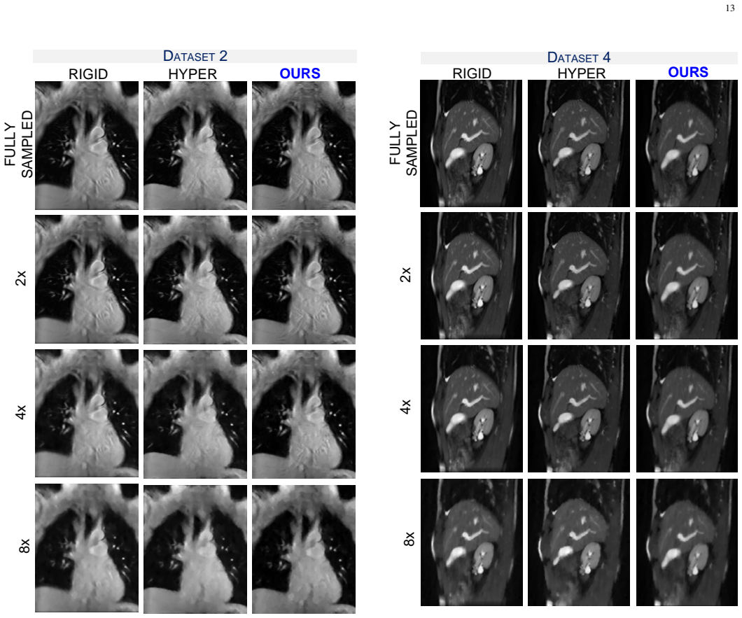
- Joint estimation reduces error propagation: details that a sequential pipeline blurs away, such as small vessels and kidney structures, survive even at acceleration factor 8 in the reported experiments.
- The framework is plug-and-play with respect to the regulariser, so the TV terms could be replaced by learned or application-specific priors without changing the joint fidelity structure.
- Because all three tasks share one fidelity term, the computational cost stays comparable to a two-task method (DC-CS) while delivering a third task, and is far below the sequential baselines.
- The existence result gives the model a guarantee that the optimisation problem is well-posed under the stated boundedness and regularity assumptions.
Reading between the lines
- Because the paper assumes a known degradation operator, a natural stress test would replace the Gaussian blur with a measured point-spread function; if the fine-detail gains persist, true super-resolution is occurring, whereas if they vanish or produce ringing, the model is partly fitting its own assumption.
- A blind variant that estimates $C$ alongside $u$ and $\varphi_t$ would remove the need to know the exact slice profile and could be tested on the same datasets.
- The expert study does not isolate the super-resolution task; a simulated ground-truth ablation would quantify how much of the quality gain comes from each of the three tasks.
- The same three-way fidelity structure could transfer to PET or ultrasound, where undersampling, motion, and limited resolution occur together.
Signed reviews
Editorial analysis
A structured set of objections, weighed in public.
Referee Report
Summary. The paper proposes a variational multi-task framework that jointly performs MRI reconstruction, registration, and super-resolution from a stack of undersampled, motion-corrupted acquisitions. The model combines an L2 fidelity term linking the three tasks, a weighted total variation term, a standard TV regularizer on the high-resolution image, and an Ogden-type hyperelastic regularizer on the deformation fields. The authors prove existence of minimizers, derive a splitting-based numerical scheme with five sub-problems, and evaluate the method on five free-breathing MRI datasets against sequential pipelines (rigid and hyperelastic registration followed by TV reconstruction and super-resolution) and two-task baselines (DC-CS and GW-CS), using an expert user study and CPU-time comparisons.
Significance. If the model and experiments were sound, this would be a valuable contribution: it is, to the authors' knowledge, the first variational framework to combine three MRI tasks in a single optimization, and the theoretical well-posedness result plus the computationally tractable splitting scheme would be useful to the community. The hyperelastic regularization is physically motivated, and the idea of sharing representation across reconstruction, registration, and super-resolution is attractive. However, the significance is conditional: the fidelity term appears to reverse the physical order of degradation and deformation, and the experimental reporting contains an inconsistency between the narrative and the displayed user-study percentages. These issues directly affect the central claims of the paper.
major comments (3)
- [Section III-B/III-C/III-D] The acquisition model in Eq. (3) is warp-then-degrade (DB W_i u), while the fidelity term in Eqs. (5)–(6) is degrade-then-warp, ((Cu)∘φ_t^{-1}) with C=DB. For nonrigid deformations, C and composition by φ_t^{-1} do not commute, so the functional minimized in Eq. (6) is not the likelihood of the measured data under the stated physical model. Sub-problem 5 confirms the reversed order, since it solves min_u (γ2/2T) Σ_t ||h_t∘φ_t - Cu||^2. Consequently, the recovered deformations φ_t are not the tissue motions that generated the acquisitions, and the claimed super-resolved, motion-free reconstruction may be an artifact of the assumed blur/downsampling model. The existence proof in the supplement establishes well-posedness of the problem as written, but not its correctness as an inverse problem.
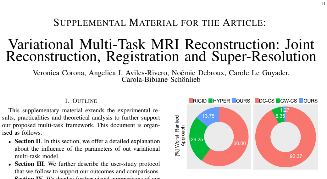
- [Fig. 3 and Section IV.D] The user-study results as presented do not support the claim that 'our approach was ranked best, with a 44.29% of agreement'. In Fig. 3, the bar for OURS in the sequential comparison is 5.89% and in the multi-task comparison is 23.38%, both the lowest in their groups, while the corresponding highest bars are 52.52% (RIGID) and 44.29% (DC-CS). The text attributes the 44.29% to OURS and to the sequential comparison, which is inconsistent with the figure. This discrepancy must be resolved; either the figure or the narrative is mislabeled, and the Friedman test should be reported with test statistics, p-values, and effect sizes before the claim of significant improvements can be assessed.
- [Section IV-B and Fig. 8] The evaluation protocol provides no ground-truth quantitative metrics for the recovered super-resolved anatomy or the deformation fields. The difference maps in Fig. 8 measure agreement between the reconstruction and the model's own registered copies h_t∘φ_t, so they cannot independently validate motion correction. The expert study uses a coarse three-point Likert scale without any inter-observer agreement statistic, and the supplement does not state whether the experts were blinded to which method produced each image. Given the model-order concern in the first comment, the current evaluation cannot substantiate that the proposed method recovers physically correct deformations or a true super-resolved anatomy.
minor comments (9)
- [Section IV.B] The phrase 'we expensively evaluate our model' should read 'we extensively evaluate our model'.
- [Section III.B] The word 'dowsampling' should be 'downsampling'.
- [Section IV.D] The word 'staircaising' should be 'staircasing'.
- [Fig. 7 and Supplement Fig. 17] The caption of Fig. 7 says 'Dataset 5' while the figure and the surrounding text refer to Dataset 3; the supplement's Fig. 17 caption similarly says 'Dataset 4' for what the text describes as Dataset 5.
- [Section III.C] The phrase 'The first term of F seeks to align the edges' should likely be 'The first term of E seeks to align the edges', since E is the functional being defined.
- [Throughout] The symbol F is used both for the undersampled Fourier operator and for the deformation gradient in the stored energy function W_O(F); this notational clash may confuse readers.
- [Sub-problem 3] The formula for h^{k+1}_t contains unbalanced parentheses, making the update rule hard to parse.
- [Supplement Section III] The user-study section should state whether the experts were blinded to the reconstruction method and should report the instructions given to the experts in full.
- [Section IV.A] The data description does not specify the acceleration factors or k-space sampling pattern used in the experiments; this should be clarified to allow reproduction.
Circularity Check
Minor circular-evidence detail in the difference-map validation; the central variational derivation and external benchmarks are independent.
-
other
[Section IV-D, paragraph on difference maps (after Fig. 8); model Eq. (7) and Table I]
"when we inspect the mean difference between our reconstruction and the individual registered acquisitions ( ht◦φt), at the middle and right sides of Fig. 8, one can see that the structures are very well-aligned resulting in a much smaller range in difference maps. Overall, our approach successfully corrects for motion even at low undersampling rates"
The evidence for 'successfully corrects for motion' is the small difference between ht◦φt and Cu, which is not an independent measurement: Eq. (7) contains the penalty (γ2/2)||ht−(Cu)◦φ_t^{-1}||^2, and Table I sets γ2=10^5, so the optimization forces ht≈(Cu)◦φ_t^{-1} and hence ht◦φt≈Cu by construction. Reporting this enforced residual as validation of motion correction is a self-consistency check rather than a test of the estimated deformations. The paper's main comparisons against DC-CS, GW-CS, RIGID and HYPER, plus the expert user study, are external to the optimized objective, so this is a minor circular-evidence step, not a collapse of the central derivation.
full rationale
Overall, the proposed method is a new variational model, not a quantity fitted from data and then renamed as a prediction. The derivation chain is self-contained: Eq. (6) defines the joint objective over u and φt, the existence of minimizers is proved in the supplement, and the numerical scheme is a standard splitting with the update equations given. The performance claim is supported by comparisons with external baselines (DC-CS, GW-CS, RIGID, HYPER) and by an expert scoring study; the paper explicitly notes 'there is not ground truth for this task,' which is a limitation of the evaluation rather than a circularity. The only self-citation, [44] for the Ogden-type hyperelastic energy, is a transparent modeling choice and is not invoked as a uniqueness theorem; the current paper verifies the coercivity and lower-semicontinuity properties needed for well-posedness. Also, the inconsistency between the warp-then-degrade order in Eq. (3) and the degrade-then-warp order in Eq. (6) is a physical-modeling correctness concern, not a circularity, because the objective is not equivalent to its own input by definition. The single minor circular-evidence item is the difference-map discussion, where small residuals are enforced by the γ2 penalty; since the central comparative claims do not depend on that discussion, the circularity is not load-bearing. Score 1 reflects that one minor self-consistent validation step while the central derivation remains independent.
Assumptions & free parameters
free parameters (11)
- a1 =
1 (all datasets)
- a2 =
50 (D1-D3), 100 (D4-D5)
- gamma1 =
5 (D1-D3), 1 (D4-D5)
- gamma2 =
1e5 (all datasets)
- gamma3 =
15 (D1-D3), 1 (D4-D5)
- theta =
5 (all datasets)
- sigma =
1.5 (D1-D3), 2 (D4-D5)
- k =
2 (all datasets)
- alpha =
0.01 (D1-D3), 0.001 (D4-D5)
- delta =
not reported
- N, n (iteration counts) =
500 (all datasets)
assumptions (5)
- domain assumption The MRI data model x = F u + η with F = S A, where S is subsampling and A is the Fourier transform, accurately represents the acquisition.
- domain assumption The blurring operator B in C = DB is a fixed Gaussian kernel and D is an averaging downsampling window.
- domain assumption Deformations φ_t are diffeomorphisms in W = Id + W^{1,4}_0(Ω,R^2) with det∇φ_t > 0 a.e., and biological tissue is modeled as an Ogden-type hyperelastic material with stored energy WOp.
- standard math Existence of minimizers for (6) follows from standard calculus of variations arguments (coercivity, weak lower semi-continuity, Ball's results).
- domain assumption The weighted total variation TV_{gt} is well-defined with weights g_t from a Canny edge detector satisfying the assumptions in [51].
Cite this review
Pith. "Pith review of Variational Multi-Task MRI Reconstruction: Joint Reconstruction, Registration and Super-Resolution." pith.science (2026). https://pith.science/paper/PAGG4CU6
@misc{pith2026190805911,
  author       = {Pith},
  title        = {Pith review of: Variational Multi-Task MRI Reconstruction: Joint Reconstruction, Registration and Super-Resolution},
  year         = {2026},
  howpublished = {\url{https://pith.science/paper/PAGG4CU6}},
  note         = {Machine review of arXiv:1908.05911}
}
abstract
Motion degradation is a central problem in Magnetic Resonance Imaging (MRI). This work addresses the problem of how to obtain higher quality, super-resolved motion-free, reconstructions from highly undersampled MRI data. In this work, we present for the first time a variational multi-task framework that allows joining three relevant tasks in MRI: reconstruction, registration and super-resolution. Our framework takes a set of multiple undersampled MR acquisitions corrupted by motion into a novel multi-task optimisation model, which is composed of an $L^2$ fidelity term that allows sharing representation between tasks, super-resolution foundations and hyperelastic deformations to model biological tissue behaviors. We demonstrate that this combination yields to significant improvements over sequential models and other bi-task methods. Our results exhibit fine details and compensate for motion producing sharp and highly textured images compared to state of the art methods.
Figures


Figures from the paper (9 more)






Reference graph
Works this paper leans on
-
[1]
M. A. Brown, R. C. Semelka, and B. M. Dale, MRI: basic principles and applications . John Wiley & Sons, 2015
work page 2015
-
[2]
Motion artifacts in MRI: a complex problem with many partial solutions,
M. Zaitsev, J. Maclaren, and M. Herbst, “Motion artifacts in MRI: a complex problem with many partial solutions,” J Magn Reson Imaging, , vol. 42, no. 4, 2015
work page 2015
-
[3]
The diminishing variance algorithm for real-time reduction of motion artifacts in MRI,
T. S. Sachs, C. H. Meyer, P. Irarrazabal, B. S. Hu, D. G. Nishimura, and A. Macovski, “The diminishing variance algorithm for real-time reduction of motion artifacts in MRI,” Magn Reson Med , vol. 34, no. 3, pp. 412–422, 1995
work page 1995
-
[4]
Are movement artifacts in magnetic resonance imaging a real problem? A narrative review,
I. Havsteen, A. Ohlhues, K. H. Madsen, J. D. Nybing, H. Christensen, and A. Christensen, “Are movement artifacts in magnetic resonance imaging a real problem? A narrative review,” Frontiers in neurology , vol. 8, p. 232, 2017
work page 2017
-
[5]
J. B. Andre, B. W. Bresnahan, M. Mossa-Basha, M. N. Hoff, C. P. Smith, Y . Anzai, and W. A. Cohen, “Toward quantifying the prevalence, severity, and cost associated with patient motion during clinical mr examinations,” Journal of the American College of Radiology , vol. 12, no. 7, pp. 689–695, 2015
work page 2015
-
[6]
Experimental designs and processing strategies for fmri studies involving overt verbal responses,
R. M. Birn, R. W. Cox, and P. A. Bandettini, “Experimental designs and processing strategies for fmri studies involving overt verbal responses,” Neuroimage, vol. 23, no. 3, pp. 1046–1058, 2004
work page 2004
-
[7]
Quantification of left ventricular function with magnetic resonance images acquired in real time,
R. M. Setser, S. E. Fischer, and C. H. Lorenz, “Quantification of left ventricular function with magnetic resonance images acquired in real time,” J Magn Reson Imaging , vol. 12, no. 3, pp. 430–438, 2000. 10
work page 2000
-
[8]
S. Plein, W. H. Smith, J. P. Ridgway, A. Kassner, D. J. Beacock, T. N. Bloomer, and M. U. Sivananthan, “Qualitative and quantitative analysis of regional left ventricular wall dynamics using real-time magnetic resonance imaging: comparison with conventional breath-hold gradient echo acquisition in volunteers and patients,” J Magn Reson Imaging
Show all 58 references
-
[9]
Rapid evaluation of left ventricular volume and mass without breath-holding using real-time interactive cardiac magnetic resonance imaging system,
S. Kaji, P. C. Yang, A. B. Kerr, W. W. Tang, C. H. Meyer, A. Macovski, J. M. Pauly, D. G. Nishimura, and B. S. Hu, “Rapid evaluation of left ventricular volume and mass without breath-holding using real-time interactive cardiac magnetic resonance imaging system,” Journal of th...
2001
-
[10]
Audio-visual biofeedback for respiratory-gated radiotherapy: Impact of audio instruction and audio-visual biofeedback on respiratory- gated radiotherapy,
“Audio-visual biofeedback for respiratory-gated radiotherapy: Impact of audio instruction and audio-visual biofeedback on respiratory- gated radiotherapy,” International Journal of Radiation Oncol- ogy*Biology*Physics, vol. 65, no. 3, pp. 924 – 933, 2006
2006
-
[11]
Technical aspects of image-guided respiration-gated radiation therapy,
“Technical aspects of image-guided respiration-gated radiation therapy,” Medical Dosimetry , vol. 31, no. 2, pp. 141 – 151, 2006
2006
-
[12]
Motion adaptive x-ray therapy: a feasibility study,
P. J. Keall, V . R. Kini, S. S. Vedam, and R. Mohan, “Motion adaptive x-ray therapy: a feasibility study,” Physics in Medicine and Biology , vol. 46, no. 1, pp. 1–10, nov 2000
2000
-
[13]
Prediction of lung tumour position based on spirometry and on ab- dominal displacement: Accuracy and reproducibility,
“Prediction of lung tumour position based on spirometry and on ab- dominal displacement: Accuracy and reproducibility,” Radiotherapy and Oncology, vol. 78, no. 3, pp. 339 – 346, 2006
2006
-
[14]
Sparse MRI: The application of compressed sensing for rapid MR imaging,
M. Lustig, D. Donoho, and J. M. Pauly, “Sparse MRI: The application of compressed sensing for rapid MR imaging,” Magn Reson Med , pp. 1182–1195, 2007
2007
-
[15]
Spatiotemporal imagingwith partially separable functions,
Z.-P. Liang, “Spatiotemporal imagingwith partially separable functions,” in IEEE International Symposium on Biomedical Imaging: From Nano to Macro, 2007, pp. 988–991
2007
-
[16]
Accelerated dynamic mri exploiting sparsity and low-rank structure: kt slr,
S. G. Lingala, Y . Hu, E. DiBella, and M. Jacob, “Accelerated dynamic mri exploiting sparsity and low-rank structure: kt slr,” IEEE Trans Med Imaging, pp. 1042–1054, 2011
2011
-
[17]
Accelerated dynamic mri exploiting sparsity and low-rank struc- ture: kt slr,
——, “Accelerated dynamic mri exploiting sparsity and low-rank struc- ture: kt slr,” IEEE Trans Med Imaging , vol. 30, no. 5, pp. 1042–1054, 2011
2011
-
[18]
Low-rank plus sparse matrix decomposition for accelerated dynamic mri with separation of background and dynamic components,
R. Otazo, E. Cand `es, and D. K. Sodickson, “Low-rank plus sparse matrix decomposition for accelerated dynamic mri with separation of background and dynamic components,” Magn Reson Med , pp. 1125– 1136, 2015
2015
-
[19]
Accelerating parameter mapping with a locally low rank constraint,
T. Zhang, J. M. Pauly, and I. R. Levesque, “Accelerating parameter mapping with a locally low rank constraint,” Magn Reson Med , vol. 73, no. 2, pp. 655–661, 2015
2015
-
[20]
Deformation corrected compressed sensing (dc-cs): A novel framework for accelerated dynamic mri,
S. G. Lingala, E. DiBella, and M. Jacob, “Deformation corrected compressed sensing (dc-cs): A novel framework for accelerated dynamic mri,” IEEE Trans Med Imaging , vol. 34, no. 1, pp. 72–85, Jan 2015
2015
-
[21]
Nonrigid groupwise registration for motion estimation and compensation in compressed sensing reconstruction of breath-hold cardiac cine mri,
J. Royuela-del Val, L. Cordero-Grande, F. Simmross-Wattenberg, M. Mart ´ın-Fern´andez, and C. Alberola-L ´opez, “Nonrigid groupwise registration for motion estimation and compensation in compressed sensing reconstruction of breath-hold cardiac cine mri,” Magn Reson Med, vol. 7...
2016
-
[22]
Compressed sensing plus motion (CS+M): A new perspective for improving undersampled MR image reconstruction,
A. I. Aviles-Rivero, G. Williams, M. J. Graves, and C.-B. Sch ¨onlieb, “Compressed sensing plus motion (CS+M): A new perspective for improving undersampled MR image reconstruction,” arXiv preprint arXiv:1810.10828, 2018
2018 arXiv
-
[23]
Joint reconstruction of multiple images and motion in MRI: Application to free-breathing myocardial T2 quantification,
F. Odille, A. Menini, J.-M. Escany ´e, P.-A. Vuissoz, P.-Y . Marie, M. Beaumont, and J. Felblinger, “Joint reconstruction of multiple images and motion in MRI: Application to free-breathing myocardial T2 quantification,” IEEE Trans Med Imaging , vol. 35, no. 1, pp. 197– 207, 2016
2016
-
[24]
Joint reconstruction of image and motion in gated positron emission tomography,
M. Blume, A. Martinez-Moller, A. Keil, N. Navab, and M. Rafecas, “Joint reconstruction of image and motion in gated positron emission tomography,”IEEE Trans Med Imaging , vol. 29, no. 11, pp. 1892–1906, 2010
1906
-
[25]
Joint estimation of image and deformation parameters in motion-corrected PET,
M. Jacobson and J. Fessler, “Joint estimation of image and deformation parameters in motion-corrected PET,” in IEEE NSS Conf , vol. 5, 2003, pp. 3290–3294
2003
-
[26]
Fast method for correcting image misregistration due to organ motion in time-series MRI data,
S. N. Gupta, M. Solaiyappan, G. M. Beache, A. E. Arai, and T. K. Foo, “Fast method for correcting image misregistration due to organ motion in time-series MRI data,” Magn Reson Med , vol. 49, no. 3
-
[27]
Model-based regis- tration for dynamic cardiac perfusion MRI,
G. Adluru, E. V . DiBella, and M. C. Schabel, “Model-based regis- tration for dynamic cardiac perfusion MRI,” J Magn Reson Imaging. 2006;24(5):1062-70
2006
-
[28]
First-pass myocardial perfusion image registration by maximization of normalized mutual information,
K. K. Wong, E. S. Yang, E. X. Wu, H.-F. Tse, and S. T. Wong, “First-pass myocardial perfusion image registration by maximization of normalized mutual information,” JMRI, vol. 27, no. 3, pp. 529–537, 2008
2008
-
[29]
Rigid-body motion correction of the liver in image reconstruction for golden-angle stack-of-stars DCE MRI,
A. Johansson, J. Balter, and Y . Cao, “Rigid-body motion correction of the liver in image reconstruction for golden-angle stack-of-stars DCE MRI,” Magn Reson Med , vol. 79, no. 3, pp. 1345–1353, 2018
2018
-
[30]
Motion corrected free-breathing delayed-enhancement imaging of myocardial infarction using nonrigid registration
H. L. A. A. M. E. Ledesma-Carbayo MJ, Kellman P, “Motion corrected free-breathing delayed-enhancement imaging of myocardial infarction using nonrigid registration.” J Magn Reson Imaging , vol. 26, no. 1, pp. 184–90., Jul. 2007
2007
-
[31]
Motion corrected free-breathing delayed-enhancement imaging of my- ocardial infarction using nonrigid registration,
M. J. Ledesma-Carbayo, P. Kellman, A. E. Arai, and E. R. McVeigh, “Motion corrected free-breathing delayed-enhancement imaging of my- ocardial infarction using nonrigid registration,” J Magn Reson Imaging. 2007;26(1):184-90, 2007
2007
-
[32]
Expiration-phase template- based motion correction of free-breathing abdominal dynamic contrast enhanced MRI,
Z. Li, J. A. Tielbeek, M. W. Caan, C. A. Puylaert, M. L. Ziech, C. Y . Nio, J. Stoker, L. J. van Vliet, and F. M. V os, “Expiration-phase template- based motion correction of free-breathing abdominal dynamic contrast enhanced MRI,” IEEE TBE , vol. 62, no. 4, pp. 1215–1225, 2015
2015
-
[33]
Evaluation of motion correction for clinical dynamic contrast enhanced MRI of the liver,
M. Jansen, H. Kuijf, W. Veldhuis, F. Wessels, M. Van Leeuwen, and J. Pluim, “Evaluation of motion correction for clinical dynamic contrast enhanced MRI of the liver,” Physics in Medicine & Biology , vol. 62, no. 19, p. 7556, 2017
2017
-
[34]
A variational model for joint motion estimation and image reconstruction,
M. Burger, H. Dirks, and C.-B. Sch ¨onlieb, “A variational model for joint motion estimation and image reconstruction,” SIAM J. Imaging Sciences, 11, 1, pp. 94-128 , 2018
2018
-
[35]
Estimation of images and nonrigid deformations in gated emission CT,
B. A. Mair, D. R. Gilland, and J. Sun, “Estimation of images and nonrigid deformations in gated emission CT,” IEEE Trans Med Imaging, vol. 25, no. 9, pp. 1130–1144, 2006
2006
-
[36]
Combined reconstruc- tion and motion correction in SPECT imaging,
H. Schumacher, J. Modersitzki, and B. Fischer, “Combined reconstruc- tion and motion correction in SPECT imaging,” ITNS 56, (1)
-
[37]
Joint image reconstruction and nonrigid motion estimation with a simple penalty that encourages local invertibility,
S. Chun and J. Fessler, “Joint image reconstruction and nonrigid motion estimation with a simple penalty that encourages local invertibility,”Proc SPIE, vol. 7258
-
[38]
Optimization transfer approach to joint registra- tion/reconstruction for motion-compensated image reconstruction,
J. A. Fessler, “Optimization transfer approach to joint registra- tion/reconstruction for motion-compensated image reconstruction,” ISBI, 2010
2010
-
[39]
Quicksilver: Fast predictive image registration–a deep learning approach,
X. Yang, R. Kwitt, M. Styner, and M. Niethammer, “Quicksilver: Fast predictive image registration–a deep learning approach,” NeuroImage, vol. 158, pp. 378–396, 2017
2017
-
[40]
End-to-end unsupervised deformable image registration with a convolutional neural network,
B. D. de V os, F. F. Berendsen, M. A. Viergever, M. Staring, and I. Iˇsgum, “End-to-end unsupervised deformable image registration with a convolutional neural network,” in DLMIA, 2017, pp. 204–212
2017
-
[41]
Nonlinear total variation based noise removal algorithms
L. I. Rudin, S. Osher, and E. Fatemi, “Nonlinear total variation based noise removal algorithms.” Physica D, vol. 60, pp. 259-268 , 1992
1992
-
[42]
Exploiting rank deficiency and transform domain sparsity for mr image reconstruction,
A. Majumdar and R. K. Ward, “Exploiting rank deficiency and transform domain sparsity for mr image reconstruction,” Magn Reson Imaging, pp. 9–18, 2012
2012
-
[43]
Global invertibility of sobolev functions and the interpen- etration of matter,
J. M. Ball, “Global invertibility of sobolev functions and the interpen- etration of matter,” Proceedings of the Royal Society of Edinburgh: Section A Mathematics , vol. 88, no. 3-4, p. 315328, 1981
1981
-
[44]
Multi-tasking to correct: motion- compensated MRI via joint reconstruction and registration,
V . Corona, A. Aviles-Rivero, N. Debroux, M.Grave, C. L. Guyader, C.-B. Schoenlieb, and G. Williams, “Multi-tasking to correct: motion- compensated MRI via joint reconstruction and registration,” Scale Space and V ariational Methods in Computer Vision LNCS conference proceed- ...
2019
-
[45]
An algorithm for total variation minimization and applications,
A. Chambolle, “An algorithm for total variation minimization and applications,” J Math Imaging Vis , vol. 20, no. 1, pp. 89–97, 2004
2004
-
[46]
A first-order primal-dual algorithm for convex problems with applications to imaging,
A. Chambolle and T. Pock, “A first-order primal-dual algorithm for convex problems with applications to imaging,” J Math Imaging Vis 40: 120
-
[47]
Deformable tem- plates using large deformation kinematics,
G. E. Christensen, R. D. Rabbitt, and M. I. Miller, “Deformable tem- plates using large deformation kinematics,” IEEE Trans Image Process , vol. 5, no. 10, pp. 1435–1447, Oct 1996
1996
-
[48]
Autoadaptive motion modelling for MR-based respiratory motion estimation,
C. F. Baumgartner, C. Kolbitsch, J. R. McClelland, D. Rueckert, and A. P. King, “Autoadaptive motion modelling for MR-based respiratory motion estimation,” Med Image Anal , vol. 35, pp. 83 – 100, 2017
2017
-
[49]
4D MR imaging of respiratory organ motion and its variability,
M. von Siebenthal, G. Szekely, U. Gamper, P. Boesiger, A. Lomax, and P. Cattin, “4D MR imaging of respiratory organ motion and its variability,” Physics in Medicine & Biology , vol. 52, 2007
2007
-
[50]
Modersitzki, F AIR: Flexible Algorithms for Image Registration
J. Modersitzki, F AIR: Flexible Algorithms for Image Registration . Philadelphia: SIAM, 2009
2009
-
[51]
Weighted BV functions,
A. Baldi, “Weighted BV functions,” Houston J. Math. , vol. 27, no. 3, pp. 683–705, 2001
2001
-
[52]
Dacorogna, Direct Methods in the Calculus of Variations, Second Edition
B. Dacorogna, Direct Methods in the Calculus of Variations, Second Edition. Springer, 2008
2008
-
[53]
Global invertibility of Sobolev functions and the interpenetration of matter,
“Global invertibility of Sobolev functions and the interpenetration of matter,” vol. 88
-
[54]
Brezis, Analyse fonctionelle: , ser
H. Brezis, Analyse fonctionelle: , ser. Collection Math ´ematiques ap- pliqu´ees pour la ma ˆıtrise, 1983
1983
-
[55]
Demengel, G
F. Demengel, G. Demengel, and R. Ern ´e, Functional Spaces for the Theory of Elliptic Partial Differential Equations , ser. Universitext. Springer London, 2012. 11 SUPPLEMENTAL MATERIAL FOR THE ARTICLE : Variational Multi-Task MRI Reconstruction: JointReconstruction, Registrat...
2012
-
[56]
As φt,n ⇀ n→+∞ ¯φt in W 1,4(Ω)⊂ c Lp(Ω, R2), there existsK∈ N∗ such that for anyn≥K,‖φt,n− ¯φt‖Lp(Ω,R2)≤ ε 3Lε
We now have : ‖αt◦φt,n−αt◦ ¯φt‖Lp(Ω)≤‖αt◦φt,n−ξN t ◦φt,n‖Lp(Ω) +‖ξN t ◦φt,n−ξN t ◦ ¯φt‖Lp(Ω) +‖ξN t ◦ ¯φt−αt◦ ¯φt‖Lp(Ω), ≤ ( ∫ Ω |αt−ξN t |p 1 |det∇φt,n(φ−1 t,n(y))|dy) 1 p +Lε‖φt,n− ¯φt‖Lp(Ω,R2) + ( ∫ Ω |ξN t −αt|p 1 |det∇¯φt(¯φ−1 t (y))|dy) 1 p, with Lε the Lipschitz constan...
-
[57]
But we also know that ‖C˜un‖Lp(Ω) = 1 |Ω′|| ∫ Ω′undx|‖C1‖Lp(Ω) ≤ 2 √ c2 2 +c2
-
[58]
We therefore have ‖un‖L1(Ω′)≤‖u0,n + 1 |Ω′|| ∫ Ω′ undx|‖L1(Ω′), ≤‖u0,n‖L1(Ω′) +| ∫ Ω′ undx|, ≤c1ν +c4|Ω′|< +∞
Since C1 ⁄= 0 , we have 1 |Ω′|| ∫ Ω′undx| ≤ 2 √ c2 2+c2 3 ‖C1‖Lp(Ω) =c4 < +∞. We therefore have ‖un‖L1(Ω′)≤‖u0,n + 1 |Ω′|| ∫ Ω′ undx|‖L1(Ω′), ≤‖u0,n‖L1(Ω′) +| ∫ Ω′ undx|, ≤c1ν +c4|Ω′|< +∞. 17 Thus un is uniformly bounded according to n in BV (Ω′) and there exists a subsequence...
Reviewed August 14, 2026 · model on record in the stance chip above.
Discussion (0). Continue with ORCID to comment.