REVIEW 3 major objections 5 minor 40 references
On-chip label-free biosensing based on active whispering gallery mode resonators pumped by a light-emitting diode
T0 review · 3 major / 5 minor · reviewed 2026-08-14 · deepseek-v4-flash
Pith's one-line read This paper claims that an LED-pumped, free-space-readout microdisk biosensor with a 25 nm slot detects unlabelled biomolecules with a reported sensitivity over 20 times higher than earlier remote-readout WGM sensors.
desk verdict The LED-pumped silicon-nanocluster WGM sensor is a real engineering advance, but its headline sensitivity number is undermined by an internal inconsistency with the paper's own kinetics fit. read the letter →
The pith
A machine-rendered reading of the paper's core claim, the machinery that carries it, and where it could break.
The reading
What carries the argument
The central mechanism is the active slot resonator: a silicon-rich silicon nitride microdisk pair supports a whispering-gallery mode (a resonance circulating around the disk rim), and annealed silicon nanoclusters make the disks photoluminescent so the mode can be excited by an unaligned top pump and read by a spectrometer through free space. The $25\ \mathrm{nm}$ slot between the disks confines $8.6\%$ of the transverse-magnetic mode energy in the sensing gap, enhancing the shift caused by bound molecules by an estimated factor of 6.5. The kinetic analysis uses the Langmuir/Hill model, including association curves and a Hill-Langmuir isotherm with $K_d = 3.8\times10^{-7}\ \mathrm{M}$, to show that binding inside the narrow slot follows normal surface kinetics.
What would settle it
Measure the equilibrium resonance shift at several streptavidin concentrations below 36 nM using a spectrometer with resolution better than 0.05 nm; if the shift per nanomolar is not constant but instead follows the saturating Hill curve with $K_d = 3.8\times10^{-7}\ \mathrm{M}$, then the reported $0.012\ \mathrm{nm/nM}$ sensitivity and $6.7\ \mathrm{nM}$ detection limit are not valid across the claimed range.
Extended reading notes
Core claim
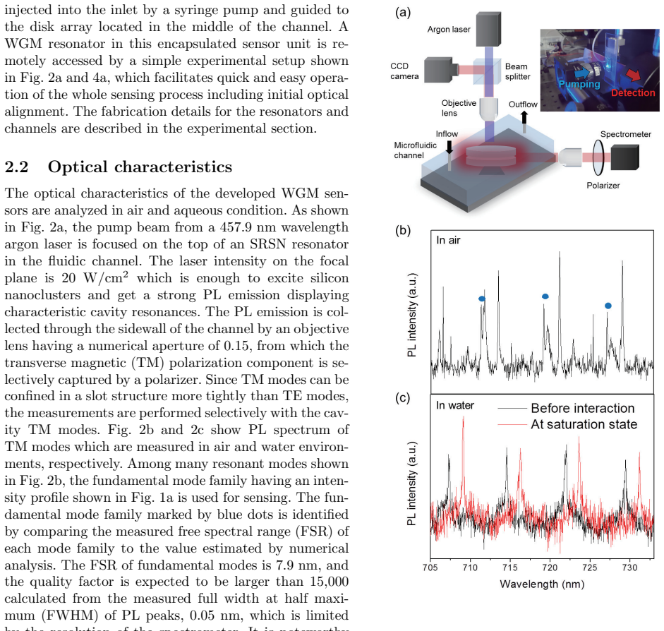
The device is a self-emitting microcavity: silicon nanoclusters in a silicon-rich nitride disk absorb pump light from above and emit a photoluminescence spectrum carrying the cavity resonance, so neither the pump nor the readout needs to physically touch the chip. A pair of disks separated by a $25\ \mathrm{nm}$ slot confines a large share of the optical mode in the solution-filled gap where binding occurs, enhancing sensitivity by an estimated factor of 6.5 over a slot-free resonator. Using the streptavidin-biotin pair, the authors report a real-time sensitivity of $0.012\ \mathrm{nm/nM}$ and a detection limit of $6.7\ \mathrm{nM}$, and with a 365 nm LED pump they demonstrate WGM sensing under direct LED illumination for the first time, measuring bulk refractive-index changes at $226.67\ \mathrm{nm/RIU}$ in agreement with numerical simulation.
Load-bearing premise
The result stands or falls on the assumption that the resonance shift grows linearly with streptavidin concentration across the measured range; the paper's own Hill-Langmuir fit with $K_d = 3.8\times10^{-7}\ \mathrm{M}$ describes a saturating response rather than a constant slope.
Editorial extensions
If this is right
- A WGM sensor chip can be operated by placing it under an LED and reading its photoluminescence through free-space optics, removing fragile tapered fibres and precision waveguide alignment from the sensing workflow.
- The reported sensitivity of $0.012\ \mathrm{nm/nM}$ for streptavidin-biotin is more than 20 times the previous remote-readout WGM result, and the detection limit of $6.7\ \mathrm{nM}$ follows from the measured mode linewidth.
- The $25\ \mathrm{nm}$ slot boosts sensitivity about 6.5-fold over an unslotted resonator while the measured association and dissociation parameters stay close to literature values, suggesting the confinement does not impede binding.
- With a 365 nm LED pump, bulk refractive-index changes are detected at $226.67\ \mathrm{nm/RIU}$, and the estimated detection limit of about 0.25% glycerol shows that the platform works without a laser.
Reading between the lines
- A testable next step is replacing the 365 nm UV LED with a visible-wavelength LED at sufficient intensity, which would let the LED-pumped scheme detect biomolecules directly rather than only refractive-index changes; the authors identify the roughly $20\ \mathrm{W/cm^2}$ visible-intensity level as the threshold.
- Reducing the slot width below $25\ \mathrm{nm}$, toward the size of the streptavidin-biotin complex, could turn the sensor into an instrument for studying how nanoconfinement alters binding kinetics, an extension the authors flag in their conclusion.
- The large-area LED illumination makes arrayed sensing natural: many top-pumped resonators could be read simultaneously by imaging their photoluminescence spectra, something the paper does not demonstrate but its geometry invites.
Signed reviews
Editorial analysis
A structured set of objections, weighed in public.
Referee Report
Summary. The manuscript demonstrates an on-chip whispering-gallery-mode (WGM) biosensor platform based on silicon nanocluster active resonators with a 25-nm slot, integrated with microfluidic channels and operated with free-space optical pumping and readout. The authors report streptavidin-biotin binding measurements with a claimed sensitivity of 0.012 nm/nM and a detection limit of 6.7 nM, kinetic analysis using Langmuir and Hill-Langmuir fits, and a demonstration of LED-pumped operation for bulk refractive-index sensing. The central claims are the first LED-pumped active WGM sensing scheme and a sensitivity improvement of over 20 times relative to prior remote-readout WGM sensors.
Significance. If fully supported, the work would be a practical advance toward label-free on-chip sensors because it eliminates fragile evanescent coupling and uses a low-cost LED for top-illumination. The COMSOL simulation for the slot field confinement is checked against measured bulk refractive-index shifts, which is a genuine strength, and the device integration with microfluidics is convincingly described. However, the headline molecular sensitivity rests on a single unsaturated measurement that contradicts the paper's own kinetic model, and the LED-pumped demonstration is limited to bulk refractometry rather than biomolecule detection. As presented, the quantitative performance claims are not yet established, although the platform concept appears promising.
major comments (3)
- [Sec. 2.3 and 2.4] The sensitivity of 0.012 nm/nM is derived from a single 1.7-nm shift recorded after exposure to 144 nM streptavidin, which the text calls 'saturation.' Yet the Hill-Langmuir fit in Sec. 2.4 reports Kd = 3.8e-7 M; at 144 nM the fitted fractional occupancy is only 144/(144+380) ≈ 0.27, i.e., the reaction is far from saturation. A single ratio Δλ/c at a nonsaturating concentration is therefore not a valid sensitivity, and the value is inconsistent with the kinetic model. Please provide a proper calibration curve, for example using the 9, 36, 54, and 90 nM equilibrium shifts already measured in Fig. 3b, and report the slope in the linear low-concentration regime with replicate devices and error bars, or explicitly state that the reported value is an apparent single-concentration response.
- [Sec. 2.5, Abstract, Conclusions] The LED-pumped operation is demonstrated only for bulk refractive-index sensing with glycerol-water mixtures; the biomolecule (streptavidin-biotin) measurements are performed with an argon-ion laser. The abstract and conclusions state that 'WGM sensing based on the direct illumination of an LED pump is demonstrated for the first time,' and the title refers to pumping by an LED, which may lead readers to infer LED-pumped biosensing. Since the paper explicitly states that UV LED light deforms biomolecules, the current device does not actually demonstrate LED-pumped label-free biosensing. Please either qualify the claims to indicate that LED pumping is shown only for refractometric sensing, or add an experiment with a visible-wavelength LED and biomolecules.
- [Sec. 2.3] The detection limit of 6.7 nM is computed as 0.08 nm (FWHM) divided by 0.012 nm/nM. This calculation inherits the unsupported linear sensitivity, and defining the resolution as a single FWHM is not a statistically meaningful detection limit; typically one uses 3σ of baseline noise or another established criterion on the calibration curve. Please report the spectral noise floor from repeated measurements of a stable resonance and recompute the limit of detection accordingly. In addition, the manuscript does not state how many devices or repeated measurements the 1.7-nm shift is based on; this information is essential for assessing the reproducibility of the headline sensitivity.
minor comments (5)
- [Sec. 2.4] The sentence 'The slightly larger kon of 9 nM streptavidin is caused by the detection limit' appears to contradict the reported kon values (1.2×10^3 M^-1 s^-1 for 9 nM versus 4.2–6.4×10^3 M^-1 s^-1 for the other concentrations). The 9 nM value is actually the smallest, not the largest; please clarify whether this is a misstatement or refers to the uncertainty of the fit.
- [Sec. 2.2 and 2.5] The FSR and Q factor for air with the argon laser are reported as 7.9 nm and greater than 15,000, while the LED measurement in air reports 9.1 nm and 15,000. The source of this discrepancy, possibly a different mode family or device, should be stated explicitly.
- [Sec. 2.3] The term 'sensitivity' is used with different units for biomolecule detection (nm/nM) and bulk sensing (nm/RIU). Please define both clearly and state the sensing area or volume normalization, since the comparison with Ref. [16] may depend on the active surface area and the binding capacity of the slot.
- [Fig. 3b] Please show the fitted Hill-Langmuir parameters with confidence intervals and state whether the fit includes the 144 nM point or is extrapolated to it. This would help reconcile the sensitivity calculation with the kinetic analysis.
- [General] The paper would benefit from a statement of the number of independent devices used for each experiment and the reproducibility across chips, data that are standard for sensor characterization.
Circularity Check
No significant circularity: the central claims are supported by direct measurements and independent simulation validation; the quoted sensitivity is a measured ratio, not a fitted prediction.
full rationale
The paper's main claims—LED-pumped active WGM sensing and slot-enhanced sensitivity—rest on directly measured photoluminescence spectra and resonance peak shifts, not on parameters that are fitted and then repackaged as predictions. The quoted sensitivity of 0.012 nm/nM is the measured 1.7 nm shift divided by the 144 nM streptavidin concentration; it is a direct experimental ratio, and no equation in the paper derives this value from an input model. The COMSOL bulk sensitivity prediction (226.67 nm/RIU) is compared with an independent glycerol-series measurement, and the agreement is used to support the slot simulation; this is validation, not circularity. The kinetics parameters (Kd, kon) are fitted to time-series data under the Langmuir model and then used for internal consistency checks, which is standard modeling rather than circular derivation. The only self-citation (Ref. [17]) is contextual background for expected WGM sensitivity and is not load-bearing. A separate concern—that the linear 'sensitivity' at 144 nM is inconsistent with the fitted Hill-Langmuir Kd = 3.8e-7 M, so the 0.012 nm/nM and 6.7 nM detection limit may be ill-defined—is a correctness or calibration issue, not circularity. No step in the derivation reduces to its own input by construction.
Assumptions & free parameters
free parameters (3)
- Kd (dissociation constant) =
3.8e-7 M
- kon (association rate constant) =
1.2e3 to 6.4e3 M^-1 s^-1
- Maximum binding shift (saturation) =
Not explicitly reported, inferred about 6.2 nm
assumptions (4)
- domain assumption Langmuir adsorption model describes the streptavidin-biotin surface binding kinetics and equilibrium.
- domain assumption COMSOL mode simulations accurately represent the fabricated resonator geometry and refractive indices.
- domain assumption Biotin immobilization via APTES on the silicon nitride surface is stable and functional.
- domain assumption The tracked PL peaks belong to the same fundamental TM mode family across conditions.
Cite this review
Pith. "Pith review of On-chip label-free biosensing based on active whispering gallery mode resonators pumped by a light-emitting diode." pith.science (2026). https://pith.science/paper/PPJMAAW5
@misc{pith2026190804506,
  author       = {Pith},
  title        = {Pith review of: On-chip label-free biosensing based on active whispering gallery mode resonators pumped by a light-emitting diode},
  year         = {2026},
  howpublished = {\url{https://pith.science/paper/PPJMAAW5}},
  note         = {Machine review of arXiv:1908.04506}
}
read the original abstract
Biosensing based on whispering-gallery mode (WGM) resonators has been continuously studied with great attention due to its excellent sensitivity guaranteeing the label-free detection. However, its practical impact is insignificant to date despite notable achievements in academic research. Here, we demonstrate a novel practical platform of on-chip WGM sensors integrated with microfluidic channels. By placing silicon nanoclusters as a stable active compound in micro-resonators, the sensor chip can be operated with a remote pump and readout, which simplifies the chip integration and connection to the external setup. In addition, silicon nanoclusters having large absorption cross-section over broad wavelength range allow active sensing for the first time with an LED pump in a top-illumination scheme which significantly reduces the complexity and cost of the measurement setup. The nano-slot structure of 25 nm gap width is embedded in the resonator where the target bio-molecules are selectively detected with the sensitivity enhanced by strongly confined mode-field. The sensitivity confirmed by real-time measurements for the streptavidin-biotin complex is 0.012 nm/nM, improved over 20 times larger than the previously reported WGM sensors with remote readout.
Figures


Figures from the paper (1 more)

Reference graph
Works this paper leans on
-
[16]
S. F. Wondimu, M. Hippler, C. Hussal, A. Hofmann, S. Kr”ammer, J. Lahann, H. Kalt, W. Freude, and C. Koos. Robust label-free biosensing using mi- crodisk laser arrays with on-chip references. Optics express, 26(3):3161–3173, 2018
work page 2018
-
[1]
E. Kim, M. D. Baaske, and F. Vollmer. To- wards next-generation label-free biosensors: recent advances in whispering gallery mode sensors. Lab on a Chip , 17(7):1190–1205, 2017
work page 2017
-
[2]
F. Vollmer and S. Arnold. Whispering-gallery- mode biosensing: label-free detection down to single molecules. Nature methods, 5(7):591, 2008
work page 2008
-
[3]
F. Vollmer and L. Yang. Review label-free detec- tion with high-q microcavities: a review of biosens- ing mechanisms for integrated devices. Nanophoton- ics, 1(3-4):267–291, 2012
work page 2012
-
[4]
S. Subramanian, H.-Y. Wu, T. Constant, J. Xavier, and F. Vollmer. Label-free optical single-molecule micro-and nanosensors. Advanced Materials , 30(51):1801246, 2018
work page 2018
-
[5]
Single-molecule nucleic acid interactions monitored on a label-free microcavity biosensor platform
Martin D Baaske, Matthew R Foreman, and Frank Vollmer. Single-molecule nucleic acid interactions monitored on a label-free microcavity biosensor platform. Nature nanotechnology, 9(11):933, 2014
work page 2014
-
[6]
L. He, S ¸. K.¨Ozdemir, J. Zhu, W. Kim, and L. Yang. Detecting single viruses and nanoparticles using whispering gallery microlasers. Nature nanotechnol- ogy, 6(7):428, 2011
work page 2011
-
[7]
A. M. Armani, R. P. Kulkarni, S. E. Fraser, R. C. Flagan, and K. J. Vahala. Label-free, single- molecule detection with optical microcavities. sci- ence, 317(5839):783–787, 2007
work page 2007
Show all 40 references
-
[8]
On-chip single nanoparticle detection and sizing by mode splitting in an ultrahigh-q microresonator
Jiangang Zhu, Sahin Kaya Ozdemir, Yun-Feng Xiao, Lin Li, Lina He, Da-Ren Chen, and Lan Yang. On-chip single nanoparticle detection and sizing by mode splitting in an ultrahigh-q microresonator. Nature photonics, 4(1):46, 2010
2010
-
[9]
Arnold, S
S. Arnold, S. I. Shopova, and S. Holler. Whisper- ing gallery mode bio-sensor for label-free detection of single molecules: thermo-optic vs. reactive mech- anism. Optics Express, 18(1):281–287, 2010
2010
-
[10]
Whispering gallery mode sensors
Matthew R Foreman, Jon D Swaim, and Frank Vollmer. Whispering gallery mode sensors. Ad- vances in optics and photonics , 7(2):168–240, 2015
2015
-
[11]
Wienhold, S
T. Wienhold, S. Kraemmer, S. F. Wondimu, T. Siegle, U. Bog, U. Weinzierl, S. Schmidt, H. Becker, H. Kalt, T. Mappes, S. Koeberaf, and C. Koos. All-polymer photonic sensing plat- form based on whispering-gallery mode microgoblet lasers. Lab on a Chip , 15(18):3800–3806, 2015
2015
-
[12]
X. Xu, W. Chen, G. Zhao, Y. Li, C. Lu, and L. Yang. Wireless whispering-gallery-mode sensor for thermal sensing and aerial mapping. Light: Science & Ap- plications, 7(1):62, 2018
2018
-
[13]
J. C. Knight, G. Cheung, F. Jacques, and T. A. Birks. Phase-matched excitation of whispering- gallery-mode resonances by a fiber taper. Optics letters, 22(15):1129–1131, 1997
1997
-
[14]
C. A. Barrios. Integrated microring resonator sensor arrays for labs-on-chips. Analytical and bioanalytical chemistry, 403(6):1467–1475, 2012
2012
-
[15]
Wienhold, S
T. Wienhold, S. Kraemmer, A. Bacher, H. Kalt, C. Koos, S. Koeber, and T. Mappes. Efficient free-space read-out of wgm lasers using circular mi- cromirrors. Optics express, 23(2):1025–1034, 2015
2015
-
[17]
T. Lu, H. Lee, T. Chen, S. Herchak, J.-H. Kim, S. E. Fraser, R. C. Flagan, and K. J. Vahala. High sen- sitivity nanoparticle detection using optical micro- cavities. Proceedings of the National Academy of Sciences, 108(15):5976–5979, 2011
2011
-
[18]
J. Su, A. FG Goldberg, and B. M. Stoltz. Label- free detection of single nanoparticles and biologi- cal molecules using microtoroid optical resonators. Light: Science & Applications , 5(1):e16001, 2016
2016
-
[19]
F. F. Lupi, D. Navarro-Urrios, J. Monserrat, C. Dominguez, P. Pellegrino, and B. Garrido. High q light-emitting si-rich si 3 n 4 microdisks. Optics Letters, 36(8):1344–1346, 2011. 7
2011
-
[20]
Kim and J
G. Kim and J. H. Shin. Luminescent silicon-rich nitride horizontal air-slot microdisk resonators for biosensing. IEEE Photonics Technology Letters , 28(21):2331–2334, 2016
2016
-
[21]
Band gap engineering of amorphous silicon quan- tum dots for light-emitting diodes
Nae-Man Park, Tae-Soo Kim, and Seong-Ju Park. Band gap engineering of amorphous silicon quan- tum dots for light-emitting diodes. Applied Physics Letters, 78(17):2575–2577, 2001
2001
-
[22]
Direct observation of quantum confine- ment of si nanocrystals in si-rich nitrides
PD Nguyen, DM Kepaptsoglou, QM Ramasse, and A Olsen. Direct observation of quantum confine- ment of si nanocrystals in si-rich nitrides. Physical Review B, 85(8):085315, 2012
2012
-
[23]
Quantum confinement in amor- phous silicon quantum dots embedded in silicon ni- tride
Nae-Man Park, Chel-Jong Choi, Tae-Yeon Seong, and Seong-Ju Park. Quantum confinement in amor- phous silicon quantum dots embedded in silicon ni- tride. Physical review letters , 86(7):1355, 2001
2001
-
[24]
V. R. Almeida, Q. Xu, C. A. Barrios, and M. Lipson. Guiding and confining light in void nanostructure. Optics letters , 29(11):1209–1211, 2004
2004
-
[25]
Scullion, T
M. Scullion, T. Krauss, and A. Di Falco. Slotted photonic crystal sensors. Sensors, 13(3):3675–3710, 2013
2013
-
[26]
W. Liu, S. K. Samanta, B. D. Smith, and L. Isaacs. Synthetic mimics of biotin/(strept) avidin.Chemical Society Reviews, 46(9):2391–2403, 2017
2017
-
[27]
Halperin, A
A. Halperin, A. Buhot, and E. B. Zhulina. On the hybridization isotherms of dna microarrays: the langmuir model and its extensions. Journal of Physics: Condensed Matter , 18(18):S463, 2006
2006
-
[28]
A. L. Washburn, L. C. Gunn, and R. C. Bailey. Label-free quantitation of a cancer biomarker in complex media using silicon photonic microring res- onators. Analytical chemistry , 81(22):9499–9506, 2009
2009
-
[29]
R. A. Latour. The langmuir isotherm: a commonly applied but misleading approach for the analysis of protein adsorption behavior. Journal of Biomedical Materials Research Part A , 103(3):949–958, 2015
2015
-
[30]
B. M. Lowe, K. Sun, I. Zeimpekis, C.-K. Skylaris, and N. G. Green. Field-effect sensors–from ph sensing to biosensing: sensitivity enhancement us- ing streptavidin–biotin as a model system. Analyst, 142(22):4173–4200, 2017
2017
-
[31]
D. A. Hanaor, M. Ghadiri, W. Chrzanowski, and Y. Gan. Scalable surface area characterization by electrokinetic analysis of complex anion adsorption. Langmuir, 30(50):15143–15152, 2014
2014
-
[32]
The adsorption of gases on plane surfaces of glass, mica and platinum
Irving Langmuir. The adsorption of gases on plane surfaces of glass, mica and platinum. Journal of the American Chemical society , 40(9):1361–1403, 1918
1918
-
[33]
Interpreting complex binding kinetics from optical biosensors: a comparison of analysis by lin- earization, the integrated rate equation, and numeri- cal integration
Thomas A Morton, David G Myszka, and Irwin M Chaiken. Interpreting complex binding kinetics from optical biosensors: a comparison of analysis by lin- earization, the integrated rate equation, and numeri- cal integration. Analytical biochemistry, 227(1):176– 185, 1995
1995
-
[34]
Crossed surface relief gratings as nanoplas- monic biosensors
Srijit Nair, Carlos Escobedo, and Ribal Georges Sabat. Crossed surface relief gratings as nanoplas- monic biosensors. ACS sensors, 2(3):379–385, 2017
2017
-
[35]
D’Agata, G
R. D’Agata, G. Grasso, and G. Spoto. Real-time binding kinetics monitored with surface plasmon resonance imaging in a diffusion-free environment. Open Spectrosc J, 2:1–9, 2008
2008
-
[36]
M. J. Eddowes. Direct immunochemical sensing: ba- sic chemical principles and fundamental limitations. Biosensors, 3(1):1–15, 1987
1987
-
[37]
S. V. Deshpande, E. Gulari, S. W. Brown, and S. C. Rand. Optical properties of silicon nitride films de- posited by hot filament chemical vapor deposition. Journal of Applied Physics , 77(12):6534–6541, 1995
1995
-
[38]
L. F. Hoyt. New table of the refractive index of pure glycerol at 20 c. Industrial & Engineering Chem- istry, 26(3):329–332, 1934
1934
-
[39]
Abdelgawad, M
M. Abdelgawad, M. W. Watson, E. W. Young, J. M. Mudrik, M. D. Ungrin, and A. R. Wheeler. Soft lithography: masters on demand. Lab on a Chip , 8(8):1379–1385, 2008
2008
-
[40]
E. H. Williams, A. V. Davydov, A. Motayed, S. G. Sundaresan, P. Bocchini, L. J. Richter, G. Stan, K. Steffens, R. Zangmeister, J. A. Schreifels, and M. V. Rao. Immobilization of streptavidin on 4h–sic for biosensor development. Applied surface science , 258(16):6056–6063, 2012. 8
2012
Reviewed August 14, 2026 · model on record in the stance chip above.
Discussion (0). Continue with ORCID to comment.