Topological Methods for Characterising Spatial Networks: A Case Study in Tumour Vasculature
Pith reviewed 2026-05-24 18:36 UTC · model grok-4.3
The pith
Topological data analysis can characterise tumour vasculature from spatial network images.
A machine-rendered reading of the paper's core claim, the machinery that carries it, and where it could break.
Core claim
TDA of spatial network structure can be used to characterise tumour vasculature, as shown in preliminary work applying these methods to vessel images.
What carries the argument
Topological data analysis (TDA), algorithmic methods that identify global and multi-scale structures in high-dimensional, noisy data sets such as vessel network images.
Load-bearing premise
Topological summaries from vessel images will capture biologically relevant differences in network function or disease state.
What would settle it
TDA summaries computed on matched sets of healthy and tumour vessel images show no statistically significant differences that align with independent measures of vessel function or disease markers.
Figures
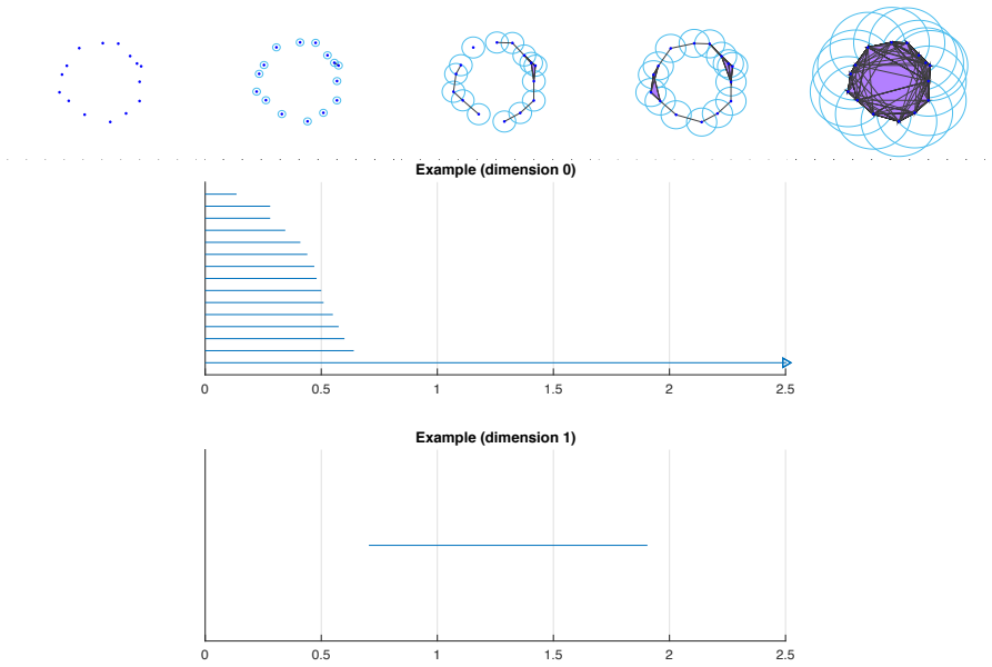


read the original abstract
Understanding how the spatial structure of blood vessel networks relates to their function in healthy and abnormal biological tissues could improve diagnosis and treatment for diseases such as cancer. New imaging techniques can generate multiple, high-resolution images of the same tissue region, and show how vessel networks evolve during disease onset and treatment. Such experimental advances have created an exciting opportunity for discovering new links between vessel structure and disease through the development of mathematical tools that can analyse these rich datasets. Here we explain how topological data analysis (TDA) can be used to study vessel network structures. TDA is a growing field in the mathematical and computational sciences, that consists of algorithmic methods for identifying global and multi-scale structures in high-dimensional data sets that may be noisy and incomplete. TDA has identified the effect of ageing on vessel networks in the brain and more recently proposed to study blood flow and stenosis. Here we present preliminary work which shows how TDA of spatial network structure can be used to characterise tumour vasculature.
Editorial analysis
A structured set of objections, weighed in public.
Referee Report
Summary. The manuscript presents preliminary work applying topological data analysis (TDA), including persistent homology on spatial graphs, to characterise the structure of tumour vasculature networks extracted from imaging data. It reviews TDA methods, notes prior applications to brain vessel networks and blood flow, and sketches their use on tumour data to identify global and multi-scale features in vessel structures.
Significance. If the pipeline produces reproducible topological summaries on vessel images, the approach could complement existing network metrics and support future studies linking vessel topology to disease states or treatment response. The modest existence claim for a preliminary demonstration carries limited immediate significance but illustrates a potentially extensible method for high-resolution biological network data.
minor comments (3)
- The abstract states the opportunity for discovering links between vessel structure and disease but provides no concrete topological features, quantitative results, or comparison to standard metrics; adding one or two illustrative examples from the case study would strengthen the claim that TDA characterises the networks.
- Section describing the vessel graph construction and filtration (likely §3 or §4) should explicitly state the choice of distance or weight function used for the spatial network and any preprocessing steps applied to the imaging data.
- The manuscript is labeled preliminary; a short concluding paragraph outlining the specific next steps (e.g., statistical comparison across disease stages) would clarify the scope and avoid overstatement of current findings.
Simulated Author's Rebuttal
We thank the referee for their constructive review and recommendation of minor revision. The report correctly identifies the work as preliminary and notes its potential extensibility. No specific major comments were provided in the report.
Circularity Check
No significant circularity identified
full rationale
The paper presents preliminary application of standard persistent homology methods from TDA to spatial vessel graphs extracted from tumour images. No derivation chain, fitted parameters, or predictions are defined; the central claim is an existence statement that the pipeline can be executed to produce topological summaries. The abstract and text invoke prior TDA applications in biology only as motivation, not as load-bearing self-citations that define the result. The argument is self-contained against external benchmarks of TDA usage on networks and requires no internal reduction of outputs to inputs by construction.
Axiom & Free-Parameter Ledger
axioms (1)
- domain assumption Betti numbers and persistence diagrams computed from spatial graphs capture multi-scale network structure relevant to biology
Lean theorems connected to this paper
-
IndisputableMonolith/Foundation/AlexanderDuality.leanalexander_duality_circle_linking unclear?
unclearRelation between the paper passage and the cited Recognition theorem.
We characterise the unique features of tumour blood vessels, in particular the loops and the high degree of tortuosity, using persistent homology. ... radial filtration ... barcodes in dimension 0 and dimension 1
-
IndisputableMonolith/Cost/FunctionalEquation.leanwashburn_uniqueness_aczel unclear?
unclearRelation between the paper passage and the cited Recognition theorem.
TDA has identified the effect of ageing on vessel networks in the brain and more recently proposed to study blood flow and stenosis.
What do these tags mean?
- matches
- The paper's claim is directly supported by a theorem in the formal canon.
- supports
- The theorem supports part of the paper's argument, but the paper may add assumptions or extra steps.
- extends
- The paper goes beyond the formal theorem; the theorem is a base layer rather than the whole result.
- uses
- The paper appears to rely on the theorem as machinery.
- contradicts
- The paper's claim conflicts with a theorem or certificate in the canon.
- unclear
- Pith found a possible connection, but the passage is too broad, indirect, or ambiguous to say the theorem truly supports the claim.
Reference graph
Works this paper leans on
-
[1]
J. W. Baish, Y. Gazit, D. a. Berk, M. Nozue, L. T. Baxter, and R. K. Jain , Role of tumor vascular architecture in nutrient and drug delivery: an invasion percolation-based network model. , Microvascular Research, 51 (1996), pp. 327–46
work page 1996
- [2]
-
[3]
P. Bendich, J. S. Marron, E. Miller, A. Pieloch, and S. Skwerer , Persistent homology analysis of brain artery trees , Annals of Applied Statistics, 10 (2016), pp. 198–218
work page 2016
-
[4]
Carlsson, Topology and data , Bulletin of the American Mathematical Society, 46 (2009), pp
G. Carlsson, Topology and data , Bulletin of the American Mathematical Society, 46 (2009), pp. 255–308
work page 2009
-
[5]
G. Carlsson and A. Zomorodian , The Theory of Multidimensional Persistence , Discrete & Computational Geometry, 42 (2009), pp. 71–93
work page 2009
-
[6]
D. Cohen-Steiner, H. Edelsbrunner, and J. Harer , Stability of persistence diagrams , Discrete & Com- putational Geometry, (2007), pp. 103–120
work page 2007
-
[7]
H. Edelsbrunner and J. Harer , Computational Topology, American Mathematical Soc., 2010
work page 2010
-
[8]
D. R. Grimes, P. Kannan, D. R. Warren, B. Markelc, R. Bates, R. Muschel, and M. Partridge , Estimating oxygen distribution from vasculature in three-dimensional tumour tissue , Journal of the Royal Society Interface, 13 (2016), p. 20160070
work page 2016
-
[9]
J. A. Grogan, A. J. Connor, B. Markelc, R. J. Muschel, P. K. Maini, H. M. Byrne, and J. M. Pitt-Francis, Microvessel chaste: an open library for spatial modeling of vascularized tissues , Biophysical Journal, 112 (2017), pp. 1767–1772
work page 2017
-
[10]
J. A. Grogan, B. Markelc, A. J. Connor, R. J. Muschel, J. M. Pitt-Francis, P. K. Maini, and H. M. Byrne , Predicting the influence of microvascular structure on tumor response to radiotherapy , IEEE Transactions on Biomedical Engineering, 64 (2016), pp. 504–511
work page 2016
-
[11]
R. K. Jain, Determinants of tumor blood flow: a review. , Cancer Res., 48 (1988), pp. 2641–58
work page 1988
-
[12]
R. K. Jain , Normalization of tumor vasculature: an emerging concept in antiangiogenic therapy. , Science, 307 (2005), pp. 58–62
work page 2005
-
[13]
M. Kahle, Topology of random simplicial complexes: a survey , AMS Contemporary Mathematics, 620 (2014), pp. 201–222
work page 2014
- [14]
-
[15]
N. V. Mantzaris, S. Webb, and H. G. Othmer , Mathematical modeling of tumor-induced angiogenesis , Journal of Mathematical Biology, 49 (2004), pp. 111–187
work page 2004
-
[16]
J. Nicponski and J.-H. Jung, Topological data analysis of vascular disease: A theoretical framework , BioRxiv, (2019), p. 637090
work page 2019
- [17]
-
[18]
S. M. Peirce, Computational and mathematical modeling of angiogenesis , Microcirculation, 15 (2008), pp. 739– 751
work page 2008
-
[19]
3D hybrid modeling of vascular network formation
H. Perfahl, B. D. Hughes, T. Alarcon, P. K. Maini, M. Lloyd, M. Reuss, and H. Byrne, 3D hybrid modelling of vascular network formation , arXiv:1610.00661, (2016)
work page internal anchor Pith review Pith/arXiv arXiv 2016
-
[20]
M. Scianna, C. Bell, and L. Preziosi , A review of mathematical models for the formation of vascular networks, Journal of theoretical biology, 333 (2013), pp. 174–209
work page 2013
- [21]
-
[22]
G. M. Tozer, S. M. Ameer-beg, J. Baker, P. R. Barber, S. A. Hill, R. J. Hodgkiss, R. Locke, et al. , Intravital imaging of tumour vascular networks using multi-photon fluorescence microscopy , Advanced Drug Delivery Reviews, 57 (2005), pp. 135–152
work page 2005
-
[23]
A. Zomorodian and G. Carlsson, Computing persistent homology , Discrete & Computational Geometry, 33 (2005), pp. 249–274
work page 2005
-
[24]
A. J. Zomorodian, Topology for Computing, Cambridge, 2005
work page 2005
discussion (0)
Sign in with ORCID, Apple, or X to comment. Anyone can read and Pith papers without signing in.