Spectral DiffuserScope: a compact snapshot hyperspectral microscope
Pith reviewed 2026-07-01 01:23 UTC · model grok-4.3
The pith
A compact device uses compressed sensing to capture higher-resolution hyperspectral images in a single snapshot on standard microscopes.
A machine-rendered reading of the paper's core claim, the machinery that carries it, and where it could break.
Core claim
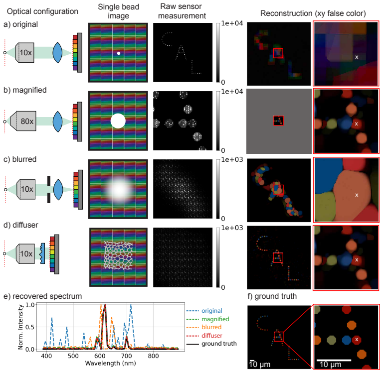
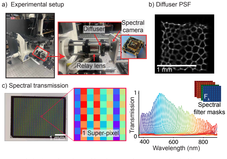
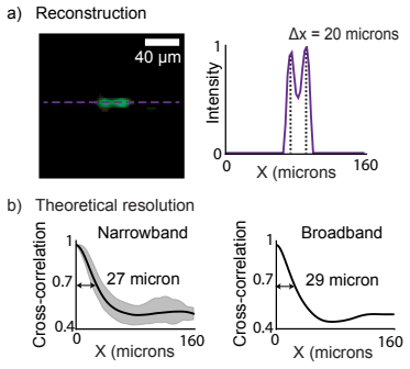
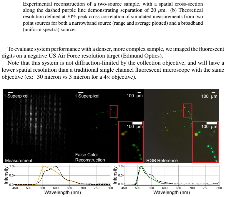
The device is a compact optical layout roughly 15 cm by 6 cm by 6 cm that encodes spectral information via a diffuser and recovers it through compressed-sensing reconstruction, delivering higher spatial-spectral resolution than conventional snapshot methods when tested on real fluorescent samples.
What carries the argument
Spectral DiffuserScope, the compact optical attachment that applies a diffuser pattern to enable compressed-sensing snapshot capture and reconstruction of hyperspectral volumes.
If this is right
- Snapshot hyperspectral imaging becomes feasible on existing fluorescence microscopes without added bulk or mechanical scanning.
- Higher spatial-spectral detail than prior snapshot systems is obtained for fluorescent bead and cell samples.
- The approach supports high-throughput biological and clinical measurements by removing the time cost of scanning.
- The compact size enables portable or integrated use in standard lab setups.
Where Pith is reading between the lines
- Similar diffuser-based encoding could be tested on other modalities such as Raman or bright-field microscopy.
- Integration with existing microscope software pipelines would need to be checked for routine lab adoption.
- Performance on thicker or more scattering samples remains an open question beyond the demonstrated thin preparations.
Load-bearing premise
The compressed-sensing algorithm recovers the stated resolution gains on actual biological samples using only the compact layout and without sample-specific extra calibration.
What would settle it
Side-by-side comparison of images from the device against scanned reference hyperspectral data on the same labeled cell samples, checking whether measured spatial and spectral resolution matches the simulation benchmarks.
Figures


read the original abstract
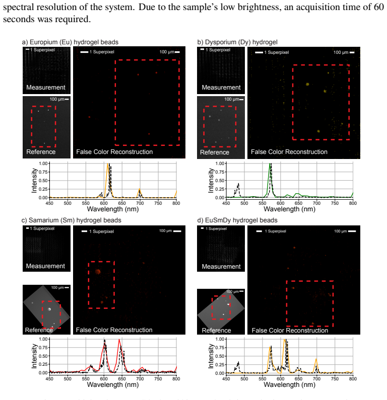
Hyperspectral fluorescence microscopy enables important biological and clinical applications, but conventional systems are bulky or require scanning, limiting temporal resolution and throughput. We introduce a computational snapshot hyperspectral microscope that uses compressed sensing to achieve higher spatial-spectral resolution than traditional snapshot systems. Our device is compact (~15 cm x 6 cm x 6 cm) and easily attaches to standard fluorescence microscopes. We benchmark our system against existing snapshot methods through simulations to evaluate its spatial and spectral performance. Experimental imaging of fluorescent beads, labeled cells, and lanthanide hydrogel beads demonstrates a practical, high-throughput solution for hyperspectral microscopy in biological and clinical applications.
Editorial analysis
A structured set of objections, weighed in public.
Referee Report
Summary. The manuscript introduces Spectral DiffuserScope, a compact (~15 cm × 6 cm × 6 cm) snapshot hyperspectral microscope that attaches to standard fluorescence microscopes and employs compressed sensing to achieve higher spatial-spectral resolution than traditional snapshot systems. It reports simulations benchmarking spatial and spectral performance plus experimental imaging of fluorescent beads, labeled cells, and lanthanide hydrogel beads as a practical high-throughput solution.
Significance. If the claimed resolution gains are quantitatively validated on real samples without sample-specific tuning, the compact, non-scanning design would offer a useful advance for biological and clinical hyperspectral fluorescence microscopy where bulk or scanning systems limit throughput.
major comments (2)
- [Abstract] Abstract: The central claim of higher spatial-spectral resolution than traditional snapshot systems is presented as demonstrated by simulations and experiments, yet the text supplies no quantitative metrics, error bars, resolution values, or reconstruction details; this absence prevents verification that the compact diffuser-based layout plus algorithm actually delivers the stated gains on the physical device.
- [Abstract] Abstract / Results: The weakest assumption—that the compressed-sensing reconstruction produces the claimed resolution improvements on fluorescent beads, cells, and hydrogels without post-hoc parameter adjustment or per-sample calibration—is not addressed by any reported controls or quantitative comparison to prior snapshot methods.
minor comments (1)
- [Abstract] The device dimensions are given but no optical layout diagram or ray-trace is referenced in the abstract; adding a figure citation would clarify the compact attachment mechanism.
Simulated Author's Rebuttal
We thank the referee for their detailed review and constructive feedback. We address each major comment below and have made revisions to strengthen the abstract and clarify the results.
read point-by-point responses
-
Referee: [Abstract] Abstract: The central claim of higher spatial-spectral resolution than traditional snapshot systems is presented as demonstrated by simulations and experiments, yet the text supplies no quantitative metrics, error bars, resolution values, or reconstruction details; this absence prevents verification that the compact diffuser-based layout plus algorithm actually delivers the stated gains on the physical device.
Authors: We agree that the abstract, as a concise summary, omitted specific quantitative metrics. In the revised manuscript we have added key resolution values (spatial and spectral) with error bars from the simulation benchmarks, along with a brief reference to the compressed-sensing reconstruction details that are fully described in the Methods and Results sections. revision: yes
-
Referee: [Abstract] Abstract / Results: The weakest assumption—that the compressed-sensing reconstruction produces the claimed resolution improvements on fluorescent beads, cells, and hydrogels without post-hoc parameter adjustment or per-sample calibration—is not addressed by any reported controls or quantitative comparison to prior snapshot methods.
Authors: The manuscript reports quantitative benchmarking against prior snapshot methods via simulations in the Results section. For the experimental demonstrations, a single fixed parameter set (determined from an initial calibration phantom) was used across all samples without per-sample retuning; this is stated in the Methods. We have added explicit language to both the abstract and Results to highlight the fixed-parameter protocol and to reference the simulation-based quantitative comparisons. Additional experimental controls comparing to other snapshot systems on the same physical samples were not performed and would require new data collection. revision: partial
Circularity Check
No circularity in derivation chain; empirical device and reconstruction treated as independent from inputs
full rationale
The paper introduces a compact hyperspectral microscope using compressed sensing, benchmarks via simulations, and demonstrates via experiments on beads and cells. No equations, fitted parameters renamed as predictions, self-definitional claims, or load-bearing self-citations appear in the provided text. The resolution gains are presented as outcomes of the hardware plus algorithm applied to data, not quantities defined by the method itself. The central claim remains falsifiable against external snapshot systems without reducing to its own inputs by construction.
Axiom & Free-Parameter Ledger
Reference graph
Works this paper leans on
-
[1]
Spectralimagingandlinearunmixing,
G.McNamara,J.M.Larson,S.A.Schwartz,andM.W.Davidson,“Spectralimagingandlinearunmixing,” https:// www.microscopyu.com/techniques/confocal/spectral-imaging-and-linear-unmixing (2024). Accessed: 2024-10-7
2024
-
[2]
A pooled cell painting CRISPR screening platform enables de novo inference of gene function by self-supervised deep learning,
S. Sivanandan, B. Leitmann, E. Lubeck, M. M. Sultan, P. Stanitsas, N. Ranu, A. Ewer, J. E. Mancuso, Z. F. Phillips, A. Kim, J. W. Bisognano, J. Cesarek, F. Ruggiu, D. Feldman, D. Koller, E. Sharon, A. Kaykas, M. R. Salick, and C. Chu, “A pooled cell painting CRISPR screening platform enables de novo inference of gene function by self-supervised deep learn...
2023
-
[3]
High content screening: seeing is believing,
F. Zanella, J. B. Lorens, and W. Link, “High content screening: seeing is believing,” Trends Biotechnol.28, 237–245 (2010)
2010
-
[4]
Hyperspectral imaging of biological targets: the difference a high resolution spectral dimension and multivariate analysis can make,
J. Timlin, M. Sinclair, D. Haaland, J. Martinez, M. Manginell, S. Brozik, J. Guzowski, and M. Werner-Washburne, “Hyperspectral imaging of biological targets: the difference a high resolution spectral dimension and multivariate analysis can make,” in2004 2nd IEEE International Symposium on Biomedical Imaging: Nano to Macro (IEEE Cat No. 04EX821),(2004), pp...
2004
-
[5]
SCOPE-seq: a scalable technology for linking live cell imaging and single-cell RNA sequencing,
J. Yuan, J. Sheng, and P. A. Sims, “SCOPE-seq: a scalable technology for linking live cell imaging and single-cell RNA sequencing,” Genome Biol.19, 227 (2018)
2018
-
[6]
MRBLES 2.0: High-throughput generation of chemicallyfunctionalizedspectrallyandmagneticallyencodedhydrogelbeadsusingasimplesingle-layermicrofluidic device,
Y. Feng, A. K. White, J. B. Hein, E. A. Appel, and P. M. Fordyce, “MRBLES 2.0: High-throughput generation of chemicallyfunctionalizedspectrallyandmagneticallyencodedhydrogelbeadsusingasimplesingle-layermicrofluidic device,” Microsyst Nanoeng6, 109 (2020)
2020
-
[7]
Programmable microfluidic synthesis of over one thousand uniquely identifiable spectral codes,
H.Q.Nguyen,B.C.Baxter,K.Brower,C.A.Diaz-Botia,J.L.DeRisi,P.M.Fordyce,andK.S.Thorn,“Programmable microfluidic synthesis of over one thousand uniquely identifiable spectral codes,” Adv Opt Mater5(2017)
2017
-
[8]
Programmable microfluidic synthesis of spectrally encoded microspheres,
R. E. Gerver, R. Gómez-Sjöberg, B. C. Baxter, K. S. Thorn, P. M. Fordyce, C. A. Diaz-Botia, B. A. Helms, and J. L. DeRisi, “Programmable microfluidic synthesis of spectrally encoded microspheres,” Lab Chip12, 4716–4723 (2012)
2012
-
[9]
Fast and robust pushbroom hyperspectral imaging via DMD-based scanning
R.Arablouei,E.Goan,S.Gensemer,andB.Kusy,“FastandrobustpushbroomhyperspectralimagingviaDMD-based scanning,” (2016), p. 99480A. ArXiv:1608.00361 [cs]
work page internal anchor Pith review Pith/arXiv arXiv 2016
-
[10]
Hyperspectral push-broom imager using a volume Bragg grating as an angular filter,
J.-H. Song and Y.-H. Kwon, “Hyperspectral push-broom imager using a volume Bragg grating as an angular filter,” Opt. Express32, 8736–8750 (2024)
2024
-
[11]
Tunablethin-filmopticalfiltersforhyperspectralmicroscopy,
P.F.Favreau,T.C.Rich,P.Prabhat,andS.J.Leavesley,“Tunablethin-filmopticalfiltersforhyperspectralmicroscopy,” inThree-Dimensional and Multidimensional Microscopy: Image Acquisition and Processing XX,vol. 8589 (SPIE, 2013), pp. 112–116
2013
-
[12]
Snapshot hyperspectral light-sheet imaging of signal transduction in live pancreatic islets,
Z. Lavagnino, J. Dwight, A. Ustione, T.-U. Nguyen, T. S. Tkaczyk, and D. W. Piston, “Snapshot hyperspectral light-sheet imaging of signal transduction in live pancreatic islets,” Biophys. J.111, 409–417 (2016)
2016
-
[13]
High throughput multichannel fluorescence microscopy with microlens arrays,
A. Orth and K. B. Crozier, “High throughput multichannel fluorescence microscopy with microlens arrays,” Opt. Express22, 18101–18112 (2014)
2014
-
[14]
Ultrahigh-throughput single-molecule spectroscopy and spectrally resolved super-resolution microscopy,
Z. Zhang, S. J. Kenny, M. Hauser, W. Li, and K. Xu, “Ultrahigh-throughput single-molecule spectroscopy and spectrally resolved super-resolution microscopy,” Nat. Methods12, 935–938 (2015)
2015
-
[15]
Snapshot hyperspectral volumetric microscopy,
J. Wu, B. Xiong, X. Lin, J. He, J. Suo, and Q. Dai, “Snapshot hyperspectral volumetric microscopy,” Sci. Rep.6, 24624 (2016)
2016
-
[16]
Identification of fluorescent beads using a coded aperture snapshot spectral imager,
C. F. Cull, K. Choi, D. J. Brady, and T. Oliver, “Identification of fluorescent beads using a coded aperture snapshot spectral imager,” Appl. Opt.49, B59–70 (2010)
2010
-
[17]
Spectral DiffuserCam: lensless snapshot hyperspectral imaging with a spectral filter array,
K. Monakhova, K. Yanny, N. Aggarwal, and L. Waller, “Spectral DiffuserCam: lensless snapshot hyperspectral imaging with a spectral filter array,” Optica7, 1298 (2020)
2020
-
[18]
Adam: A method for stochastic optimization,
D. P. Kingma and J. Ba, “Adam: A method for stochastic optimization,” arXiv [cs.LG] (2014)
2014
-
[19]
Design and single-shot fabrication of lensless cameras with arbitrary point spread functions,
K. C. Lee, J. Bae, N. Baek, J. Jung, W. Park, and S. A. H. Lee, “Design and single-shot fabrication of lensless cameras with arbitrary point spread functions,” Optica10, 72–80 (2023)
2023
-
[20]
Recapitulating complex biological signaling environments using a multiplexed, DNA-patterning approach,
O.J.Scheideler,C.Yang,M.Kozminsky,K.I.Mosher,R.Falcón-Banchs,E.C.Ciminelli,A.W.Bremer,S.A.Chern, D. V. Schaffer, and L. L. Sohn, “Recapitulating complex biological signaling environments using a multiplexed, DNA-patterning approach,” Sci. Adv.6, eaay5696 (2020)
2020
-
[21]
Improved fabrication and calibration for snapshot computational hyperspectral imaging,
Y. Raniwala, N. Aggarwal, and L. Waller, “Improved fabrication and calibration for snapshot computational hyperspectral imaging,” inSPIE Proceedings,vol. 12363 (2023), pp. 1236306–1236306–7
2023
-
[22]
Exploring alternative designs for a computational hyperspectral imager for microscopy,
Y. Raniwala, “Exploring alternative designs for a computational hyperspectral imager for microscopy,” Master’s thesis, EECS Department, University of California at Berkeley (2023)
2023
-
[23]
Phasor-based hyperspectral snapshot microscopy allows fast imaging of live, three-dimensional tissues for biomedical applications,
P. N. Hedde, R. Cinco, L. Malacrida, A. Kamaid, and E. Gratton, “Phasor-based hyperspectral snapshot microscopy allows fast imaging of live, three-dimensional tissues for biomedical applications,” Commun Biol4, 721 (2021). Supplementary Information for Spectral DiffuserScope S1. Supplementary methods S1.1. Bill of materials Item Part Number Quantity Ident...
2021
-
[24]
Assemble the spectral camera according to the instructions in main text Section 3. Fig. S1. Drawing for the camera adapter plate. All dimensions in millimeters
-
[26]
Assemble using the instructions in Supplementary Info S1.2.1
-
[27]
Calibrate the diffuser point spread function using the instructions in main text Section 3.2
-
[28]
Slidethespectralcamerausingthe60mmtranslationplate(partK)sothatthepointspread function now lands on the center of the spectral filter array
-
[29]
Place a fluorescent sample in the microscope and acquire image using TIS software, Micromanager, or other software
The setup is now ready for imaging. Place a fluorescent sample in the microscope and acquire image using TIS software, Micromanager, or other software. Make sure to also collect a background image ideally from an empty part of the sample slide for subtraction
-
[30]
Preprocess the calibration data using the instructions in main text 3.2 and in the spectral calibration notebooks in the Github repo referenced
-
[31]
Run the hyperspectral datacube reconstruction code in the Github repo referenced. S1.2.1. Optical setup assembly instructions
-
[32]
Assemble the spectral camera according to the instructions in main text Section 3
-
[33]
Calibration the spectral filter matrix according to instructions in main text Section 3.2 and Supplementary Info S1.3
-
[34]
Attach the Zeiss microscope sideport c-mount adapter (part A) to the microscope sideport. Fig. S2. Annotated image of the optical setup with labeled parts. Note, there are variations of parts B and M in the image
-
[35]
Attach the C-mount internal to SM1 external adapter (part B) to part A
-
[36]
Insert the 500 nm long pass filter (part D) into the 30mm cage plate (part C) and attach to part B
-
[37]
Attach the achromatic scan lens (part G) to the XY translation cage plate (part E) using the M25x0.75 to SM1 adapter (part F)
-
[38]
Attach part EFG to part C using cage rods (part L)
-
[39]
The microscope adapter and cage system should already be aligned to the optical axis
Align along x and y. The microscope adapter and cage system should already be aligned to the optical axis. But if not, align the scan lens to the center of the optical axis by adjusting the XY translation cage plate. This can be done by imaging a bright object in the microscope and select sideport for output. Remove the scan lens. Focus the object 1 meter...
-
[40]
This can be done by imaging a bright object
Align the scan lens along z (optical axis) so it is one focal length away from the original sideport imaging plane. This can be done by imaging a bright object. Focus on the object using the eyepiece and then switch to the sideport. Place the scan lens roughly 2 focal lengths away. This should make a 4f imaging system. Place a monohcrome camera temporaril...
-
[41]
If using a custom engineer diffuser (part Q), you can use a piece of tape with a square aperture to attach the diffuser to the mounted pinhole (part J) and then insert into the lens tube (part I) If using an off the shelf mounted diffuser, insert directly into part I
-
[42]
Attach part I to the 30mm to 60mm cage plate adapter (part H)
-
[43]
Keep the post holder screw loose until the next step
Attach a post (part N) and post holder (part O) to the bottom of part H. Keep the post holder screw loose until the next step
-
[44]
It’s important that the cage rods don’t stick out too far past part H (hence best to use 3" rods)
Attach the part assembly HIJQ to part E using the previously attached cage rods (part L). It’s important that the cage rods don’t stick out too far past part H (hence best to use 3" rods). Tighten the post holder screw on part O and fasten part O to the optical table
-
[45]
Attach the board level image sensor (part S) with the glued spectral filter to the camera adapter plate (part P) using 1" cage rods (part M)
-
[46]
Use four of the 1" cage rods (part M) to attach the camera adapter plate (part P) to the 60mm translating cage segment plate (part K)
-
[47]
Use the remaining four 1" cage rods (part M) to attach part assembly KPS to part H
-
[48]
Align the camera along the optical axis to the focal plane of the diffuser. This can be done by imaging a 5 micron fluorescent bead on the side of the image sensor unoccluded by the spectral filter and moving the camera until diffuser’s caustic ridges are in focus. We used The Imaging Source’s IC Capture software to view the camera feed. S1.3. Spectral ca...
2016
discussion (0)
Sign in with ORCID, Apple, or X to comment. Anyone can read and Pith papers without signing in.